Journal List > Ann Lab Med > v.39(6) > 1129436

Kim, Kim, Yoo, Lee, Yun, Kim, Kim, and Park: Molecular Genetics of von Willebrand Disease in Korean Patients: Novel Variants and Limited Diagnostic Utility of Multiplex Ligation-Dependent Probe Amplification Analyses

Abstract

Background

von Willebrand disease (VWD), characterized by quantitative or qualitative defects of von Willebrand factor (VWF), is the most common inheritable bleeding disorder. Data regarding the genetic background of VWD in Korean patients is limited. To our knowledge, this is the first comprehensive molecular genetic investigation of Korean patients with VWD.

Methods

Twenty-two unrelated patients with VWD were recruited from August 2014 to December 2017 (age range 28 months–64 years; male:female ratio 1.2:1). Fifteen patients had type 1, six had type 2, and one had type 3 VWD. Blood samples were collected for coagulation analyses and molecular genetic analyses from each patient. Direct sequencing of all exons, flanking intronic sequences, and the promoter of VWF was performed. In patients without sequence variants, multiplex ligation-dependent probe amplification (MLPA) was performed to detect dosage variants. We adapted the American College of Medical Genetics and Genomics guidelines for variant interpretation and considered variants of uncertain significance, likely pathogenic variants, and pathogenic variants as putative disease-causing variants.

Results

VWF variants were identified in 15 patients (68%): 14 patients with a single heterozygous variant and one patient with two heterozygous variants. The variants consisted of 13 missense variants, one small insertion, and one splicing variant. Four variants were novel: p.S764Efs*16, p.C889R, p.C1130Y, and p.W2193C. MLPA analysis in seven patients without reportable variants revealed no dosage variants.

Conclusions

This study revealed the spectrum of VWF variants, including novel ones, and limited diagnostic utility of MLPA analyses in Korean patients with VWD.

INTRODUCTION

von Willebrand disease (VWD) is the most common inherited bleeding disorder and is caused by deficient or defective plasma von Willebrand factor (VWF) [1]. High-molecular-weight (HMW) VWF is essential for platelet-dependent primary hemostasis. It protects factor VIII (FVIII) from degradation and delivers it to sites of vascular damage for secondary hemostasis [2]. The human VWF gene is located over 178 kb on chromosome 12p13.3 and comprises 52 exons [3]. The translated VWF molecule contains 2,813 amino acids, comprising a signal peptide, a propeptide, and a mature subunit of 2,050 amino acids [2]. The protein has four different domains arranged in the order of D1-D2-D′-D3-A1-A2-A3-D4-C1-C2-C3-C4-C5-C6-CK [4].
VWD is classified into type 1, 2, or 3. Types 1 and 3 are quantitative defects of VWF, in which VWF levels are partially reduced (type 1) or undetectable (type 3) [5]. Type 2 includes qualitative defects and is divided into 2A, 2B, 2M, and 2N. Correct diagnosis and classification of VWD is important to provide these patients with the best therapeutic approaches [6]. Molecular analysis of VWF is useful for the diagnosis and classification of VWD [7]. Most VWF variants can be detected by sequencing analyses; they occur throughout the VWF gene in type 1 and 3 VWD, while type 2 variants tend to be localized to particular functional domains [27]. Recently, the clinical use of multiplex ligation-dependent probe amplification (MLPA) analysis has been suggested for detecting dosage variants in sequence variant-negative cases of VWD and other bleeding disorders [789].
Recent studies on population-based sequencing data have demonstrated considerable ethnic diversity in the coding sequence of VWF (http://exac.broadinstitute.org, last updated in August 2016, http://evs.gs.washington.edu/EVS, last updated in May 2015) [10]. So far, only Song, et al. [11] have examined the genetic background of VWD in Korean patients. They performed direct sequencing of limited exons in VWF, focusing on locations containing known variants. We delineated the variant spectrum of the VWF gene in Korean patients with VWD, through a comprehensive molecular genetic investigation involving the whole coding/junction sequences of VWF and MLPA analysis.

METHODS

Patients

Twenty-two unrelated Korean patients with VWD were prospectively recruited from August 2014 to December 2017 from the Korea Hemophilia Foundation Clinic (Seoul), Ulsan University Hospital (Ulsan), Inha University Hospital (Incheon), and Kyungpook National University Hospital (Daegu) (Table 1). Their median age was 23 years (range, 28 months–64 years), and the male: female ratio was 1.2:1. VWD was diagnosed based on clinical and laboratory investigation following the International Society on Thrombosis and Haemostasis-Scientific and Standardization Committee VWF guidelines [5].
Bleeding score was assessed, and coagulation tests were performed at the institutions where the patients were recruited [12]. For each patient, two 3 mL dipotassium EDTA (K2 EDTA) tubes and three 2 mL 3.2% sodium citrate tubes were obtained. Complete blood count was performed within four hrs at room temperature. If plasma testing was not possible within four hrs at room temperature, then samples were frozen, at or below −20℃, and analyzed within three months. VWF antigen (VWF:Ag) and VWF activity were measured using an immunoturbidimetric assay with HemosIL von Willebrand Factor Antigen (Instrumentation Laboratories, Bedford, MA, USA) and HemosIL von Willebrand Factor Activity (Instrumentation Laboratories), according to the manufacturer's instructions. Ristocetin-induced platelet aggregation was measured in platelet rich plasma using an aggregometer (Chrono-Log, Havertown, PA, USA) with a ristocetin concentration of 1.0 mg/mL. For multimer analysis, non-reduced plasma samples were analyzed using sodium dodecyl sulfate-agarose electrophoresis (0.7 and 1.2%), and multimer was detected after blotting using anti-VWF antibody, biotinylated secondary antibody, avidin horseradish peroxidase, and bromophenol-blue. FVIII activity (FVIII:C) was measured using a standard one-stage clotting assay with the ACL 9000 Analyzer (Instrumentation Laboratories).

Molecular genetics analyses

Direct sequencing analyses

Peripheral blood samples were collected after obtaining written informed consent from each patient or his/her parent(s) according to the Declaration of Helsinki. The study protocol was approved by the Institutional Review Board of Samsung Medical Center, Seoul, Korea (# 2011-12-023). Molecular genetic analyses were performed at Samsung Medical Center. Genomic DNA was extracted from leukocytes using the Wizard Genomic DNA Purification Kit (Promega, Madison, WI, USA) following the manufacturer's instructions, within three days when blood was stored at room temperature or within 10 days when blood was stored at 4℃. Direct sequencing analyses were performed on all exons and exon/intron boundary sequences, as well as the promoter region of the VWF gene, using the BigDye Terminator Cycle Sequencing V1.1 Ready Reaction kit and an ABI 3130 DNA sequencer (Applied Biosystems, Foster City, CA, USA). To ensure specific amplification, we used previously reported primers considering the differences between the VWF genomic sequence and the highly homologous pseudogene sequence [13]. To identify sequence variations, patient sequences were compared with the reference sequence (GenBank accession number NM_000552.4) using the DNA sequence assembly software Sequencher 4.10.1 (Gene Codes Corporation, Ann Arbor, Michigan, USA). Any variations detected were described according to the guidelines of the Human Genome Variation Society [14].

MLPA analyses

When no sequence variants were observed in sequencing or when PCR failure was observed in one or more exons, large dosage variants were searched using MLPA with a commercially available kit (SALSA MLPA PO11-B1 and PO12-B1 kit, MRC-Holland, Amsterdam, The Netherlands), according to the manufacturer's protocols [15]. Data were analyzed using the GeneMarker software (SoftGenetics, LLC, State College, PA, USA).

Variant interpretation and classification

Variants detected were interpreted and classified according to the American College of Medical Genetics and Genomics/Association for Molecular Pathology standards and guidelines [16]. To interpret sequence variants in the VWF gene, we referred to the following public variant/variation databases: VWFdb (https://grenada.lumc.nl/LOVD2/VWF, last updated in March 2017), HemoBase (http://www.hemobase.com/vwf, last updated in 2012), dbSNP (https://www.ncbi.nlm.nih.gov/projects/SNP, last updated in April 2018), 1000 Genomes database (https://www.ncbi.nlm.nih.gov/variation/tools/1000genomes, last updated in May 2013), Exome Aggregation Consortium ExAC (http://exac.broadinstitute.org, last updated in August 2016), and the National Heart Lung and Blood Institute's Exome Sequencing Project (http://evs.gs.washington.edu/EVS, last updated in May 2015). We also referred to variation data from 622 ethnicity-matched control subjects of Korean descent in the Korean Reference Genome Database (http://coda.nih.go.kr/coda/KRGDB/index.jsp, last updated in September 2018). In addition, we performed in silico analysis for novel candidate missense variants, using Sorting Intolerant From Tolerant (SIFT, http://sift.jcvi.org, last updated in January 2015), Polymorphism Phenotyping v2 (PolyPhen-2, http://genetics.bwh.harvard.edu/pph2/, last updated in January 2016), and Align-GVGD (http://agvgd.hci.utah.edu/agvgd_input.php, last updated in September 2014). Collectively, variants were classified as pathogenic variant (PV), likely pathogenic variant (LPV), variant of uncertain significance (VUS), likely benign variant, or benign variant [16]. We considered PV, LPV, and VUS as putative disease-causing variants.

Statistical analyses

We compared VWF:Ag level between patients harboring one or more variants and patients without an identified variant, using an independent t-test and SPSS 18.0 (PASW Statistics, Chicago, IL, USA).

RESULTS

Fifteen patients met the criteria of type 1 VWD. Six patients were compatible with type 2 VWD (five 2A and one 2M). One patient was diagnosed as having type 3 VWD (Table 2). No patients had type 2B or 2N VWD. A family history of bleeding diathesis was documented in nine patients (41%): four with type 1 and five with type 2 (four with 2A and one with 2M).
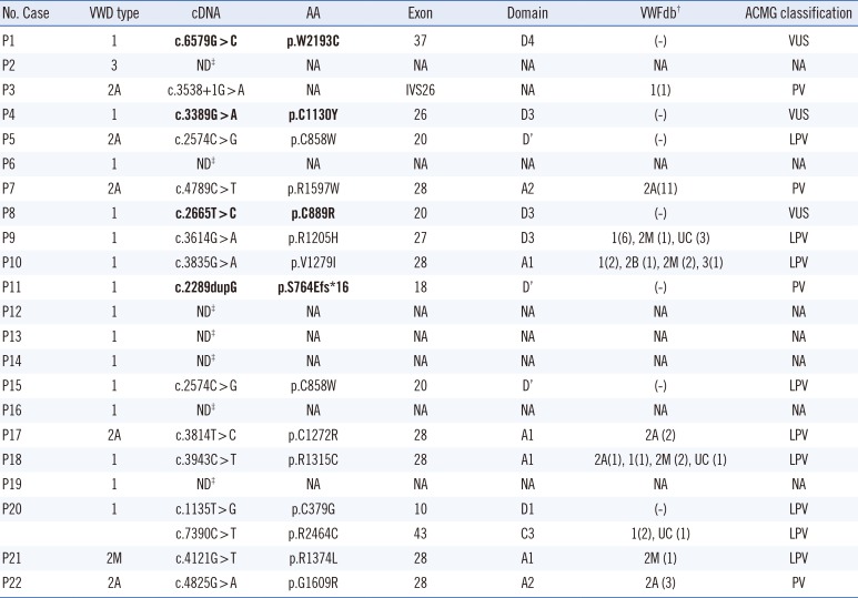
We identified putative disease-causing variants in 15 patients (68%): 14 patients with a single heterozygous variant and one patient with two heterozygous variants. The variants included 13 missense variants, one small insertion, and one splicing variant. All variants were unique, except for p.C858W, which was detected in two patients. MLPA analysis in seven patients without sequence variants by sequencing did not reveal any dosage variants. Four variants were novel: p.S764Efs*16, p.C889R, p.C1130Y, and p.W2193C.
Of the 15 patients with type 1 VWD, nine patients had VWF variants (variant detection rate 60%). Eight patients had a single heterozygous variant, and one patient (P20) had two known missense variants. The variants in type 1 VWD were distributed throughout the VWF region with the highest frequency in the D3 domain. The majority were missense variants (N=9; 90%), including three novel variants (p.C889R, p.C1130Y, and p.W2193C). One novel small insertion variant (c.2289dupG) in one patient (P11) was predicted to cause a reading frame shift and subsequently, premature termination of the VWF protein. All six patients with type 2 VWD were heterozygous for a known VWF variant (variant detection rate 100%): five missense variants and one splicing variant. No sequence or dosage variants were detected in one patient with type 3 VWD (P2).
In silico analyses were performed to predict the pathogenicity of three novel putative missense variants (p.C889R, p.C1130Y, and p.W2193C; Table 3). Both SIFT and PolyPhen algorithms showed all novel missense variants most likely cause damage. The Align-GVGD software predicted that all novel missense variants most likely interfere with protein function.

DISCUSSION

We investigated molecular defects of VWF in 22 unrelated Korean patients with VWD recruited from multiple institutions in Korea with a nationwide distribution. The patients consisted of 15 type 1 (68%), six type 2 (27%), and one type 3 (5%), which is largely compatible with the distribution in previous studies in western countries and Korea [1117]. Song, et al. [11] screened exons 12, 14, 16, 18, 19, 20, 24, 26, 27, 28, and 52 in Korean patients with VWD. They reported variant detection rates of 11%, 30%, and 0% for type 1, type 2, and type 3 VWD, respectively. We increased the variant detection rate by involving whole exons of VWF. Our results revealed the molecular genetic profile and genotype-phenotype correlations in Korean patients with VWD, albeit in a limited number of patients.
Except for one splicing variant and one small insertion variant, all remaining variants were missense variants. The variants in type 1 VWD were spread throughout the VWF gene, in line with previous reports that the variants responsible for type 1 VWD include missense (70%), splice (9%), transcription (8%), and other deleterious variants (13%) [218]. The VWF:Ag level was variable in our patients with type 1 VWD, ranging from 6 to 73 IU/dL (mean VWF:Ag 28.7 IU/dL). The VWF:Ag level in the patients without variants was higher than that in patients with variants (39.0 vs. 22.2 IU/dL), but not significantly (P=0.113). This is also in line with previous studies demonstrating that the genetic determinants in milder cases of VWD are more complex and might involve other factors outside the VWF locus [181920].
According to a French study and the VWFdb, variants in type 2A VWD are mostly located in A domains (~80%) [21]. Of the five patients with type 2A VWD in our study, three had a variant typically found in domains A1/A2 (P7, P17, and P22), while the variants in the remaining two patients, P3 and P5, were a splicing variant in the donor site of IVS26 and a missense variant in the D′ domain (p.C858W), respectively. p.C858W was the only recurrent variant in our study, detected in two patients, type 1 (P15) and type 2A (P5) VWD (Table 2). Of note, p.C858W was previously reported in a Korean patient with type 1 VWD [11]. Other missense variants affecting the C858 residue, p.C858S and p.C858F, are well known for their association with type 2N VWD (VWFdb). Type 2N VWD is inherited in an autosomal recessive manner and heterozygous carriers of type 2N VWD are often asymptomatic and most of laboratory findings could be normal except for the VWF:FVIIIB/VWF:Ag ratio [22]. Thus, the presence of p.C858W in Korean patients with VWD demonstrates a pleiotropic effect of missense variants involving the C858 residue. To our knowledge, the IVS26+1G>A variant in P3 is the first splicing variant in type 2 VWD. As other forms of premature termination variants, such as frameshift and nonsense in type 2, have been reported, it is plausible that deleterious variants due to a splicing defect could also cause type 2A VWD (VWFdb).
Type 2M accounts for a relatively small proportion of VWD patients, with a limited number of variants being reported to date. Most variants are missense or in-frame deletion variants in exon 28 (D3-A1) (VWFdb). We identified only one patient with type 2M, carrying a missense variant in the A1 domain. Finally, we identified one type 3 VWD patient (P2). While the variant detection rate for type 3 VWD has been approximately 90% [2], we did not detect any variants in this patient by sequencing or MLPA. Possible explanations include deep intronic variants affecting the expression of the VWF protein or large rearrangement variants without dosage aberrations. Furthermore, as the patient had no family history of bleeding diathesis, we could not rule out the possibility of acquired VWD.
All in silico analysis tools predicted that the three novel missense variants (p.C889R, p.C1130Y, and p.W2193C) detected in patients with type 1 VWD exert damaging effects on protein function (Table 3). p.C889R is the first missense variant reported to affect the C889 residue. According to recent studies, VWF contains free thiols, associated with nine cysteines, including C889. These free thiols may be important for lateral self-association of VWF via thiol-disulfide exchange after secretion, thus increasing VWF size and consequently, platelet binding ability [2324]. Other missense variants affecting the C1130 (C1130F/G/R) and W2193 (W2193R) residues have been reported in type 1/2 VWD [252627]. Missense variants at position C1130 in the D3-domain can induce intracellular retention and impaired multimerization, as well as increased protein clearance [2829]. A rare variant, p.N901K, was observed in patient P15 with type 1 VWD due to a known variant, p.C858W. p.N901K is registered in the Single Nucleotide Polymorphism Database (dbSNP; rs753545906) with a frequency of 0.00009 in East Asian populations and 0.00007 in combined populations. We regard p.N901K as a rare single nucleotide polymorphism (SNP) in East Asian populations rather than a variant in this patient. In addition, we could not rule out the possibility that p.C858W and p.N901K are on the same allele. A novel small insertion variant, c.2289dupG (p.S764Efs*16), was also identified in a patient with type 1 VWD (P11).
Recent studies have shown that exonic deletion or duplication variants contribute to a variety of coagulation disorders; the clinical utility of MLPA analyses for detecting these large dosage variants has also been demonstrated in type 3 VWD [7893031]. However, our 2nd-line MLPA analyses in sequence variant-negative patients using sequencing did not reveal any additional large dosage variants. This could be due to the small number of patients in this study, but might also indicate that MPLA has limited diagnostic utility in Korean patients with VWD.
In summary, this study represents the first comprehensive molecular genetic investigation involving the whole coding/junction sequences of VWF and MLPA analyses in Korean patients with VWD. The genotype-phenotype correlations were largely in line with previous findings; however, our data provide additional evidence on the pleiotropic effect of VWF variants. Furthermore, the results suggested limited diagnostic utility of MLPA analyses in Korean patients with VWD.

Acknowledgements

This study was supported by a grant from the Korea Hemophilia Foundation.

Notes

Authors' Disclosures of Potential Conflicts of Interest: No potential conflicts of interest relevant to this article were reported.

References

1. Rodeghiero F, Castaman G, Dini E. Epidemiological investigation of the prevalence of von Willebrand's disease. Blood. 1987; 69:454–459. PMID: 3492222.
2. Goodeve AC. The genetic basis of von Willebrand disease. Blood Rev. 2010; 24:123–134. PMID: 20409624.
3. Mancuso DJ, Tuley EA, Westfield LA, Worrall NK, Shelton-Inloes BB, Sorace JM, et al. Structure of the gene for human von Willebrand factor. J Biol Chem. 1989; 264:19514–19527. PMID: 2584182.
4. Zhou YF, Eng ET, Zhu J, Lu C, Walz T, Springer TA. Sequence and structure relationships within von Willebrand factor. Blood. 2012; 120:449–458. PMID: 22490677.
5. Sadler JE, Budde U, Eikenboom JC, Favaloro EJ, Hill FG, Holmberg L, et al. Update on the pathophysiology and classification of von Willebrand disease: a report of the Subcommittee on von Willebrand Factor. J Thromb Haemost. 2006; 4:2103–2114. PMID: 16889557.
6. Schneppenheim R. The pathophysiology of von Willebrand disease: therapeutic implications. Thromb Res. 2011; 128(S1):S3–S7. PMID: 22221847.
7. Baronciani L, Goodeve A, Peyvandi F. Molecular diagnosis of von Willebrand disease. Haemophilia. 2017; 23:188–197. PMID: 28220580.
8. Kim HJ, Chung HS, Kim SK, Yoo KY, Jung SY, Park IA, et al. Mutation spectrum and inhibitor risk in 100 Korean patients with severe haemophilia A. Haemophilia. 2012; 18:1008–1013. PMID: 22741565.
9. Yadegari H, Driesen J, Hass M, Budde U, Pavlova A, Oldenburg J. Large deletions identified in patients with von Willebrand disease using multiple ligation-dependent probe amplification. J Thromb Haemost. 2011; 9:1083–1086. PMID: 21410641.
10. Wang QY, Song J, Gibbs RA, Boerwinkle E, Dong JF, Yu FL. Characterizing polymorphisms and allelic diversity of von Willebrand factor gene in the 1000 Genomes. J Thromb Haemost. 2013; 11:261–269. PMID: 23216583.
11. Song J, Choi JR, Song KS. Investigation of von Willebrand factor gene mutations in Korean von Willebrand disease patients. Korean J Lab Med. 2007; 27:169–176. PMID: 18094571.
12. Rodeghiero F, Castaman G, Tosetto A, Batlle J, Baudo F, Cappelletti A, et al. The discriminant power of bleeding history for the diagnosis of type 1 von Willebrand disease: an international, multicenter study. J Thromb Haemost. 2005; 3:2619–2626. PMID: 16359502.
13. Corrales I, Ramírez L, Altisent C, Parra R, Vidal F. Rapid molecular diagnosis of von Willebrand disease by direct sequencing. Detection of 12 novel putative mutations in VWF gene. Thromb Haemost. 2009; 101:570–576. PMID: 19277422.
14. den Dunnen JT, Dalgleish R, Maglott DR, Hart RK, Greenblatt MS, McGowan-Jordan J, et al. HGVS Recommendations for the Description of Sequence Variants: 2016 Update. Hum Mutat. 2016; 37:564–569. PMID: 26931183.
15. Cabrera N, Casaña P, Cid AR, Moret A, Moreno M, Palomo A, et al. First application of MLPA method in severe von Willebrand disease. Confirmation of a new large VWF gene deletion and identification of heterozygous carriers. Br J Haematol. 2011; 152:240–242. PMID: 20955405.
16. Richards S, Aziz N, Bale S, Bick D, Das S, Gastier-Foster J, et al. Standards and guidelines for the interpretation of sequence variants: a joint consensus recommendation of the American College of Medical Genetics and Genomics and the Association for Molecular Pathology. Genet Med. 2015; 17:405–424. PMID: 25741868.
17. Mikhail S, Aldin ES, Streiff M, Zeidan A. An update on type 2B von Willebrand disease. Expert Rev Hematol. 2014; 7:217–231. PMID: 24521271.
18. Yadegari H, Driesen J, Pavlova A, Biswas A, Hertfelder HJ, Oldenburg J. Mutation distribution in the von Willebrand factor gene related to the different von Willebrand disease (VWD) types in a cohort of VWD patients. Thromb Haemost. 2012; 108:662–671. PMID: 22871923.
19. Eikenboom J, Van Marion V, Putter H, Goodeve A, Rodeghiero F, Castaman G, et al. Linkage analysis in families diagnosed with type 1 von Willebrand disease in the European study, molecular and clinical markers for the diagnosis and management of type 1 VWD. J Thromb Haemost. 2006; 4:774–782. PMID: 16634746.
20. James PD, Paterson AD, Notley C, Cameron C, Hegadorn C, Tinlin S, et al. Genetic linkage and association analysis in type 1 von Willebrand disease: results from the Canadian type 1 VWD study. J Thromb Haemost. 2006; 4:783–792. PMID: 16634747.
21. Meyer D, Fressinaud E, Gaucher C, Lavergne JM, Hilbert L, Ribba AS, et al. Gene defects in 150 unrelated French cases with type 2 von Willebrand disease: from the patient to the gene. INSERM Network on Molecular Abnormalities in von Willebrand disease. Thromb Haemost. 1997; 78:451–456. PMID: 9198195.
22. Casonato A, Pontara E, Sartorello F, Cattini MG, Perutelli P, Bertomoro A, et al. Identifying carriers of type 2N von Willebrand disease: procedures and significance. Clin Appl Thromb Hemost. 2007; 13:194–200. PMID: 17456630.
23. Choi H, Aboulfatova K, Pownall HJ, Cook R, Dong JF. Shear-induced disulfide bond formation regulates adhesion activity of von Willebrand factor. J Biol Chem. 2007; 282:35604–35611. PMID: 17925407.
24. Shapiro SE, Nowak AA, Wooding C, Birdsey G, Laffan MA, McKinnon TA. The von Willebrand factor predicted unpaired cysteines are essential for secretion. J Thromb Haemost. 2014; 12:246–254. PMID: 24283831.
25. Eikenboom JC, Matsushita T, Reitsma PH, Tuley EA, Castaman G, Briët E, et al. Dominant type 1 von Willebrand disease caused by mutated cysteine residues in the D3 domain of von Willebrand factor. Blood. 1996; 88:2433–2441. PMID: 8839833.
26. Goodeve A, Eikenboom J, Castaman G, Rodeghiero F, Federici AB, Batlle J, et al. Phenotype and genotype of a cohort of families historically diagnosed with type 1 von Willebrand disease in the European study, Molecular and Clinical Markers for the Diagnosis and Management of Type 1 von Willebrand disease (MCMDM-1VWD). Blood. 2007; 109:112–121. PMID: 16985174.
27. Eikenboom JC, Castaman G, Vos HL, Bertina RM, Rodeghiero F. Characterization of the genetic defects in recessive type 1 and type 3 von Willebrand disease patients of Italian origin. Thromb Haemost. 1998; 79:709–717. PMID: 9569178.
28. Tjernberg P, Vos HL, Castaman G, Bertina RM, Eikenboom JC. Dimerization and multimerization defects of von Willebrand factor due to mutated cysteine residues. J Thromb Haemost. 2004; 2:257–265. PMID: 14995987.
29. Casari C, Lenting PJ, Wohner N, Christophe OD, Denis CV. Clearance of von Willebrand factor. J Thromb Haemost. 2013; 11(S1):202–211. PMID: 23809124.
30. Choung HS, Kim HJ, Gwak GY, Kim SH, Kim DK. Inherited protein S deficiency as a result of a large duplication mutation of the PROS1 gene detected by multiplex ligation-dependent probe amplification. J Thromb Haemost. 2008; 6:1430–1432. PMID: 18489710.
31. Kim HJ, Kim DK, Yoo KY, You CW, Yoo JH, Lee KO, et al. Heterogeneous lengths of copy number mutations in human coagulopathy revealed by genome-wide high-density SNP array. Haematologica. 2012; 97:304–309. PMID: 21993689.
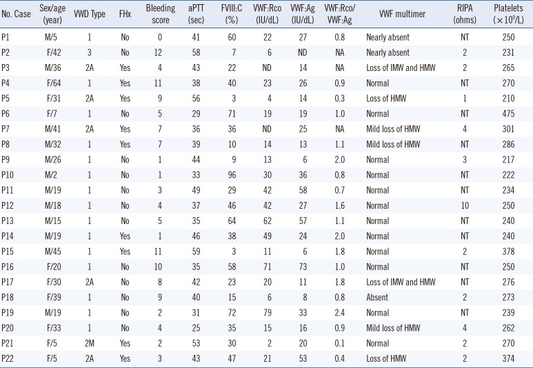
Table 1

Clinical and laboratory characteristics of 22 Korean patients with VWD

alm-39-545-i001
No. Case Sex/age (year) VWD Type FHx Bleeding score aPTT (sec) FVIII:C (%) VWF:Rco (IU/dL) VWF:Ag (IU/dL) VWF:Rco/ VWF:Ag VWF multimer RIPA (ohms) Platelets (×109/L)
P1 M/5 1 No 0 41 60 22 27 0.8 Nearly absent NT 250
P2 F/42 3 No 12 58 7 6 ND NA Nearly absent 2 231
P3 M/36 2A Yes 4 43 22 ND 14 NA Loss of IMW and HMW 2 265
P4 F/64 1 Yes 11 38 40 23 26 0.9 Normal NT 270
P5 F/31 2A Yes 9 56 3 4 14 0.3 Loss of HMW 1 210
P6 F/7 1 No 5 29 71 19 19 1 Normal NT 475
P7 M/41 2A Yes 7 36 36 ND 25 NA Mild loss of HMW 4 301
P8 M/32 1 Yes 7 39 10 14 13 1.1 Mild loss of HMW NT 286
P9 M/26 1 No 1 44 9 13 6 2 Normal 3 217
P10 M/2 1 No 1 33 96 30 36 0.8 Normal NT 222
P11 M/19 1 No 3 49 29 42 58 0.7 Normal NT 234
P12 M/18 1 No 4 37 46 42 27 1.6 Normal 10 250
P13 M/15 1 No 5 35 64 62 57 1.1 Normal NT 240
P14 M/19 1 Yes 1 46 38 49 24 2 Normal NT 240
P15 M/45 1 Yes 11 59 3 11 6 1.8 Normal 2 378
P16 F/20 1 No 10 35 58 71 73 1 Normal NT 250
P17 F/30 2A No 8 42 23 20 11 1.8 Loss of IMW and HMW NT 276
P18 F/39 1 No 9 40 15 6 8 0.8 Absent 2 273
P19 M/19 1 No 2 31 72 79 33 2.4 Normal NT 239
P20 F/33 1 No 4 25 35 15 16 0.9 Mild loss of HMW 4 262
P21 F/5 2M Yes 2 53 30 2 20 0.1 Normal 2 270
P22 F/5 2A Yes 3 43 47 21 53 0.4 Loss of HMW 2 374

Abbreviations: P, patient; VWD, von Willebrand disease; FHx, family history; FVIII:C, FVIII coagulant activity; VWF, von Willebrand factor; VWF:Rco, VWF ristocetin cofactor activity; RIPA, ristocetin-induced platelet aggregation; AA, amino acid; ND, not detected; NT, not tested; NA, not applicable; HMW, high molecular weight multimers; IMW, intermediate molecular weight multimers.

Table 2

VWF gene variants identified in 22 Korean patients with VWD

alm-39-545-i002
No. Case VWD type cDNA AA Exon Domain VWFdb ACMG classification
P1 1 c.6579G > C p.W2193C 37 D4 (-) VUS
P2 3 ND NA NA NA NA NA
P3 2A c.3538+1G > A NA IVS26 NA 1(1) PV
P4 1 c.3389G > A p.C1130Y 26 D3 (-) VUS
P5 2A c.2574C > G p.C858W 20 D′ (-) LPV
P6 1 ND NA NA NA NA NA
P7 2A c.4789C > T p.R1597W 28 A2 2A(11) PV
P8 1 c.2665T > C p.C889R 20 D3 (-) VUS
P9 1 c.3614G > A p.R1205H 27 D3 1(6), 2M (1), UC (3) LPV
P10 1 c.3835G > A p.V1279I 28 A1 1(2), 2B (1), 2M (2), 3(1) LPV
P11 1 c.2289dupG p.S764Efs*16 18 D′ (-) PV
P12 1 ND NA NA NA NA NA
P13 1 ND NA NA NA NA NA
P14 1 ND NA NA NA NA NA
P15 1 c.2574C > G p.C858W 20 D′ (-) LPV
P16 1 ND NA NA NA NA NA
P17 2A c.3814T > C p.C1272R 28 A1 2A (2) LPV
P18 1 c.3943C > T p.R1315C 28 A1 2A(1), 1(1), 2M (2), UC (1) LPV
P19 1 ND NA NA NA NA NA
P20 1 c.1135T > G p.C379G 10 D1 (-) LPV
c.7390C > T p.R2464C 43 C3 1(2), UC (1) LPV
P21 2M c.4121G > T p.R1374L 28 A1 2M (1) LPV
P22 2A c.4825G > A p.G1609R 28 A2 2A (3) PV

VWD type registered in VWFdb. The number of reported patient(s) is presented in parentheses; Multiplex-ligation dependent probe amplification analysis was performed in the seven patients without identifiable sequence variants by sequencing; no dosage variants were detected.

Novel variants are indicated in bold.

Abbreviations: P, patient; VWD, von Willebrand disease; VWF, von Willebrand factor; AA, amino acid; ND, not detected; NA, not applicable; UC, unclassified; ACMG, American College of Medical Genetics standards and guidelines; VUS, variant of uncertain significance; PV, pathogenic variant; LPV, likely pathogenic variant.

Table 3

In silico analysis and allele frequencies of three novel missense VUS of the VWF gene

alm-39-545-i003
cDNA change AA change Exon Domain VWD type SIFT PolyPhen AGVGD dbSNP ESP ExAc KRGDB
c.2665T>C p.C889R 20 D3 1 Damaging Probably damaging Most likely interferes with function (-) (-) (-) (-)
c.3389G>A p.C1130Y 26 D3 1 Damaging Probably damaging Most likely interferes with function (-) (-) (-) (-)
c.6579G>C p.W2193C 37 D4 1 Damaging Probably damaging Most likely interferes with function (-) (-) (-) (-)

Abbreviations: VUS, variant of uncertain significance; AA, amino acid; VWD, von Willebrand disease; SIFT, Sorting Intolerant From Tolerant; PolyPhen, Polymorphism Phenotyping; AGVGD, Align GVGD; ESP, Exome Sequencing Project; ExAC, Exome Aggregation Consortium; KRGDB, Korean Reference Genome Database.

TOOLS
Similar articles