This article has been
cited by other articles in ScienceCentral.
Dear Editor:
Erythrokeratodermia variabilis (EKV) is a rare, heterogenous, autosomal dominant genodermatosis1. It is characterized by two distinct features: transient erythematous patches of various size and morphology and fixed hyperkeratotic plaques
12. Here, we describe a rare case of EKV of a Korean male patient.
A 39-year-old male was presented with pruritic erythematous patches and keratotic plaques on the whole body, which had persisted since birth. The keratotic lesions of the knee, axilla, buttock, palm and sole were consistently maintained on the same location, but erythematous patches transiently showed depending on stress and weather. The elder brother had a history of similar skin lesions since birth and his father also had palmoplantar keratoderma.
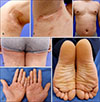
Physical examinations revealed symmetrical keratotic plaques on axilla, inguinal and buttock area with mild erythematous patches on the trunk (
Fig. 1A~D). It was accompanied by symmetric palmoplantar keratoderma (
Fig. 1E, F).
A histopathological examination of the axilla and flank demonstrated hyperkeratosis and moderate acanthosis in the epidermis with mild perivascular lymphocyte infiltrate in the upper dermis (
Fig. 2). After the diagnosis of EKV, skin lesions progressively improved with oral acitretin in 4 weeks.
First described in 1907, EKV which is the disorder of keratinization, have rarely been reported. It has been associated with mutation in GJB3 and GJB4 encoding Connexin 31 and Connexin 30.3
12. Connexins are the protein constituents of gap junctions in the stratum granulosum of the epidermis which provide a mechanism for synchronized cellular response facilitating metabolic and electronic functions of the cell. The Connexin 31 and 30.3 are involved in keratinocyte differentiation. Based on the genetic factor, EKV present itself usually within the first years of life as in our patient but rarely arise later in childhood
3.
Clinically, it is characterized by the existence of erythematous patches with fixed hyperkeratotic plaques
1245. Transient, bizarre, figurate, bright red patches develop and changes frequently. In contrast, fixed, sharply demarcated geographic hyperkeratotic plaques mainly located symmetrically on the extensor surface of the limbs, axilla, inguinal and buttock appear. Approximately 50% of EKV patients have palmoplantar keratoderma.
The histological features of EKV are non-specific; hyperkeratosis, moderate papillomatosis and acanthosis with mild perivascular inflammatory infiltration in the dermis
1. The diagnosis therefore depends on the clinical features and family history. EKV generally responds well to retinoids which restores the deficient keratinosome. Emollients, topical retinoid acid, intralesional steroids have also been used
3. The clinical differential diagnosis of EKV includes plaque psoriasis, CHILD syndrome, progressive symmetric erythrokeratoderma, and familial pityriasis rubra pilaris
134. The main feature distinguishing EKV from others is the transient lesions and possibly lack of facial lesions in most cases.
EKV is an extremely rare disease and so far, only two cases have been reported in Korea. However, one case was reported in child, the other from a foreigner
45. This study is the first case of EKV in a Korean adult. It's also worth noting that the patient has a familial history and was treated successfully with retinoid.
Therefore, dermatologists should keep in mind that EKV occurs rarely in Koreans with a favorable outcome using acitretin.
Figures and Tables
Fig. 1
(A~D) Well demarcated, symmetric erythematous keratotic plaques on both axilla, trunk and buttock, and thigh. (E, F) Thick keratoderma on palm and soles are shown. We received the patient's consent form about publishing all photographic material.

Fig. 2
(A) Marked hyperkeratosis and mild acanthosis with mild inflammatory infiltration in upper dermis. Mild increase in number of eccrine glands is shown (left axilla: H&E, ×40). (B) Close up view, compact hyperkeratosis, normal-appearing granular layer and irregular acanthosis in the epidermis with mild perivascular lymphocyte infiltration in the papillary to upper dermis (left axilla: H&E, ×200).


 PDF
PDF ePub
ePub Citation
 Print
Print


 XML Download