

 PDF
PDF ePub
ePub Citation
 Print
Print


INTRODUCTION
Celiac disease (CD) is a systemic, immune mediated disorder that occurs in individuals with a genetic predisposition in response to ingestion of dietary gluten [1]. CD is a common but markedly under-diagnosed condition. It may lead to serious long-term complications if untreated [2]. It has a wide range of clinical manifestations, including the “classic” presentation of malabsorption with diarrhea and abdominal cramps and atypical symptoms such as headache, joint pain, and seizures. Patients may present CD asymptomatically due to a comorbid condition or family history of CD [23]. Compared to the general public, first-degree relatives (FDRs) of patients with CD have higher risk (up to 20%) for CD development [456].
Several studies have shown that obese or overweight children diagnosed with CD are becoming more common. The prevalence of excess weight in CD is 8.8% to 20.8% for overweight and up to 6% for obesity [78910111213141516]. In Italian studies, the coexistence of CD and overweight or obesity has been reported in 11% to 12% of patients diagnosed with CD [1013]. Despite increased calorie, fat, and sugar content of gluten free food items, 75% of patients with high body mass index (BMI) at diagnosis tend to lose weight on gluten-free diet (GFD) [11121314].
Here we describe 2 siblings with excess weight and attenuated gastrointestinal and atypical symptoms are diagnosed with CD in the absence of a known family history of CD. After these children's diagnosis, CD was also investigated in their parents. It was identified in their father affected by overweight.
CASE REPORT
Case 1
The subject was born at 41 weeks and 3 days of gestation by normal delivery with a normal birth weight of 3,700 g. The newborn received mixed lactation for the first 4 months and began weaning by that time. At 2 years and 3 months of age, she was examined in our out-patient clinic for a general endocrinological assessment concerning unilateral breast enlargement that was present at birth but regressed over the following few weeks. At clinical examination, she showed good general clinical conditions and growth parameters, including height of 90 cm (75th percentile) [17], weight of 17.2 kg (>97th percentile) [17], and BMI of 21.2 kg/m2 (95th–97th percentile) [17]. According to Marshall and Tanner [18] Pubertal Scale, she showed breast stage 2, pubic hair stage 1, and negative axillary hair.
Hematological and biochemical tests were then performed. Results were compatible with premature thelarche (no increase in the level of any sexual hormone, correct bone age according to chronological age; prepubertal uterus and ovarian aspect volume <2 mL at pelvic ultrasound examination). At 3 years and 2 months of age, her thelarche spontaneously regressed.
During follow-up, persistent weight excess was noted. Diet history revealed excessive energy consumption with an increased intake of lipids, carbohydrates, and simple sugars. At 9 years and 6 months of age, the patient presented moderate epigastric pain. Her auxological examination showed height of 138 cm (75th–90th percentile) [17], weight of 45.5 kg (90th–97th percentile) [17], and BMI of 23.9 kg/m2 (95th percentile) [17]. A gastroesophageal reflux disease (GERD) was suspected. Thus, she was sent to a gastroenterologist for assessing GERD. Although treatment therapy (magaldrate, pantoprazole, and omeprazole) was started, no results were obtained. She then developed constipation due to ongoing symptoms. We decided to investigate her further, suspecting a CD.
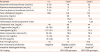
Her parameters according to screening of CD were positive for autoantibodies anti-tissue transglutaminase (TTG) immunoglobulin A (IgA) 13 U/mL (n.v.<10 U/mL). Her anti-endomysium (EMA) results were slightly positive. Polymerase chain reaction revealed a DR3-DQ2 genotype. Subsequently, she underwent an esophagogastroduodenoscopy (EGD) that elicited a clinical picture compatible with CD (Marsh Score System grade 3b). No significant biochemical alterations were recorded (Table 1).
Table 1
Biochemical parameters, intestinal histological findings, and genetic profile of cases 1 and 2
Physiologically balanced GFD plus lifestyle modifications including structured physical activity were started. TTG-IgA was normalized with clinical improvement for epigastric pain and constipation over the following 4 months. The girl maintained excess weight at 18 months of follow-up (BMI, 24.3 kg/m2 equivalent to 90th percentile [17]) at the age of 11 years.
Case 2
The brother of case 1 came to our clinic at 6 years and 9 months of age for endocrinological evaluation concerning obesity. Assessment of dietary intake revealed an excessive energy intake with an increased consumption of lipids, carbohydrates, and simple sugars. At clinical examination, he showed BMI of 23.19 kg/m2 (>97th percentile) [17] with height of 126.3 cm (75th–90th percentile) [17] and weight of 37 kg (>97th percentile) [17]. He presented with sporadic light headache.
He was born at 41 weeks and 4 days of gestation by normal delivery with a normal birth weight of 3,650 g. The newborn received exclusive breastfeeding for the first 6 months and began weaning by that time. He did not receive any medication nor showed any allergy.
Considering his sister's clinical diagnosis, we decided to perform the same assessment. Results revealed TTG-IgA value higher than 100 U/mL (n.v.<10 U/mL) with positive EMA. Human leukocyte antigen-DQ typing was then performed, showing DR3-DQ2 genotype. EGD showed the same clinical picture as his sister: CD grade 3b Marsh Score System.
The patient started GFD without normalization of autoantibodies due to poor dietary adherence. The boy continued to maintain excess weight at 24 months of follow-up (BMI, 23.59 kg/m2, equivalent to >97th percentile [17]) at the age of 8 years and 9 months despite lifestyle modification treatment, including physiological balanced diet and structured physical activity. Biochemical parameters, intestinal histological findings, and genetic profile of the 2 cases are shown in Table 1.
Family screening was also performed to assess if there was any correlation with their FDRs. Positivity for TTG-IgA and EMA was found in the father affected by overweight who was otherwise asymptomatic. EGD was then performed for the father and the result was positive for CD. Parents gave their consent after being informed about the nature of the report.
DISCUSSION
Several studies on adults and children with CD have reported that obesity/overweight at disease onset is not unusual [78910111213141516]. In the report by Reilly et al. [11], 19% of patients had a high BMI at CD diagnosis (12.6% overweight and 6% obese) while 74.5% had a normal BMI.
Overweight/obesity has been reported to be more frequent in newly diagnosed CD patients if abdominal pain is present [811]. However, previous studies [813] also suggest that identifying CD patients based on screening tests, not symptoms, may increase the probability of finding overweight or obese subjects at CD diagnosis [14].
We described a familial condition in which the diagnosis of CD in one of family members allowed us to diagnose the same disease in 2 other family members who were otherwise asymptomatic with excess weight. Clinical review did not uncover any biochemical alterations such as hypertransaminasemia, low iron level, or anemia [1920].
The pathogenesis and clinical implications of the coexistence of CD and overweight/obesity remain unclear. Possible reasons of such findings include early serological diagnosis, predominant expression of the disease outside the GI, and distal small bowel compensatory absorption [10212223]. In the present study, considering family history of obesity, genetics as well as unbalanced diet and incorrect lifestyle habits must also be considered.
Several studies have also reported a correlation between GFD and normalization of BMI in patients who are underweight and overweight at diagnosis [14]. Thus, improved absorption could play a significant role. Furthermore, weight gain persisted in our subjects over follow up due to persistent unhealthy lifestyles, including energy dense and nutrient poor diets, reduced physical activity, poor adherence to GFD particularly by the boy who kept consuming foods containing gluten (bread, pizza, and pasta). Additionally, nutritional imbalance and hypercaloric content of commercial gluten-free food items can contribute to excess weight.
In conclusion, the presentation of patients with CD has changed. Nowadays, patients are more likely to have normal weight, overweight, or obesity than underweight. While patients with overweight and obesity commonly have symptoms such as abdominal pain, reflux, headache, and constipation due to lifestyle factors, CD should also be considered in patients with or without a family history of CD. Careful nutritional status assessment and follow-up monitoring after the diagnosis of CD are mandatory, especially in subjects who are already overweight at the presentation of this disease.
 XML Download