

 PDF
PDF ePub
ePub Citation
 Print
Print


Dear Editor:
Atopic dermatitis (AD) is a chronic inflammatory skin disease characterized by altered skin barrier function and immune dysregulation. Previously, we demonstrated that AD may be associated with defective basement membrane (BM) and low regenerative capacity1.
The regenerative capacity of the skin is related to the ability of the stem cells to self-renew and appropriately differentiate. To localize the interfollicular epidermal stem cells (IFESCs), p63 expression has conventionally been used, which has also been expressed in the upper layers of the epidermis, suggesting that it may not be a specific stem cell marker. Recently, we reported that combined staining of p63 and histone deacetylase 1 (HDAC1) can be a new method to distinguish IFESCs2. We showed that a p63-positive/HDAC1-negative expression pattern may be a better indicator of stem cells than p63-positive pattern alone. Meanwhile, sirtuin 1 (SIRT1) is a nicotinamide adenine dinucleotide-dependent protein with multiple roles in the process of aging and disease development3. It promotes the regeneration of tissues in the process of age-related degeneration or wound healing45. In the present study, we aimed to elucidate the expression of IFESC marker and SIRT1 in AD and discuss the regenerative capacity of AD. Tissue samples from 15 AD patients (11 males, 4 females; mean age=17.33±9.84 years) and 11 normal volunteers (7 males, 4 females; mean age=15.18±7.37 years) were collected. The mean eczema area and severity index score of AD patients was 12.34±9.24; all samples were harvested from the chronic eczematous skin lesions. This study was approved by the Institutional Review Board of Seoul National University Bundang Hospital (B-1002/094-301) and Keimyung University Hospital (DSMC 2014-11-039-007).
Immunohistochemical staining of p63 with HDAC1 and SIRT1 was performed. Furthermore, type IV collagen staining was performed to study the status of BM, a niche for IFESCs. After staining with DAPI, images were obtained by Confocal Laser Scanning Microscope (#LSM710; Carl Zeiss, Jena, Germany) and analyzed using ZEN 2011 microscope software (Carl Zeiss). To evaluate the epidermal stem cells, six fields with the same size were randomly chosen in each sample, at 200× magnification. Then, the percentages of epidermal cells with a p63-positive/HDAC1-negative expression pattern were calculated. To evaluate the expression of SIRT1 and type IV collagen, six circular areas were also selected, and the average area stained above a certain intensity was determined using an image analysis program (MetaMorphâ Microscopy Automation & Image Analysis Software; Molecular Devices, Sunnyvale, CA, USA). The Kruskal-Wallis test using IBM SPSS Statistics ver. 20.0 (IBM Corp., Armonk, NY, USA) was used to compare the expression of each marker between the two groups.
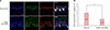
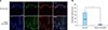
In normal skin, HDAC1 showed strong localization in the outer, differentiating cell layers, whereas p63 was most strongly expressed in the basal layer (Fig. 1A). In a merged image, there was a subpopulation showing a p63-positive/HDAC1-negative expression pattern in the basal layer (Fig. 1A). In AD skin, HDAC1 expression was different between AD skin and normal skin. HDAC1 was strongly expressed in some cells of the basal layer, and the cells with completely negative HDAC1 were rarely observed (Fig. 1A). The ratio of p63-positive to HDAC1-negative cells within the epidermis was significantly lower in AD skin compared with normal skin (Fig. 1B, p<0.01). Meanwhile, SIRT1 was clearly expressed in the basal and suprabasal layers (Fig. 2A) of normal skin. In contrast, it was not expressed in the epidermis in almost all AD cases. The calculated stained area for SIRT1 was significantly smaller in AD skin compared with normal skin (Fig. 2B, p<0.01). The expression of type IV collagen was significantly decreased in AD skin compared with normal skin, which is similar with our previous report1. The calculated stained area was significantly smaller in AD skin compared with normal skin (15.82±3.14 in AD, 25.15±5.54 in normal, p<0.05).
In the present study, our data suggests that the epidermal regenerative capacity is decreased in AD based on the decreased number of IFESCs in atopic skin. This might be related with the disruption of BM. The result of this study showed that type IV collagen, a main component of BM, was significantly decreased, which is consistent with our previous results1. BM is a niche for IFESCs: it is a specific microenvironment that regulates the activities and fate of the residing stem cells6. Interleukin (IL)-13, one of the cytokines known for its involvement in the development of AD, may possibly play an important role in these defects via the stimulation of keratinocytes to produce matrix metalloproteinase (MMP)-97, degrading the type IV collagen, which is a major component of BM. Furthermore, in a previous study, IL-17—recently revealed to play a role in the pathogenesis of AD—significantly increased the secretion of MMP-9 in the peripheral blood mononuclear cells and polymorphonuclear cells8. Recently, BM has been reported to be involved in skin morphogenesis, homeostasis, and differentiation by controlling the dermal-epidermal junction (DEJ) interactions9. In this previous study, the skin equivalent model lacked heparan sulfate at DEJ, showing abnormal expression of filaggrin. Decreased SIRT1 expression in AD can be understood in the same context. As mentioned earlier, SIRT1 expression is known to be related to the regenerative potential of the skin and retina45. In line with this, Ming et al.10, through a mouse model, showed that SIRT1 plays a critical role in skin barrier maintenance. In that study, the SIRT1 protein level was shown to be down-regulated in human AD and non-AD, which is consistent with our results. In summary, regenerative potential and barrier dysfunction are closely related with one another in AD. Further investigation, however, regarding the role of BM and SIRT1 in AD and non-AD is necessary.
In conclusion, our data showed that the number of IFECSs and expression of SIRT1 are decreased in AD. These findings suggest that the regenerative potential of the skin is decreased in AD. Chronic and recurrent course of AD may be explained by reduced regenerative potential. Furthermore, it affords novel therapeutic target for AD that focuses on the restoration of regenerative capacity.
 XML Download