

 PDF
PDF ePub
ePub Citation
 Print
Print


INTRODUCTION
Alopecia areata (AA) is a chronic inflammatory disease characterized by well-circumscribed oval or round bald patches1. Although the pathogenesis of AA has yet to be elucidated, it is considered to be an autoimmune disease caused by autoreactive CD8+ T-cells with hair follicle immune privilege collapse12. Previous studies have reported an association between AA and various autoimmune diseases, such as autoimmune thyroiditis, lupus erythematosus, and rheumatoid arthritis (RA)345. Moreover, there is accumulating evidence that AA is a tissue-specific autoimmune disease67.
Cold-inducible RNA-binding protein (CIRP) belongs to the family of cold-shock proteins that respond to cold stresses. CIRP is a 172-amino-acid molecule that consists of one amino-terminal consensus sequence RNA-binding domain, and is known to be constitutively but weakly expressed in various tissues891011. Under stress conditions, such as hypothermia, hypoxia, and exposure to ultraviolet irradiation, CIRP is upregulated and migrates from the nucleus to the cytoplasm, where it promotes the translation of target messenger RNAs121314. Recent studies have focused on the emerging role of CIRP, as a damage-associated molecular pattern molecule (DAMP), in triggering the inflammatory response815. Qiang et al.8 reported that CIRP induces inflammation by promoting secretion of tumor necrosis factor alpha (TNF-α) and the high-mobility group box 1 (HMGB1) protein in sepsis and hemorrhagic shock. Fein and Calalang-Colucci15 demonstrated that CIRP increases caspase-1 and interleukin (IL)-1β, and induces pyroptosis in mouse lung vascular endothelial cells. Recently, the role of these DAMPs and cytokines was highlighted in the pathogenesis of AA161718. In addition, the serum and synovial levels of CIRP were increased, and correlated well with disease activity in patients with RA, an autoimmune disease19. Therefore, it could be hypothesized that CIRP plays a significant role in the pathogenesis of AA.
However, the involvement of CIRP in AA has not yet been elucidated. The aim of this study was to compare CIRP levels in patients with AA and healthy controls, and to investigate the clinical significance of serum CIRP levels in AA.
MATERIALS AND METHODS
Patients
Blood samples were collected from 68 patients with AA and 20 healthy controls. The control group had no history of AA. To assess the correlation between clinical characteristics and CIRP levels, a medical chart review was conducted to collect clinical data such as patient age, disease type, treatment history, disease duration, and disease activity at the time of blood sampling. The disease activity of AA patients was determined based on hair pull test results. To perform this test, the examiner grasped 50 to 60 hairs and then pulled firmly on the bundle from the base of the hairs using slow traction. When more than 10% of the hairs were pulled away from the scalp, the hair pull test was considered positive. To analyze the serum level of CIRP according to disease activity, the hair pull test was performed at the same time as the serum samples were taken. Patients with a history of systemic disease known to increase CIRP levels, such as autoimmune disease, infectious disease, and malignancy, were excluded from the study. Informed consent was obtained from each participant. This study received ethical approval from the Institutional Review Board at Chungnam National University, Daejeon, Korea (CNUH-IRB-2018-07-065).
Sample preparation
Venous blood samples were obtained under sterile conditions from all subjects and centrifuged for 15 minutes at approximately 1,000 g (3,000 rpm) to separate the serum. Serum samples were stored at −20℃ or −80℃ before assaying.
Immunohistochemistry
Tissue samples were taken from the patients of AA and healthy controls. Tissue samples were fixed with 10% formaldehyde, embedded in paraffin, and cut into 4-µmthick sections. The sections were deparaffinized in xylene and then rehydrated using an alcohol series. The primary antibody was diluted at a ratio of 1:100 (CIRP antibody; Abcam, Cambridge, UK) and incubated overnight at 4℃. The sections were then incubated with a secondary antibody at room temperature for 30 minutes. Finally, the sections were incubated with a diaminobenzidine tetrachloride solution at room temperature for 30 seconds and counterstained with Mayer's hematoxylin.
Enzyme-linked immunosorbent assay analysis of serum CIRP levels
CIRP levels were measured using an enzyme-linked immunosorbent assay kit (Cusabio Biotech, Wuhan, China). Measurements were performed according to the manufacturer's instructions.
Statistical analysis
All statistical analyses were performed using IBM SPSS Statistics for Windows software (ver. 20.0; IBM Corp., Armonk, NY, USA). The Mann–Whitney U test was used to compare serum CIRP levels between the AA and healthy control groups, and to analyze the correlation with various clinical characteristics, such as age, disease duration, hair pull test results, and treatment history. The Kruskal-Wallis test was used to examine the relationship between the AA types and serum CIRP levels. A p-value <0.05 was considered to indicate statistical significance.
RESULTS
Characteristics of patients
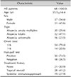
In total, 68 AA patients and 20 healthy individuals were enrolled in this study. The mean age of the patients was 35.5 years (range, 11~71 years). The patients were treated with intralesional corticosteroid, topical glucocorticoids, systemic immunosuppressant, or ultraviolet phototherapy. Twelve patients were diagnosed with AA at the time of blood collection. The demographic data and characteristics of the enrolled AA patients are illustrated in Table 1.
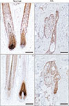
Expression of CIRP in AA scalp specimens
The expression of CIRP in the scalp lesions of patients with AA was compared to that in normal scalp skin. CIRP was mainly expressed in the nuclei of hair follicle cells in both normal hair and AA scalp specimens. There was no significant difference in CIRP expression between normal hair and AA scalp specimens (Fig. 1).
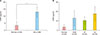
Elevated serum CIRP levels in AA patients
Serum CIRP levels were measured using an enzyme-linked immunosorbent assay kit. The serum CIRP level in AA patients was significantly higher than that in healthy controls (AA patients, 13.58±3.52 pg/ml; controls, 3.40±2.79 pg/ml; p=0.029) (Fig. 2A). Additionally, the differences in serum CIRP level according to type of AA (AA multiplex, alopecia totalis, and alopecia universalis) were examined and no significant difference was found (p=0.707) (Fig. 2B).
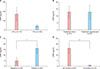
Correlations between serum CIRP levels and clinical characteristics
The relationship between serum CIRP levels and clinical markers such as age, treatment history, disease activity, and duration was analyzed in patients with AA. The mean serum CIRP level was elevated in patients younger than 18 years of age, but not significantly (33.67±14.54 pg/ml vs. 8.09±2.22 pg/ml, p=0.275) (Fig. 3A). Moreover, there was no significant difference in serum CIRP level between the treatment-naïve and treatment-experienced groups (12.94±4.06 pg/ml vs. 12.99±4.69 pg/ml, p=0.188) (Fig. 3B). However, the mean serum CIRP level was significantly higher in patients with a positive hair pull test (15.96±4.51 pg/ml vs. 4.70±3.11 pg/ml, p=0.048) (Fig. 3C). In addition, the serum CIRP level was significantly elevated in patients experiencing acute disease. The mean serum CIRP level in patients with a disease duration of less than 6 months was significantly higher than that in patients with a disease duration of more than 6 months (16.12±4.26 pg/ml vs. 0.84±0.67 pg/ml, p=0.013) (Fig. 3D).
DISCUSSION
Although the pathogenesis of AA remains incompletely understood, it is believed to result from autoreactive T-cell-mediated hair follicle destruction12. A “swarm of bees” clustering of inflammatory cells, mainly T-cells, is observed in and around the bulb region of hair follicles20 and there is a correlation between CD8+ T-cell density and disease severity in AA21. Moreover, multiple lines of evidence have shown that various cytokines play an important role in the cascade of pathogenesis in AA22. CIRP, as a novel DAMP molecule, is believed to induce inflammatory responses leading to tissue injury. Recent studies reported that CIRP binds to the TLR4/MD2 complex on both CD8+ and CD4+ T-cells to induce activation23 and stimulates the release of TNF-α and HMGB1 from antigen-presenting cells in sepsis8. To obtain new insight into the role of CIRP as a possible key molecule in AA, we compared the serum CIRP levels of patients with AA and healthy controls, and investigated the correlation between the serum CIRP level and clinical characteristics in AA.
In this study, we showed that patients with AA had a significantly higher serum CIRP level than the healthy controls. A previous study showed that CIRP activates inflammation through the NF-κB protein complex and induces IL-1β, IL-8, and TNF-α in cultured fibroblasts24. Moreover, recombinant CIRP can induce the release of TNF-α, IL-6, and HMGB1 from cultured macrophages8. TNF-α and IL-1 are thought to play a key role in the innate immune system and were significantly elevated in the serum samples of AA patients2225. Recent genetic studies of AA have identified a number of genes that regulate acquired and innate immunity, suggesting that multiple factors work together to induce immune dysregulation in the pathogenesis of AA2627. Thus, CIRP might participate in dysregulation of the innate immune system in AA. Our previous report also revealed that the expression of HMGB1 increases in both the skin and serum samples of AA patients and correlates well with disease severity18. Taken together, our results suggest that CIRP is involved in the inflammatory response in the pathogenesis of AA. Unfortunately, we did not check the serum CIRP level after treatment. However, we expect that the serum CIRP level will decline in patients after treatment because healthy controls have a low serum CIRP levels than patients of AA significantly. More studies are needed to verify this.
With regard to the association between the mean serum CIRP level and various clinical characteristics in AA, we found that the serum CIRP level was significantly elevated in groups with more acute disease and positive hair pull test results. Yoo et al.19 reported that the synovial CIRP levels in RA patients closely correlated with disease activity, suggesting that CIRP is a potential marker for disease activity in RA. RA is an autoimmune disorder and its association with AA has been reported in several epidemiological and genetic studies526. Thus, our results imply that CIRP is a promising marker of disease activity in AA.
Recent studies showed that CIRP induces caspase-1 and IL-1β expression through activation of the NLRP3 inflammasome assembly in mouse lung vascular endothelial cells, and causes significant lung damage in pulmonary inflammation1528. Previously, we investigated the constitutive expression of NLRP3 inflammasomes, and secretion of IL-1β and HMGB1, in human outer root sheath cells of hair follicles29. Thus, we hypothesize that elevated serum CIRP might activate NLRP3 inflammasomes in the hair follicle, and then induce secretion of inflammatory cytokines such as IL-1β and HMGB1, contributing to the pathogenesis of AA. Further studies are warranted to confirm our results in skin samples, and to elucidate the location of CIRP expression in the hair follicle, as well as the particular pathway through which CIRP could mediate inflammation and induce hair loss.
In conclusion, this is the first study to investigate the role of CIRP in AA. The serum CIRP levels were higher in patients with AA and were significantly correlated with disease activity. These results suggest that CIRP serves as an inflammatory mediator in AA. To elucidate the exact role of CIRP in the pathogenesis of AA, further studies with larger numbers of patients, and in vitro studies, are needed to clarify the clinical significance of CIRP levels in AA. This study could result in the identification of a useful marker for disease activity in AA and identification of a new therapeutic target for AA.
 XML Download