

 PDF
PDF ePub
ePub Citation
 Print
Print


I. Introduction
Management of temporomandibular joint (TMJ) ankylosis varies from gap arthroplasty to advanced interpositional arthroplasty and reconstructive procedures. The main objectives for managing TMJ ankylosis are joint function and movement, prevention of relapse, facilitation of normal jaw growth (particularly in children), and facial aesthetics. In 1986, Sawhney1 was the first to classify TMJ ankylosis, dividing it into four types on the basis of anatomical relationships, as shown via computed tomography (CT) of the joint. Type III ankylosis involves an improperly treated or displaced condylar process fracture; in this condition, a clear bridge of bone is present between the ramus and zygomatic arch, and after the bony bridge is excised, the upper articular surface and the articular disc on the deeper surface are intact. A condyle of reduced size located slightly medial to the normal anatomical position is present and potentially functional1. Nitzan et al.2 hypothesized that management of type III cases should involve preservation rather than elimination of both the condyle and disc and that both should fulfill their assigned roles in mandibular function and growth, even with their awkward shape and medial position. In patients with type III ankylosis, the objectives of TMJ surgery can be achieved by using the novel and simple technique of lateral arthroplasty along with buccal fat pad (BFP) interpositioning, thus avoiding complex and advanced reconstructive procedures. The goal of this surgical procedure is to attain a well-functioning short condyle in a medial location after resection of the lateral bony ankylotic mass. The BFP interpositioned on the lateral aspect prevents reformation of the lateral bony bridge.
II. Materials and Methods
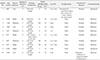
A retrospective study was performed of 10 patients with TMJ ankylosis (7 unilateral and 3 bilateral; 13 joints) who were confirmed as having Sawhney type III ankylosis using coronal sections of cone-beam computed tomography (CBCT) and were treated with lateral arthroplasty and BFP interposition in the Department of Dentistry, Shaheed Hasan Khan Mewati (SHKM), Govt. Medical College (Mewat, India), between 2013 to 2016. This retrospective chart review was exempted from institutional review board approval. In all patients, various demographic and clinical information was recorded.(Table 1) Patients with a history of previous TMJ surgery were excluded.
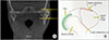
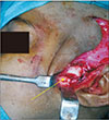
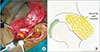
Coronal sections of the CBCT were used to orient the surgeon regarding the angulation and design of the osteotomy, ensuring that the medially placed condyle and disc were not damaged.(Fig. 1. A) All surgical procedures were performed under general anesthesia, and a fiber optic endoscope was used for nasal intubation. The bony bridge of the ankylotic mass on the lateral aspect was resected, leaving a distance of 1.5 to 2.0 cm from the base of the skull to the neck of the condyle.(Fig. 2. A) The condyle was left intact.(Fig. 2, 3) Coronoidectomy was performed on the ipsilateral side through the same approach in all cases. The inter-incisal opening was measured at that time, and if it was less than 35 mm, contralateral coronoidectomy was performed with an intra-oral approach. After satisfactory inter-incisal mouth opening (≥35 mm) was achieved, the TMJ surgical site was revisited, and the BFP was retrieved and used to cover the lateral aspect of the medially placed condyle.(Fig. 4. A) The BFP was sutured to the soft tissue covering the anterior aspect of the external auditory meatus. The surgical site was then closed as conventional.
Aggressive physiotherapy was started from the first postoperative day with the help of Heister's mouth opener (provided to each patient). The patients were asked to use the device for 5 minutes every hour while awake, every day. The patients were also asked to chew gum 8 to 10 times a day. These exercises were continued for 3 months.
III. Results
The present study included 10 patients with a mean age of 11.8 years (range, 8–16 years); there were 5 males and 5 females. Trauma was the etiological factor in all patients. Bilateral coronoidectomy was performed in 8 cases, and unilateral coronoidectomy was performed in 2 cases. The BFP was harvested and interpositioned in all cases. The mean preoperative maximum inter-incisal mouth opening (MIMO) was 5 mm (range, 0–14 mm). The mean MIMO at the last follow-up was 34.7 mm (range, 30–40 mm). None of the patients complained of any pain while chewing or at rest at the last follow-up. All patients were satisfied with their results. No signs of facial paresis were present in any patients at the last follow-up. At 6 months after surgery, all patients were advised to undergo CBCT scans. The scans showed a well-maintained intra-articular space in all patients.(Fig. 2)
IV. Discussion
Multiple operative procedures are used to manage TMJ ankylosis, but none are universally accepted. Operative procedures include gap arthroplasty with or without interpositional grafts; and resection of the ankylotic mass and reconstruction of the ramus-condyle unit with autogenous or alloplastic grafts3. In a study of 791 cases of TMJ ankylosis, Mehrotra et al.4 concluded that sternoclavicular graft reconstruction in children and dermal fat interposition arthroplasty in adults should be the treatment of choice for the management of TMJ ankylosis. In a variety of studies, gap or interpositional arthroplasty has been performed for TMJ type III ankylosis. Zhi et al.5 treated 19 cases of type III ankylosis with gap or interpositional arthroplasty. Elgazzar et al.6 treated 101 cases of ankylosis with gap or interpositional arthroplasty, and Dimitroulis7, in a retrospective study, presented the clinical experience of using a dermis-fat interpositional graft in the surgical management of TMJ ankylosis in 11 adult patients. None of these studies provided a description of the type of ankylosis. All patients in these studies underwent the same treatment strategy; no adaptations were made for type III cases.
When the condylar process is fractured below the insertion of the lateral pterygoid muscle, it is displaced along with pterygoid muscle in an antero-inferio-medial direction. This type of trauma is treated either by open or closed reduction, but in children, the condition is usually managed conservatively8.
In closed treatment, maxilla-mandibular fixation for 10 to14 days is used to re-establish normal occlusion, followed by intensive physiotherapy for rehabilitation of normal symmetrical mandibular movement. During the period of repeated exercises, the stump with the attached medially displaced condyle grows and remodels in young individuals, creating a double hump-shaped condyle that can perform all mandibular movements and functions normally as a site of growth8.
Improper treatment of a displaced condylar process fracture results in the stump ankylosing to the fossa, producing type III ankylosis. In India, because of negligence and parents' lack of awareness, these cases are not typically treated at an early stage, making such clinical situations more common8.
Preoperative diagnosis of TMJ ankylosis with the help of CBCT (coronal view) is very important to detect Sawhney type III cases. Although CT is a standard investigative procedure for TMJ ankylosis, clinicians working in resource-scarce environments may rely on plain radiographs alone9. However, plain radiographs alone cannot detect Sawhney type III cases, and as a result, these cases are managed improperly as type IV cases. The importance of getting a preoperative CT or CBCT scan cannot be over-emphasized9.
In type III ankylosis where CBCT clearly shows a medially displaced condyle and a lateral bony bridge, one should try to preserve the condyle rather than eliminating it because advanced reconstructive surgical procedures are required when both the condyle and disc cannot be retained. Although located in an awkward position, the medially displaced condyle should function exactly as it would after a properly treated displaced condylar fracture. In type III ankylosis, reconstruction is not required because the medially displaced condyle maintains the ramus height even after lateral arthroplasty8. To prevent reformation of the lateral bony bridge, the BFP is positioned lateral to the medially displaced condyle. This placement of the BFP not only helps in achieving haemostasis by reducing dead space, but also acts as a barrier in reformation of the lateral bony bridge, thus preventing re-ankylosis.
Harvold et al.10, in treating hemifacial microsomia, argued that even a deformed condyle shows function and growth when released, and therefore, the condyle and disc should be used rather than eliminated and replaced with a suboptimal solution. Caldwell11 maintained that restoration of joint function at an early age, rather than removal of the condyle, is necessary to activate as much growth potential as possible.
In a study of 15 patients, Singh et al.8 concluded that lateral arthroplasty is the treatment of choice in Sawhney type III TMJ ankylosis and aggressive resection of the ankylosed joints is strongly contraindicated in these cases. Compared to surgical procedures that require complete resection of the ankylotic mass, lateral arthroplasty has many advantages. First, removal of the medially displaced condylar stump increases the risk of injury to deep vessels, which can cause intraoperative hemorrhage. Since lateral arthroplasty involves removal of the superficial bony bridge only, the risk of injury to deep vessels is very small. Second, lateral arthroplasty is associated with reduced postoperative discomfort and a faster recovery because the procedure is less traumatic. Third, as there is no need for joint reconstruction, the operative time is reduced, and thus, the time for which the patient requires general anesthesia is also reduced.
On the other hand, if the condylar stump is completely resected, joint reconstruction is required, especially in cases of bilateral TMJ ankylosis, in order to maintain the vertical height of the ramus and avoid an anterior open bite in occlusion. In such cases, opting for TMJ reconstruction with autogenous grafts such as costochondral or sternoclavicular grafts will lead to additional surgical site morbidity. Alternatively, opting for alloplastic TMJ reconstruction will lead to an additional financial burden for patients. In bilateral TMJ ankylosis, these additional problems can be avoided, even if one is able to perform lateral arthroplasty on only one side.
The first clinical use of the BFP was described by Egyedi12 in 1977. There was a time when the BFP was considered a surgical nuisance131415. However, use of the BFP has become a preferred option in oral and maxillofacial applications because of its relative ease of use, anatomically favorable position, minimal dissection required for harvesting, and low rate of failure16.
The BFP is a simple lobulated mass consisting of a central body and four extensions: buccal, pterygoid, pterygopalatine, and temporal. The body consists of three different lobes: anterior, intermediate, and posterior. Each lobe is encapsulated by an independent membrane and separated by a natural space17. The mean volume of the BFP is 10 square centimeter18. Regardless of overall body weight and fat distribution, the size of the BFP is fairly constant. Even in cachectic patients, the BFP is of normal weight and volume19.
The success of using the BFP is attributed to its rich vascular supply, reduced donor site morbidity, ease of harvest, and low rate of complications20. Although rare, complications associated with use of the BFP include complete or partial loss of the flap, limitation of mouth opening, hematoma, hemorrhage, postoperative infection, and depressed cheek21222324. None of these rare complications were encountered in our study.
Rattan17 used the BFP as a useful adjunct to autologous or alloplastic TMJ reconstruction after ankylosis release. Singh et al.25 evaluated the usefulness and feasibility of the BFP as an interposition graft in the treatment of TMJ ankylosis and found it successful. Gagnani et al.26 have advocated for use of the BFP for interposition after gap arthroplasty. According to a literature review by Singh et al.16, elimination of dead space is the main goal in TMJ operations. The BFP helps in achieving this goal. In addition, isolation of the joint by the BFP reduces the risk of formation of fibrosis and bone in the area. Gaba et al.27 used magnetic resonance imaging to assess the fate of the BFP when used as an interpositional graft in TMJ ankylosis. They concluded that the BFP is viable after 1 year and prevents heterotopic bone formation after TMJ ankylosis release.
In our experience, BFP interposition on the lateral aspect of the medially displaced condyle after lateral arthroplasty in type III ankylosis is a simple and effective method for managing this condition. The treatment is also less traumatic than autogenous joint reconstruction and more cost efficient than alloplastic reconstructive procedures.

 XML Download