

 PDF
PDF ePub
ePub Citation
 Print
Print


Introduction
The face of a human is a complex morphological structure [1] mainly consisting of the muscles of facial expression which function in conveying the communicating intentions within a social context through the production of facial expression. The facial expression muscles are composed of several muscles arranged in functional groups over the face; the orbicularis oculi muscle, however, is the familiar representation among them owing to the characteristic feature in the movement of eyes. The orbicularis oculi muscle has been generally classified into the orbital, palpebral and lacrimal parts [2]. The orbicularis oculi muscle has been frequently reported to play a significant role in movement production around the orbital region in relationship to facial aging appearance [345]. The orbital part generally originates from the medial palpebral ligament, nasal process of the frontal bone and frontal process of the maxilla [2678]. In addition, the orbital part usually extends the outer muscle fibers beyond the orbital rim to the periorbital regions including the cheeks, forehead and temples [8], and interdigitates with surrounding muscles especially in the region inferior to the orbital part [2679], including the levator labii superioris alaeque nasi, levator labii superioris, zygomaticus minor and zygomaticus major muscles. The region among the inferior extending fibers of the orbital part of the orbicularis oculi muscle and mentioned adjacent facial muscles, however, distinctive muscle bundles were found. These muscle bundles were first observed and defined as the malaris muscle in the study of Henle [10]. The malaris muscle described by Henle [10] is divided into the medial and lateral parts corresponding with the following study of Lightoller [11] that also examined the malaris muscle consisting of the medial and lateral heads. Thenceforth numerous studies have attempted to investigate the existence of the malaris muscle according to the studies of Henle [10] and Lightoller [11] as well as its aesthetic facial clinical implications. Although the studies each reported a similar relationship between the malaris muscle and facial aging appearance [111213141516]; in the anatomical context, however, the understanding of the malaris muscle remains inconclusive. Many studies have explained the malaris muscle that is the muscle bundle located lateral to the orbital part of the orbicularis oculi muscle [1415], while some have stated that the malaris muscle is merely the variation or part of the orbicularis oculi muscle [12131718]. However, a recent study of the malaris muscle mentioned the medial and lateral bundles of the malaris muscle together [16].
Evidently, these research findings reveal different morphological definitions of the malaris muscle. Therefore, this review of the literature was undertaken with the purpose of reconsidering the anatomical concept of the malaris muscle such as attachment sites, innervation patterns including its plausible function related to facial aging appearance that may be useful for facial rejuvenation.
Anatomical Concept of Malaris Muscle
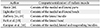
The morphological studies of the orbicularis oculi muscle have allowed the further studies to expose the distinct muscle bundles adhering to the orbital part of the orbicularis oculi muscle especially the inferior region. The distinct muscle bundles found inferior to the orbital part of the orbicularis oculi muscle were first examined and described by Henle [10] as the malaris muscle consisting of the medial and lateral parts. Likewise, Lightoller [11] was able to investigate the muscle bundles inferior to the orbital part of the orbicularis oculi muscle and defined these muscle bundles as the medial and lateral heads of the malaris muscle and referred to the study of Henle [10]. Afterwards, several studies attempted to examine more clearly the malaris muscle; however, the results provided a variety of anatomical descriptions (Table 1). Nevertheless, some studies also described these identical muscle bundle as the part of the orbicularis oculi muscle [12131718]. Even though there are miscellaneous concepts of the malaris muscle, current anatomical textbooks described the malaris muscle as the considerable variation that is merely a thin muscle sheet continuing with the inferior part of the orbicularis oculi muscle and extending the fibers downwards medially [2]. It is apparent that in general the malaris muscle is currently perceived comprising the lateral muscle bundle inclining downwards medially [2141519], the medial muscle bundle inclining downwards laterally mentioned in the studies of Henle [10] and Lightoller [11]. The suspending bundle has not been mentioned until the latest study of Kampan et al. [16].
Attachment Sites
Medial bundle of malaris muscle
The detail of the medial bundle is scarce, however the studies that revealed the anatomical description of the medial bundle exist [101116]. The medial bundle of the malaris muscle was described as the muscle bundle adhering medially to the orbital part of the orbicularis oculi muscle and running downwards laterally. All reviewed studies noted the medial bundle of the malaris muscle attaching from the frontal process of the maxilla [101116]. In addition, the medial palpebral ligament and the frontal belly of the occipitofrontalis muscle were indicated as the common attachments of the medial bundle of the malaris muscle [1116]. The medial bundle of the malaris muscle runs inferiorly and is usually attached to the superficial fascia of cheek [1011], levator labii superioris alaeque nasi [16] and levator labii superioris [16] muscles.
Although studies showing the medial bundle of the malaris muscle are few, studies regarding the muscle bundle resemble the medial bundle of the malaris muscle. Some studies claimed that the muscle bundle found medial to the orbital part of the orbicularis oculi muscle is merely the part of it [131718], while another study named the same muscle bundle as the orbitozygomatic muscle (OZM) and stated that it simply is a variation of the orbicularis oculi muscle [12]. These studies did not define the muscle bundle found medial to the orbital part of the orbicularis oculi muscle as the medial bundle of the malaris muscle, nevertheless the description regarding the attachments of this muscle bundle were similar to the medial bundle of the malaris muscle. According to the studies describing the medial bundle of the malaris muscle as the part of the orbital part of the orbicularis oculi muscle, the muscle bundles were found arising from the inferior margin of the orbital part of the orbicularis oculi muscle, which are well developed [1317]. Moreover, the muscle bundle was observed attaching from the frontal belly of the occipitofrontalis muscle [1718] and the medial palpebral ligament [1218]. In addition, this muscle bundle also runs downward and attaches to the superficial fascia of cheek [121318] as well as the levator labii superioris alaeque nasi and levator labii superioris muscles [17], similar to the medial bundle of the malaris muscle.
Lateral bundle of the malaris muscle
According to the current understanding on the morphology, the malaris muscle is the muscle sheet located lateral to the orbicularis oculi muscle. Most of the studies regarding the malaris muscle pursued and reported various anatomical details of the lateral bundle of the malaris muscle. In general, the lateral bundle of the malaris muscle was recognized as the muscle bundle adjacent to the orbital part of the orbicularis oculi muscle laterally and running downward medially. The lateral bundle of the malaris muscle was apparently found arising from the superficial temporal fascia [210111415161719], while the distal attachments were diverse. From the superficial temporal fascia, the lateral bundle of the malaris muscle runs downward medially and is generally attached to the superficial fascia of cheek [21011141915], zygomaticus minor [2141516] and zygomaticus major [2101114151619] muscles. In addition, some studies indicated that the lateral bundle of the malaris muscle extends towards the angle of the mouth [21115] and the platysma muscle [16]. Even the study explaining the identical muscle bundle as the part of the orbicularis oculi muscle also noticed the similar attachment sites arising from the superficial temporal fascia and extending to the zygomaticus major muscle [17] and the superficial fascia of cheek [13].
Suspending bundle of malaris muscle
The suspending bundle of the malaris muscle was first described in a recent study [16]. Apart from the medial and lateral bundles of the malaris muscle, the U-shaped muscle bundle was observed inferior to the orbital part of the orbicularis oculi muscle. Interestingly, the attachment sites of this U-shaped muscle bundle in the medial and lateral sides were found in common with the proximal attachments of the medial and lateral muscle bundles of the malaris muscle, respectively. The medial attachments of the suspending bundle were derived from the frontal process of the maxilla, medial palpebral ligament and some adjoined with the frontal belly of the occipitofrontalis muscle, whereas the lateral attachment of the suspending bundle was derived from the superficial temporal fascia [16].
Innervation Patterns
Typically, the muscles of facial expression are innervated by the five terminal branches of the facial nerve [2]. In the periorbital region, the facial expression muscles are reported to be innervated mainly by the temporal and zygomatic branches [2202122232425], and partly by the buccal branch [21232425]. Throughout the studies that commonly investigated the malaris muscle in the periorbital region, however the innervation of the malaris muscle was barely informed. The innervation of the malaris muscle was first thoroughly examined in the most recent study of Kampan et al. [16]. With regard to the distribution of the facial nerve branches, the malaris muscle consisting of the medial, lateral and suspending bundles were chiefly innervated by the temporal and zygomatic branches. After passing through the parotid gland, the temporal branches run upward medially to innervate the periorbital muscles on the lateral side as well as the lateral bundle and lateral part of the suspending bundle of the malaris muscle. Meanwhile, the zygomatic branches ran medially to innervate the periorbital muscles located on the medial side including the medial bundle and medial part of the suspending bundle of the malaris muscle. Interestingly, some temporal and zygomatic branches of the facial nerve innervating the malaris muscle were observed penetrating the facial muscles along their courses from the deep to superficial facial layers.
Functions Related to Facial Aging Appearance
With aging, the facial soft tissues gradually descend and lose the volume as well as the elasticity [2627]. For this reason, the periorbital muscles especially the orbicularis oculi muscle located in the midface is reported to become atrophic, stretched or thin [34572627282930313233], resulting in the appearance of aging in the midface. Moreover, together with the repetitive motion from the facial muscle contraction and gravity [26], the midface aging could seem more obvious. The characteristics of typical midface aging are sagging of the malar fat pads [34], formation of the malar bags [30], laxity of the periorbital skin and the external canthus [19]. Although the orbicularis oculi muscle is the principal of the midface aging [345], the malaris muscle located in the periorbital region was determined to be related to facial aging as well [11121314151619].
Regarding previous studies, the malaris muscle comprises the medial [2101116], lateral [2101114151619] and suspending bundles [16]; however, only the lateral bundle is mentioned in relationship to the midface aging [2141519]. The lateral bundle of the malaris muscle is believed to be the assistant of the zygomaticus major muscle to raise the upper lip and angle of the mouth during smiling and laughing, according to the attachment of the lateral bundle to the zygomaticus major muscle [215]. Duchenne de Boulogne [35] reported that “enjoyment” smiles move not only the angle of the mouth but also the neighboring muscles of the eye, simultaneously contracting the orbicularis oculi and zygomaticus major muscles. The lateral bundle of malaris muscle located lateral to the orbicularis oculi muscle would therefore cause the well-known wrinkle at the corners of the eyes [11] named crow's feet during the smiling or laughing. Apart from the significant role in facial animation, the lateral bundle of the malaris muscle also participates in dimple formation together with the risorius muscle [15]. In the studies of Zufferey [1419], the lateral bundle of the malaris muscle was concluded to have function in preventing the midface aging. Regarding the location and dermal attachments of the lateral bundle of the malaris muscle in the midface, it is considered as the muscular component of the superficial musculoaponeurotic system (SMAS) that enhance the SMAS to maintain and to prevent the laxity of the soft tissues including the skin and superficial fascia in the midface [1419]. In other words, the midface aging is a consequence of loss of dynamic superficial structure like the lateral bundle of the malaris muscle, which is the essential muscular component of the SMAS that retains the midface region by contracting and pulling up the soft tissues where the lateral bundle of the malaris muscle attaches to. This finding is consistent with the study of Wassef [36] that was able to observe the unique muscle sheet on the fascia between the superficial temporal fascia and the platysma muscle in the fetus.
The suspending bundle of the malaris muscle identified in the study of Kampan et al. [16] is also described as related to the midface aging especially in the periorbital region. Based on the positional relationships between the suspending bundle and the intraorbital structures, the role of the suspending bundle could be to sustain the intraorbital structures. Moreover, the deep groove usually found on the medial side of the eyes or the palpebro-malar groove might be caused by the simultaneous factors comprising the laxity of the lateral attachment of the suspending bundle together with the thin orbicularis oculi muscle resulting in the infraorbital fat protrusion [16].
The studies of the medial bundle of the malaris muscle are limited, the relation to the midface aging was still revealed in the studies of the muscle bundle resembling the medial bundle of the malaris muscle [25]. The muscle band resembling the medial bundle of the malaris muscle in the study of Hwang [25] was named the OZM and described to cause the unpleasing nasojugal fold. In addition, the OZM was accepted to have function in preventing the infraorbital fat herniation as well as drooping of the suborbicularis oculi fat pad.
Discussion
The purpose of this review was to reconsider and summarize the available literature regarding the anatomical concepts of the malaris muscle. Initially, it is apparent that there is an exact definition of the malaris muscle provided by the current anatomical textbooks and the primary literature; however, the recent research of the malaris muscle was reintroduced [16], thus the morphological understanding of the malaris muscle should be reconsidered.
There were previous studies presenting the various anatomical definitions of the malaris muscle such as the attachment sites and its function but the main controversy among these studies was that the precise compartmentalization of the malaris muscle remains ambiguous. Accordingly, it is difficult to consider the function of the malaris muscle influencing the facial aging gradation. The diverse compartmentalization of the malaris muscle obtained from a number of the examinations might be consequent upon the differences in methods and techniques; however, the descriptions of the malaris muscle such as the attachment sites are relatively similar across the studies. For example, the studies dividing the malaris muscle into the medial and lateral bundles [1011] explained the attachment sites of the lateral bundle similar to the studies stating the malaris muscle merely consisting of the lateral bundle [2141519]. Consequently, the controversial issue about the compartmentalization of the malaris muscle might be inconsiderable as much as its function.
The lack of study concerning about the innervation patterns of the malaris muscle leads to difficulty in comparison across studies. Nevertheless, only one study thoroughly reported the innervation patterns of the malaris muscle which allow us to perceive that the malaris muscle might occupy the transitional layer between the superficial and deep facial muscles according to the innervation pattern of facial nerve branches penetrating from the deep to superficial layers of face [16].
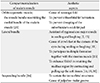
Although various morphological details of the malaris muscle especially the compartmentalization leads to the different extrapolation of their possible functions (Table 2), the importance of having a precise understanding of anatomical descriptions of the malaris muscle is still essential when considering clinical applications in the aspect of facial rejuvenation associated with morphologies as well as changes of the malaris muscle. Hence, the practical clinical applications for facial rejuvenation should be considered to be associated with the diverse anatomical knowledge of the malaris muscle for effectively correcting any midface aging. For example, in the study of Zufferey [19], the subject with the sagging of the soft tissues in the midface received the experimental injection of Botox in the superficial temporal fascia, which is the proximal attachment of the lateral bundle of the malaris muscle. After the injection, the soft tissues in the midface region no longer sagged. It is obvious that although any bundle of the malaris muscle may cause the midface aging, the exact location of pathology in the midfacial aging, relative to known anatomical descriptions of the malaris muscle bundles, such understanding could provide the effective clinical assessment, differential diagnosis including practical management for facial rejuvenation.
Conclusion
This review provides the summarized anatomical concepts of the malaris muscle regarding the attachment sites, innervation patterns and functions related to midface aging appearance from the available literature. However, it could be seen that the existing anatomical details of the malaris muscle remain discrepant and lacking owing to the differences of the sample, or other methodologies. This review, therefore, suggests the need for further study into the anatomical aspects including the morphological compartmentalization, innervation as well as the possible functions of the malaris muscle so that the comprehensive understanding of the malaris muscle related to the midface aging can effectively enhance practical clinical applications in facial rejuvenation.
 XML Download