Journal List > J Adv Prosthodont > v.11(3) > 1128691

Alfrisany, Shokati, Tam, and De Souza: Simulated occlusal adjustments and their effects on zirconia and antagonist artificial enamel

Abstract

PURPOSE

The aim of this study was to evaluate the effect of occlusal adjustments on the surface roughness of yttria-tetragonal zirconia polycrystal (Y-TZP) and wear of opposing artificial enamel.

MATERIALS AND METHODS

Twenty-five Y-TZP slabs from each brand (Lava, 3M and Bruxzir, Glidewell Laboratories) with different surface conditions (Control polished - CPZ; Polished/ground - GRZ; Polished/ground/repolished - RPZ; Glazed - GZ; Porcelain-veneered - PVZ; n=5) were abraded (500,000 cycles, 80 N) against artificial enamel (6 mm diameter steatite). Y-TZP roughness (in µm) before and after chewing simulation (CS) and antagonist steatite volume loss (in mm3) were evaluated using a contact surface profilometer. Y-TZP roughness was analyzed by three-way analysis of variance (ANOVA) and teatite wear by two-way ANOVA and Tukey Honest Difference (HSD) (P=.05).

RESULTS

There was no effect of Y-TZP brand on surface roughness (P=.216) and steatite loss (P=.064). A significant interaction effect (P<.001) between surface condition and CS on Y-TZP roughness was observed. GZ specimens showed higher roughness after CS (before CS − 3.7 ± 1.8 µm; after CS − 13.54 ± 3.11 µm), with partial removal of the glaze layer. Indenters abraded against CPZ (0.09 ± 0.03 mm3) were worn more than those abraded against PVZ (0.02 ± 0.01 mm3) and GZ (0.02 ± 0.01 mm3). Higher wear caused by direct abrasion against zirconia was confirmed by SEM.

CONCLUSION

Polishing with an intraoral polishing system did not reduce the roughness of zirconia. Wear of the opposing artificial enamel was affected by the material on the surface rather than the finishing technique applied, indicating that polished zirconia is more deleterious to artificial enamel than are glazed and porcelain-veneered restorations.

INTRODUCTION

Yttria-partially stabilized tetragonal zirconia polycrystal (Y-TZP) is frequently referred to as zirconia. Zirconia-based crowns and prostheses have been used as an alternative to porcelain-fused-to-metal restorations due to the absence of the metal and the white color.1 Metal-free dental restorations made of zirconia are one of the most attractive treatments for extensively compromised teeth also because of zirconia's biocompatibility and high mechanical properties.23 However, zirconia's opacity still demands the coping to be veneered with porcelain in cases with high esthetic demand, therefore resulting in a bilayer restoration. One of the most common clinical failures found in veneered zirconia (bilayer) restorations is the cohesive fracture within the veneering porcelain.4 An alternative way to avoid veneer fracture is to exclude the veneering material and to manufacture monolithic full contour Y-TZP crowns,56 by milling blocks with different levels of translucency.7 The milling of monolithic crowns has other advantages such as reduced manufacturing time and improved cost-effectiveness. Instead of building up the porcelain in several layers and firing in multiple firing cycles, the esthetics of monolithic restorations can be improved by using some staining techniques.8 Optimized surface finishing is achieved through glazing or polishing,9 but glazing seems to result in a smoother surface.10
Monolithic zirconia restorations have been gaining popularity as opposed to the use of bilayer restorations due to their mechanical predictability combined with improved esthetic properties.1112 Nevertheless, the high hardness and wear resistance of zirconia13 may cause wear on opposing natural dentition14 and indirect restorative materials,15 especially when the restoration's surface is not perfectly smooth.16 A correlation between surface roughness and wear has been demonstrated.17 Under clinical conditions, monolithic zirconia crowns seem to cause more wear to the opposing enamel than the human enamel itself.18 This is even more problematic when occlusal adjustments of monolithic zirconia crowns are required after installation of the prosthesis, since grinding with a diamond bur is known to significantly increase the surface roughness of ceramics19 and wear of the antagonist surface.20 Inadequate surface polishing is known to result in cracks that are densely distributed throughout the surface and that may compromise the mechanical performance of not only zirconia21 but also other glass ceramics.22 Nonetheless, there is no standard method for polishing monolithic zirconia crowns yet.23
The data currently available on wear and roughness of Y-TZP based restorations is not conclusive. The application of low number of cycles and142425 low loading forces,1626 or the use of different wear simulation devices27 may lead to false predictions, indicating that zirconia is a “wear-friendly” material28 when the conditions employed do not actually represent the in vivo conditions. Lack of correlation between number of cycles and time of clinical use may also make the interpretation difficult.6 The use of clinically relevant scenarios for the analysis of failure29 and wear3031 and the selection of the ideal counter sample material32 has been strongly advocated. Therefore, the aim of this study is to evaluate the roughness on Y-TZP surface submitted to different grinding/polishing protocols before and after chewing simulation and the consequent wear of the opposing artificial enamel. The hypothesis of this study is that surface condition has an effect on the roughness of zirconia and, as a consequence, that surface condition has an effect on wear of opposing artificial enamel.

MATERIALS AND METHODS

Y-TZP computer-aided-design/computer-aided-machining (CAD/CAM) blocks (BruxZir - Lot #B 0633325, expiry date 10/2017 - Glidewell Laboratories, Newport Beach, CA, USA; Lava Plus - Lot #520217, expiry date 9/2016 - 3M/ESPE, Seefeld, Germany) were cut in the pre-sintered stage with a diamond-embedded blade (Buehler - Series 15LC Diamond, Buehler, Lake Bluff, IL, USA) under water cooling to obtain 25 slices (6 × 6 × 2.0 mm3) from each material. The specimens were sintered following the manufacturers' instructions and then were randomly distributed among the experimental groups (n = 5) by blinded selection. The number of samples was defined by a power analysis, which indicated that n = 4 would be the minimum ideal number to identify the effect of treatment in a study design of five experimental groups per material.
Surface treatments were applied according to the experimental group as follows:
  • - Control polished zirconia (CPZ): specimens were polished using a dental laboratory technique according to instructions from the zirconia's manufacturers. Polishing was performed with an extraoral polishing kit (K0238 Dialite ZR Extra-Oral Zirconia Polishing System, Brasseler USA Dental, Savannah, GA, USA), which was composed of epoxy-based diamond impregnated polishers. The sequential polishing was performed as follows: prepolishing with a medium grit disc-shaped tip (⅒ mm × 2 mm - H8MZR.HP, Brasseler USA Dental) applying regular manual pressure in a parallel direction until the entire surface presented similar surface finishing; then, final polishing was performed by using the fine grit disc-shaped tip (H8FZR.HP, Brasseler USA Dental) with increased manual pressure in a parallel direction until a smooth or glossy surface could be observed by visual inspection. The same polishing procedure was applied to samples from groups GRZ and RPZ as the initial treatment.

  • - Ground zirconia (GRZ): polished zirconia specimens were subsequently ground with a diamond bur (837LF FG 014, 27 - 76 µm, Meisinger, Centennial, CO, USA) under water cooling. Grinding was applied to the surface of zirconia in a parallel direction for 10 seconds (two strokes, 5 seconds each). The same grinding procedure was applied to samples in RPZ group.

  • - Repolished zirconia (RPZ): zirconia specimens, polished and ground as described above, were subsequently repolished using an intraoral polishing system (eZr intraoral Adjustment Finishing & Polishing System, Garrison Dental Solutions, Spring Lake, MI, USA), which is a diamond-based polishing system with medium and fine grits indicated for repolishing. The procedure was performed as follows: a medium grit flame-shaped tip (4 mm × 10 mm) (FPZM020, Garrison Dental Solutions) in a high-speed handpiece was applied in a parallel direction and under regular manual pressure to the entire zirconia surface; final high-gloss polishing was performed by using the fine grit flame-shaped tip with increased manual pressure in a parallel direction until a smooth surface could be observed by visual inspection.

  • - Glazed zirconia (GZ): sintered unpolished zirconia slices were glazed with the Zenostar glaze system (Ivoclar Vivadent Inc., Amherst, NY, USA) and fired according to the manufacturer's instructions.

  • - Porcelain veneered zirconia (PVZ): sintered unpolished zirconia slices were veneered using the powder build-up technique with IPS e.max Ceram veneer material (Ivoclar Vivadent Inc.) following the manufacturer's instructions.

The specimens were cleaned (isopropanol solution in ultrasonic bath), air-dried, and stored in deionized water (~22℃) until analyses were performed.
Initial roughness of the specimens (before chewing simulation - CS) was assessed using a contact surface profilometer (Alpha-Step D-600, KLA Tencor Corp., Milpitas, CA, USA) with a dedicated software (KLA Tencor Apex 3D Mountains, KLA Tencor Corp.). An area of 2 × 2 mm2 on the center of each specimen was analyzed to determine mean roughness33 (Ra in µm). After CS specimens were cleaned ultrasonically, and the area that presented visible signs of abrasion was analyzed again using the same profilometer. Data was analyzed by three-way ANOVA and Tukey Honest Significance Difference (HSD) and an overall significance of 5% was pre-set.
Occlusal wear was artificially induced in a chewing simulator (CS-4.4, SD Mechatronik GMBH, Feldkirchen-Westerham, Germany). The bottom of the zirconia specimens was embedded in polymethylmethacrylate (PMMA) and inserted into metallic rings connected to the base of the equipment. The chewing simulation compartments were filled with artificial saliva34 at 37℃. Spherical steatite indenters (6 mm diameter - SD Mechatronik GMBH) were placed in the upper arm of the chewing simulator to simulate the antagonist tooth, and 80 N load was applied (60 mm/sec in a 2 mm horizontal motion).35 Four specimens were cycled simultaneously and 500,000 cycles were performed. The specimens were cleaned (isopropanol solution in ultrasonic bath) and air-dried, and roughness and wear were analyzed.
For analysis of wear, one baseline steatite indenter was scanned using the surface contact profilometer previously mentioned (Alpha-Step D-600, KLA Tencor Corp.) with the dedicated software (KLA Tencor Apex 3D Mountains) to register the baseline dimensions of the antagonists. Following chewing simulation, the worn steatite indenters were scanned and the diameter and height of the worn surfaces were obtained for the calculation of volumetric loss (in mm3).36 Wear data was analyzed by two-way ANOVA and Tukey Honest Significance Difference (HSD) and an overall significance of 5% was pre-set.
One zirconia substrate and steatite indenter from each experimental group were cleaned in acetone in an ultrasonic bath, and mounted on stubs with carbon adhesive tape and colloidal silver paint. The specimens were gold sputtered and observed under Scanning Electron Microscopy (SEM) (JEOL-SEM 6400, Peabody, MA, USA) with high vacuum mode under different magnifications.

RESULTS

Three-way ANOVA showed no effect of zirconia brand on surface roughness (P = .216) and volume loss of the opposing steatite (P = .064). Chewing simulation (P < .001) and the interaction between chewing simulation and surface condition (P < .001) had significant effect on roughness. Therefore, due to the interaction effect, roughness data was compiled regardless of the material brand and results were analyzed by a Tukey HSD test (Table 1). Porcelain veneered (15.22 µm ± 2.9) and glazed (13.54 µm ± 3.11) samples presented significantly higher roughness after chewing simulation when compared to the initial values (PVZ -13.43 µm ± 3.64; GZ - 3.7 µm ± 1.8). Before chewing simulation, the lowest roughness was presented by the control samples (1.3 µm ± 0.51), which was similar to the repolished samples (3.37 µm ± 1.63). After chewing simulation, ground (4.02 µm ± 2.20), repolished (4.14 µm ± 1.85), and control (1.43 µm ± 0.41) samples presented similar roughness values, which were significantly lower than porcelain veneered and glazed samples.
There was an effect of surface condition on the volume loss of the opposing steatite (P = .025) after chewing simulation (Fig. 1). However, ANOVA showed no effect of material on wear (P = .064). Samples abraded against the control group (0.09 mm3 ± 0.03) presented significantly higher volume loss than samples abraded against glazed (0.02 mm3 ± 0.01) and porcelain veneered (0.02 mm3 ± 0.01) zirconia. Intermediate values were presented by samples abraded against ground (0.07 mm3 ± 0.02) and repolished (0.06 mm3 ± 0.02) zirconia.
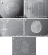
Surface characterization by SEM showed Y-TZP abraded areas that were not always compatible with the results provided by the surface profilometry. Ground (Fig. 2B) and repolished (Fig. 2C) zirconia presented smoother surface topography under the abraded area, whilst a significantly higher roughness could be found for the glazed samples (Fig. 2D). Figure 3 illustrates the level of damage to the structure of porcelain (Fig. 3A) and glaze (Fig. 3B) materials caused by chewing simulation. For the wear of the steatite, worn areas were more pronounced for samples abraded against control (Fig. 4B), ground (Fig. 4C), and repolished (Fig. 4D) zirconia. Glazed and porcelain veneered groups caused the least volume loss to the opposing enamel, and these findings were corroborated by the SEM findings, which show a significantly smaller abrasion area for steatite abraded against glazed (Fig. 4E) and porcelain veneered (Fig. 4F) zirconia. The SEM of the opposing surfaces (Fig. 2D and 2E respectively), however, showed that the material applied on zirconia was partially removed by the abrasion against the steatite.

DISCUSSION

The results of analysis of variance (ANOVA) showed that there was no significant effect of zirconia brand either on surface roughness (P = .216) or on the opposing steatite volume loss (P = .064), which can be explained by the similarities in the chemical composition and mechanical properties between the two brands.3 Irrespective of grain size, Y-TZP materials with similar chemical composition present similar surface hardness.7 A significant factor for the mechanical properties of zirconia is their level of translucency, due to the chemical changes needed to improve light transmission,37 but the materials used in the current study were both of medium opacity, indicating similar optical, surface and mechanical properties between them.
Occlusal adjustment of the zirconia surface is required in most cases after installation of the prosthesis, and this can result in a rougher surface and/or removal of the glaze layer. The resulting roughness may be reduced depending on the type of material and the technique applied for polishing,2123 but no standard method has been defined for polishing monolithic zirconia restorations.23 In the case of glazed surfaces, previous researchers recommended reglazing the restoration after clinical adjustments due to the easy removal of this layer.38 In the current study, a combination of a clinically significant surface treatment and chewing simulation was observed. Before chewing simulation, the highest surface roughness was presented by the porcelain veneered specimens, which was significantly higher than all of the other four groups (P < .0001). Therefore, the first hypothesis, which stated that surface condition has an effect on the roughness of zirconia, is accepted. The porcelain veneered surface did not receive a glaze layer or any additional surface treatment (Fig. 2E) to avoid having the same surface material among groups. We also aimed at investigating the effect of the porcelain on the opposing enamel. The veneering material used in this study is a fluorapatite veneering ceramic that is composed of glass powder, fused silica dioxide (SiO2) (60%) and alumina trioxide (Al2O3) (40%) crystals (IPS e.max Ceram, 2005).39 The presence of small particles like nano-fluorapatite (300 to 500 nm) extruding from the glassy matrix led to greater roughness in comparison to the glazed group, since the latter is basically a glass matrix with no fillers.6 Therefore, the veneer layer investigated in this study was overall rougher than in the clinical scenario, in which it would be covered with a glaze layer. The smoothness of the glazed surface was confirmed by the similar roughness values between glazed zirconia and polished (control) zirconia before CS, and this finding is in agreement with previous studies.10 After the application of chewing simulation, the surface roughness of the glazed zirconia specimens presented values that were similar to the porcelain veneered specimens, and the values were significantly higher (P < .0001) than the control, repolished, and ground specimens. This increase in surface roughness was due to the removal of the most superficial and smooth surface, which exposed the inner structure of the glaze layer, with voids, bubbles, and irregularities (Fig. 2D). Figure 3B shows evidence of voids and bubbles spread throughout the glaze layer, keeping roughness values at high levels for the lifetime of the restoration or until the glaze material is completely removed. A clinical study has previously shown that the glaze layer can be removed within the first six months after the installation of the restoration.22
The similar roughness values between ground and repolished groups were possibly due to the simplicity of the polishing procedure in the in vitro condition. The specimens were flat and fully accessible, and the operator could control the pressure applied. An intraoral occlusal adjustment may show different results, due to all the limitations associated with an in vivo procedure. Therefore, one should not assume that the intraoral polishing would be able to generate a level of surface polishing similar to the polishing provided by this study when occlusal adjustments are performed in the clinical scenario.
For a more comprehensive analysis, comparison of the roughness values before and after the application of chewing simulation for the ground and repolished groups can be combined with the SEM images of the surfaces (Fig. 2B and 2C, respectively). Different from the similar roughness values (Table 1), SEM indicated the smoothening of the abraded area after chewing simulation for both ground and repolished specimens. These conflicting results are a consequence of the limitations of the surface profilometry technique: it scanned a pre-set area of 2 × 2 mm2 instead of remaining within the boundaries of the abraded area, which was of approximately 1 mm2. Therefore, the roughness reading incorporated both abraded and non-abraded areas. Only a technique sensitive enough to particularly scan the abraded surface would be able to characterize changes in surface topography of this magnitude. In the absence of such technique, it is recommended to combine quantitative analyses with the qualitative assessment of the surface through higher magnification imaging techniques.
The volumetric loss measurement is considered the most effective way to assess wear of the opposing surface.3031 In the present study, ANOVA showed a significant effect of surface condition on the wear of the steatite (P = .025). Also, the wear of the steatite was rather affected by the surface material - zirconia, glaze or porcelain - than by the surface finishing technique - grinding or polishing (Fig. 1). Therefore, the second hypothesis, which stated that surface condition has an effect on wear of opposing artificial enamel is accepted. Interestingly, the wear values obtained were in agreement with the micrographs of the steatite obtained after chewing simulation (Fig. 4). The highest volume loss was caused by the control (polished) zirconia specimens (Fig. 4B), which was similar to ground (Fig. 4C) and repolished (Fig. 4C) specimens, but significantly higher than the wear caused by glazed (Fig. 2E) and porcelain veneered (Fig. 3E) specimens (P = .008). These results are in agreement with a previous study that reported higher wear of stainless steel indenters abraded against polished zirconia as opposed to those abraded against glazed zirconia.2 However, zirconia has been considered “wear-friendly” due to significantly lower human enamel wear26 and glass-ceramic antagonists25 when their surfaces are abraded against zirconia in comparison to glazed and porcelain-veneer materials. These contradictory results may be explained by the methods employed in other studies. While Janyavula et al.26 used only 10 N to simulate masticatory forces and a mix of glycerin/distilled water for humidifying the surfaces, the present study used a considerably higher load (80 N) and artificial saliva to simulate the oral environment. The 80 N load was applied because, based on previous studies, this is considered a high masticatory load that is still within the daily average for an adult without parafunctional habits.40 It is possible that the higher loading forces between zirconia and steatite increased the damaging effect of the significantly harder zirconia on the artificial enamel substrate.1341 This, combined with the absence of glycerin to act as a lubricating agent during the chewing cycles, may have maximized the wear caused by zirconia. Therefore, the present study indicates that the use of zirconia as a monolithic material under clinically relevant masticatory conditions may maximize the wear of the antagonist tooth.
The impact of the zirconia polishing technique on zirconia roughness and wear of opposing surface has been demonstrated.915202628 Nonetheless, the current study showed absence of effect of zirconia surface finishing technique on the volume loss of artificial enamel, in agreement with Preis et al.1920 Interestingly, hardness had a more significant effect than the roughness of the opposing substrate on the wear dynamics between zirconia and artificial enamel. The lower surface hardness of both veneering porcelain and glaze, when compared to the zirconia substrate and to the steatite, implied that glazed (Fig. 2D) and veneered (Fig. 2E) zirconia presented larger wear facets than the other zirconia samples.1732 It is possible that longer chewing cycles (e.g. 2 × 10⁶ cycles) would result in the total removal of the surface material (glaze or porcelain), with exposure of the underneath unpolished zirconia, which might have an impact on the dynamic wear process of the opposing enamel, but this hypothesis is far beyond the scope of the current study.
The metastability of Y-TZP tetragonal grains in mouth or room temperature is well known by researchers and clinicians.42 Yttria-doped zirconia maintains its high mechanical properties at low temperatures by stabilizing the tetragonal crystals upon cooling.2 However, some factors may trigger the return of the tetragonal crystals to their natural monoclinic state,43 challenging the longevity of Y-TZP devices. The application of 1 million masticatory cycles (vertical and horizontal loading) on the surface of some Y-TZP-based materials has caused significant changes in the materials' mechanical properties in the nanoscale.43 Atomic force microscopy was also able to show changes in surface morphology after mastication against stainless steel indenters.43 We hypothesize that any surface changes as a consequence of phase transformation in the current study would be overshadowed by the macro-morphological changes caused by the abrasion during the wear simulation. Analysis of the cross-section of samples also did not indicate the existence of a micro-cracked layer (Fig. 3C), which would be a consequence of crystalline re-arrangements after tetragonal-tomonoclinic phase transformation.
The information currently available in the literature on zirconia roughness and subsequent tooth wear is generally associated with either low number of cycles91424 or low loading.91626 Therefore, it fails to predict the performance of zirconia-based prostheses in the long-term. 500,000 cycles were applied in the current study, corresponding to two to five years of clinical service2729 which can be considered more representative of the long-term performance of the material in the oral environment. However, this study still presents limitations. Due to the nature of an in vitro design, variables such as temperature and pH cycles, often present in a clinical scenario, could not be simulated. Additionally, this study employed artificial enamel indenters instead of human enamel cusps in an attempt to minimize anatomic variability, which could have an impact on the measurement of wear. Further studies of the interaction between zirconia-based prostheses and human enamel are encouraged so that the effect of one surface on another can be realistically estimated.

CONCLUSION

Within the limitations of this in vitro study, we concluded that materials with similar composition presented similar roughness values and showed similar degree of wear of opposing artificial enamel after chewing simulation. The surface finishing technique has a significant effect on roughness of monolithic zirconia, even though it does not affect the wear of opposing artificial enamel. The material applied on zirconia surface affects the wear of opposing artificial enamel when compared to polished zirconia, and the interaction of surface condition and chewing simulation affects the roughness of zirconia.

Figures and Tables

Fig. 1

Bar chart illustrating opposing steatite wear (mm3) abraded against Y-TZP surface with different surface finishing conditions. Different lowercase letters above the bars indicate significant difference (P = .25).

jap-11-162-g001
Fig. 2

SEM micrographs overview of the surface after chewing simulation: (A) polished zirconia; (B) ground zirconia; (C) repolished zirconia; (D) glazed zirconia; (E) porcelain veneered zirconia. (B) and (C) show a smoother condition within the abraded surface.

jap-11-162-g002
Fig. 3

SEM micrographs of cross-sections of (A) porcelain veneered zirconia; (B) glazed zirconia; (C) ground zirconia. All images were taken after chewing simulation. Control and repolished samples presented a smooth surface similar to (C) ground zirconia.

jap-11-162-g003
Fig. 4

SEM micrographs for the steatite indenters: (A) baseline; (B) abraded against the control zirconia; (C) abraded against ground zirconia; (D) abraded against repolished zirconia; (E) abraded against glazed zirconia; (F) abraded against porcelain veneered zirconia. Larger abrasion areas are shown in (B), (C), and (D).

jap-11-162-g004
Table 1

Mean, standard deviation (SD) and Tukey HSD test results* for zirconia surface roughness (µm) before and after chewing simulation (CS)

jap-11-162-i001
Treatment Roughness
Before CS After CS
Porcelain veneered 13 43 (3.64) Aa 15.22 (2.90) Aa
Ground 4.04 (1.64) Bb 4.02 (2.20) Bb
Glazed 3.70 (1.80) Bbc 13.54 (3.11) Aa
Repolished 3.37 (1.63) Bbc 4.14 (1.85) Bb
Control 1.30 (0.51) Bc 1.43 (0.41) Bb

*Dissimilar lowercase letters within the same column and uppercase letters within the same row indicate significant difference (P < .01).

Notes

The authors are thankful to King Saud University for the first authors's MSc scholarship (NMA) and for funding research expenses related to the current study. Lava zirconia was kindly donated by 3M Canada.

References

1. Tuncel I, Eroglu E, Sari T, Usumez A. The effect of coloring liquids on the translucency of zirconia framework. J Adv Prosthodont. 2013; 5:448–451.
crossref
2. Beuer F, Stimmelmayr M, Gueth JF, Edelhoff D, Naumann M. In vitro performance of full-contour zirconia single crowns. Dent Mater. 2012; 28:449–456.
crossref
3. Alghazzawi TF, Janowski GM. Correlation of flexural strength of coupons versus strength of crowns fabricated with different zirconia materials with and without aging. J Am Dent Assoc. 2015; 146:904–912.
crossref
4. Guess PC, Kulis A, Witkowski S, Wolkewitz M, Zhang Y, Strub JR. Shear bond strengths between different zirconia cores and veneering ceramics and their susceptibility to thermocycling. Dent Mater. 2008; 24:1556–1567.
crossref
5. Long HA. Monolithic zirconia crowns and bridges. Inside Dent. 2012; 8:60–66.
6. Stawarczyk B, Frevert K, Ender A, Roos M, Sener B, Wimmer T. Comparison of four monolithic zirconia materials with conventional ones: Contrast ratio, grain size, four-point flexural strength and two-body wear. J Mech Behav Biomed Mater. 2016; 59:128–138.
crossref
7. Tong H, Tanaka CB, Kaizer MR, Zhang Y. Characterization of three commercial Y-TZP ceramics produced for their high-translucency, high-strength and high-surface area. Ceram Int. 2016; 42:1077–1085.
crossref
8. Johansson C, Kmet G, Rivera J, Larsson C, Vult Von Steyern P. Fracture strength of monolithic all-ceramic crowns made of high translucent yttrium oxide-stabilized zirconium dioxide compared to porcelain-veneered crowns and lithium disilicate crowns. Acta Odontol Scand. 2014; 72:145–153.
crossref
9. Kontos L, Schille C, Schweizer E, Geis-Gerstorfer J. Influence of surface treatment on the wear of solid zirconia. Acta Odontol Scand. 2013; 71:482–487.
crossref
10. Tholt de Vasconcellos B, Miranda-Júnior WG, Prioli R, Thompson J, Oda M. Surface roughness in ceramics with different finishing techniques using atomic force microscope and profilometer. Oper Dent. 2006; 31:442–449.
11. Raigrodski AJ. Concepts of design for contemporary anterior all-ceramic restorations. J Cosme Dent. 2013; 28:46–58.
12. Sorrentino R, De Simone G, Tetè S, Russo S, Zarone F. Five-year prospective clinical study of posterior three-unit zirconia-based fixed dental prostheses. Clin Oral Investig. 2012; 16:977–985.
crossref
13. Candido L, Fais L, Reis JMdSN, Pinelli L. Surface roughness and hardness of yttria stabilized zirconia (Y-TZP) after 10 years of simulated brushing. Rev Odontol UNESP. 2014; 43:379–383.
crossref
14. Kwon MS, Oh SY, Cho SA. Two-body wear comparison of zirconia crown, gold crown, and enamel against zirconia. J Mech Behav Biomed Mater. 2015; 47:21–28.
crossref
15. Albashaireh ZS, Ghazal M, Kern M. Two-body wear of different ceramic materials opposed to zirconia ceramic. J Prosthet Dent. 2010; 104:105–113.
crossref
16. Stawarczyk B, Özcan M, Schmutz F, Trottmann A, Roos M, Hämmerle CH. Two-body wear of monolithic, veneered and glazed zirconia and their corresponding enamel antagonists. Acta Odontol Scand. 2013; 71:102–112.
crossref
17. Ghazal M, Kern M. The influence of antagonistic surface roughness on the wear of human enamel and nanofilled composite resin artificial teeth. J Prosthet Dent. 2009; 101:342–349.
crossref
18. Stober T, Bermejo JL, Rammelsberg P, Schmitter M. Enamel wear caused by monolithic zirconia crowns after 6 months of clinical use. J Oral Rehabil. 2014; 41:314–322.
19. Preis V, Weiser F, Handel G, Rosentritt M. Wear performance of monolithic dental ceramics with different surface treatments. Quintessence Int. 2013; 44:393–405.
20. Preis V, Behr M, Handel G, Schneider-Feyrer S, Hahnel S, Rosentritt M. Wear performance of dental ceramics after grinding and polishing treatments. J Mech Behav Biomed Mater. 2012; 10:13–22.
crossref
21. Rashid H. The effect of surface roughness on ceramics used in dentistry: A review of literature. Eur J Dent. 2014; 8:571–579.
crossref
22. Etman MK. Confocal examination of subsurface cracking in ceramic materials. J Prosthodont. 2009; 18:550–559.
crossref
23. Lawson NC, Janyavula S, Syklawer S, McLaren EA, Burgess JO. Wear of enamel opposing zirconia and lithium disilicate after adjustment, polishing and glazing. J Dent. 2014; 42:1586–1591.
crossref
24. Jung YS, Lee JW, Choi YJ, Ahn JS, Shin SW, Huh JB. A study on the in-vitro wear of the natural tooth structure by opposing zirconia or dental porcelain. J Adv Prosthodont. 2010; 2:111–115.
25. Luangruangrong P, Cook NB, Sabrah AH, Hara AT, Bottino MC. Influence of full-contour zirconia surface roughness on wear of glass-ceramics. J Prosthodont. 2014; 23:198–205.
crossref
26. Janyavula S, Lawson N, Cakir D, Beck P, Ramp LC, Burgess JO. The wear of polished and glazed zirconia against enamel. J Prosthet Dent. 2013; 109:22–29.
crossref
27. Steiner M, Mitsias ME, Ludwig K, Kern M. In vitro evaluation of a mechanical testing chewing simulator. Dent Mater. 2009; 25:494–499.
crossref
28. Kim MJ, Oh SH, Kim JH, Ju SW, Seo DG, Jun SH, Ahn JS, Ryu JJ. Wear evaluation of the human enamel opposing different Y-TZP dental ceramics and other porcelains. J Dent. 2012; 40:979–988.
crossref
29. Kelly JR. Clinically relevant approach to failure testing of all-ceramic restorations. J Prosthet Dent. 1999; 81:652–661.
crossref
30. DeLong R, Douglas WH, Sakaguchi RL, Pintado MR. The wear of dental porcelain in an artificial mouth. Dent Mater. 1986; 2:214–219.
crossref
31. Magne P, Oh WS, Pintado MR, DeLong R. Wear of enamel and veneering ceramics after laboratory and chairside finishing procedures. J Prosthet Dent. 1999; 82:669–679.
crossref
32. Shortall AC, Hu XQ, Marquis PM. Potential countersample materials for in vitro simulation wear testing. Dent Mater. 2002; 18:246–254.
crossref
33. Hmaidouch R, Müller WD, Lauer HC, Weigl P. Surface roughness of zirconia for full-contour crowns after clinically simulated grinding and polishing. Int J Oral Sci. 2014; 6:241–246.
crossref
34. Ablal MA, Kaur JS, Cooper L, Jarad FD, Milosevic A, Higham SM, Preston AJ. The erosive potential of some alcopops using bovine enamel: an in vitro study. J Dent. 2009; 37:835–839.
crossref
35. Lameira DP, Buarque e Silva WA, Andrade e Silva F, De Souza GM. Fracture strength of aged monolithic and bilayer zirconia-based crowns. Biomed Res Int. 2015; 2015:418641.
crossref
36. Ludovichetti FS, Trindade FZ, Werner A, Kleverlaan CJ, Fonseca RG. Wear resistance and abrasiveness of CAD-CAM monolithic materials. J Prosthet Dent. 2018; 120:318.e1–318.e8.
crossref
37. Pereira GKR, Guilardi LF, Dapieve KS, Kleverlaan CJ, Rippe MP, Valandro LF. Mechanical reliability, fatigue strength and survival analysis of new polycrystalline translucent zirconia ceramics for monolithic restorations. J Mech Behav Biomed Mater. 2018; 85:57–65.
crossref
38. Barghi N, Alexander L, Draughn RA. When to glaze - An electron microscope study. J Prosthet Dent. 1976; 35:648–653.
39. Roselino Lde M, Cruvinel DR, Chinelatti MA, Pires-de-Souza Fde C. Effect of brushing and accelerated ageing on color stability and surface roughness of composites. J Dent. 2013; 41:e54–e61.
40. Tortopidis D, Lyons MF, Baxendale RH, Gilmour WH. The variability of bite force measurement between sessions, in different positions within the dental arch. J Oral Rehabil. 1998; 25:681–686.
crossref
41. Aydın B, Pamir T, Baltaci A, Orman MN, Turk T. Effect of storage solutions on microhardness of crown enamel and dentin. Eur J Dent. 2015; 9:262–266.
crossref
42. Camposilvan E, Leone R, Gremillard L, Sorrentino R, Zarone F, Ferrari M, Chevalier J. Aging resistance, mechanical properties and translucency of different yttria-stabilized zirconia ceramics for monolithic dental crown applications. Dent Mater. 2018; 34:879–890.
crossref
43. Pinto PA, Colas G, Filleter T, De Souza GM. Surface and mechanical characterization of dental yttria-stabilized tetragonal zirconia polycrystals (3Y-TZP) after different aging processes. Microsc Microanal. 2016; 22:1179–1188.
crossref
TOOLS
ORCID iDs

Najm Mohsen Alfrisany
https://orcid.org/0000-0003-1258-9863

Babak Shokati
https://orcid.org/0000-0003-2752-2530

Laura Eva Tam
https://orcid.org/0000-0002-4670-6952

Grace Mendonca De Souza
https://orcid.org/0000-0002-7521-9723

Similar articles