

 PDF
PDF ePub
ePub Citation
 Print
Print


INTRODUCTION
Acute leukemia is the most common pediatric malignancy and accounts for approximately 29% of all childhood malignancies. Continuing along this line, acute lymphoblastic leukemia (ALL) accounts for approximately 76% of acute leukemia in children, while the subtype called B-cell ALL (B-ALL) constitutes more than 80% of ALL cases in children [1]. B-ALL is a malignant clonal tumor originating from a hematopoietic stem cell with chromosomal aberrations. It is associated with massive proliferation and accumulation of lymphoblasts that exhibit maturation arrest at the early stages of differentiating into lymphocytes. Furthermore, the disease has been associated with a wide range of molecular and clinical changes that might aggravate disease progression [2]. These changes include epigenetic alterations such as DNA methylation, which play a key role in regulating gene expression. This DNA methylation mechanism is characterized by the addition of a methyl group to the 5' position of the cytosine ring in cytosine-phosphate diester-guanine (CpG) islands [34].
Ikaros is a transcription factor of the zinc finger family encoded by the Ikaros family zinc finger 1 (IKZF1) gene and it is crucial in regulating lymphoid development and immunoglobulin rearrangement. Although Ikaros plays an important role in the normal development of the immune system, excessive has been linked to various cancers, such as bladder, breast, blood, glial, head and neck cancers. This highlights the multi-dimensional character of Ikaros [5] and its role in the initiation and progression of several cancer types [6]. In addition, aberrant expression of Ikaros affects the prognosis of several unique cancer types [5]. Ikaros is primarily involved in ALL biology by attenuating the normal repression of target genes and inhibiting the regulation of genes expression involved in lymphoid differentiation. However, the intricacies of the molecular mechanisms underlying the functionality of Ikaros remain unclear [7].
Although a large number of molecular and genetic studies have explored the pathogenesis and biology of lymphoid malignancies, a detailed analysis of DNA methylation patterns might help explain the mechanisms underlying such diseases [8]. As stated above, Ikaros has a significant role in the incidence of cancers, especially B-ALL. Altered expression levels of IKZF1 and aberrant DNA methylation patterns have been associated with different types of leukemia [9]. Notably, lymphoid B-cell differentiation is disrupted in B-ALL, and IKZF1 is a major regulator of B-cell differentiation. Thus, we hypothesized that aberrant methylation (hypomethylation) of the IKZF1 promoter region results in excessive IKZF1 expression. Overexpression of IKZF1 is a potential cause of B-cell differentiation arrest and proliferation induction in B-ALL patients. Therefore, to further elucidate the role of IKZF1 in B-ALL, we investigated the methylation status of CpG islands in the promoter region in children who were recently diagnosed with B-ALL.
MATERIALS AND METHODS
Patient information
We included 25 cases with recently diagnosed ALL and an age of 15 years or younger. Ninety percent of the patients were in the 1–9 years group, whereas only 10% were part of the other two age groups. A similar age distribution was observed for the 25 age- and gender-matched controls. According to the complete blood count (CBC) and clinical history, all of these control children were healthy. Diagnosis of all patients, who were in a pre-therapy state, was suggested and confirmed according to clinical, biochemical, morphological and flow cytometry findings made by the same medical doctor. We collected 2 mL of peripheral blood per subject in ethylenediaminetetraacetic acid (EDTA)-containing tubes. Patient blood samples were collected in the Pediatric Hospital of Tabriz, while blood samples of the control group were collected in the Dey Clinical Laboratory up to one year after having obtained informed consent from the parents. The study was supervised by the Medical Ethics Committee of Tabriz University of Medical Sciences, Iran, under the code 5/4/6065 in 2015.
Lymphocyte separation and DNA extraction
We isolated lymphocytes from whole blood with Ficoll 1.077 Lymphosep (Biowest, Nuaillé, France), followed by phosphate-buffered saline (PBS) multiple washes. Next, genomic DNA was extracted using the phenol/chloroform method [10]. In turn, extracted DNA was dissolved in 20 µL of RNase- and DNase-free water and frozen (−80℃) until required. We measured DNA concentrations using a NanoDrop 2000c spectrophotometer (Thermo Fisher Scientific, Lenexa, KS, USA).
Sodium bisulfite treatment
Of each sample, we treated 2 µg of DNA with sodium bisulfite from the EpiTect Bisulfite Kit (QIAGEN, Hilden, Germany), according to the manufacturer's instructions. After this procedure, the processed DNA samples were dissolved in 20 µL of elution buffer and stored at −70℃. A whole-genome amplified DNA sample, where all CpG sites were unmethylated, and a DNA sample treated with SssI methyltransferase (New England BioLabs, Beverly, MA, USA) (i.e. all CpG sites were methylated) were used as negative and positive controls, respectively [11].
DNA methylation analysis
We assessed DNA methylation by polymerase chain reaction (PCR) analysis of bisulfite-modified genomic DNA. 150 µg treated DNA per sample was utilized for methylation-specific PCR (MS-PCR) and the total PCR reaction volume was 25 µL. We designed the forward and reverse primers for methylated and unmethylated statuses with MethPrimer (The Li Lab, China) (Table 1).
Statistical analysis
We used a chi-square test to compare the abundances of the three gene methylation statuses (methylated, unmethylated and partial methylation) using SPSS statistical software version 16.0 (SPSS Inc., Chicago, IL, USA). Chi-square and t-tests were used to compare between age and gender. Statistical significance was set at P<0.05.
RESULTS
The patient and control group age medians were 6.5 and 4.5 years, respectively (P=0.077). In addition, the two groups were appropriately matched in terms of sex and there was no significant difference between the two groups (P=0.08). Clinical characteristics are shown in Table 2.
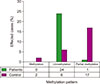
The principle of the MS-PCR assay is based on the conversion of cytosine (C) to uracil (U) via single base substitution (SBS), when the C in CpG is unmethylated. Contrastingly, when the C in CpG is methylated, then SBS has no effect on the methylated cytosine. Therefore, following bisulfite modification, two specific primers (targeting unmethylated and methylated states) were used in MS-PCR assay. To assess promoter methylation status via PCR, we designed our primers to specifically amplify methylated or unmethylated sequences in the CpG islands of the IKZF1 promoter. Amplification with the methylated or unmethylated primers resulted in the presence or absence of PCR product, depending on the methylation status of the CpG dinucleotides assayed by primers. We did not detect IKZF1 DNA methylation in the ALL cases (24 patients out of 25 ALL cases or 96% of the cases were unmethylated), while in the control group two subjects out of 25 (8%) were methylated, six out of 25 controls (24%) were unmethylated and 17 out of 25 controls (68%) were partially methylated. DNA methylation patterns of IKZF1 are presented in Fig. 1.
DISCUSSION
IKZF1, located at 7p12, is essential for B-cell lineage development. IKZF1 biology is complex. In 1992, Ikaros was identified as a zinc finger protein with an important role in the development and maturation of T-lymphocytes. Ikaros can activate or inhibit target genes' expression by binding to their regulatory regions and subsequently recruiting chromatin modifier complexes, thus initiating differentiation and inhibiting proliferation [1213]. Furthermore, Ikaros has been extensively implicated in regulating the development of the immune system [141516]. In addition, some studies have attributed the role of tumor suppression to this transcription factor [5]. Therefore, the deletion or aberrant methylation of the IKZF1 promoter, which results in gene silencing, has been associated with a wide variety of malignancies. Conversely, other studies have shown that an increased expression of the gene is also related to cancers such as leukemia, bladder, lung, and glioma [57]. In general, it appears that the optimal expression levels of Ikaros vary per tissue and disease condition. Moreover, its deletion or overexpression may contribute to the development of various cancer types. We hypothesized that aberrant methylation (hypomethylation) of the IKZF1 promoter increases IKZF1 expression, and that excessive expression of IKZF1 is a potential cause of B-cell differentiation arrest and proliferation induction in B-ALL patients. Considering the above, we investigated the methylation pattern of the IKZF1 promoter in childhood B-ALL.
Durchdewald et al. [6] reported that FOS family members, including c-Fos, GATA-1, Elk-1, and NKX6-B, have a binding site upstream of the IKZF1 transcription site. These family members dimerize with c-Jun and form AP-1; an important transcription factor in the initiation and progression of cancer.
Although aberrant DNA methylation is a common phenomenon in child and adult ALL patients, it appears that Ikaros gene silencing through this mechanism remains a rare phenomenon in ALL [17]. Our study supports these findings, as we observed a hypomethylation pattern of the IKZF1 gene promoter in 96% of our B-ALL samples. It is possible that this hypomethylation might increase the expression of Ikaros in B-ALL patients. This hypothesis is supported by a publication of Zhang et al. [7], which demonstrated an increased expression of Ikaros gene, due to increased gene demethylation, in lung cancer patients.
Ikaros has 8 different exons that encode 8 different isoforms [18]. Long isoforms of this protein (IK1 to 3), with over 3 zinc fingers at their N terminal, are functional and have a high DNA-binding affinity. However, the short isoforms (IK4 to 8), which possess fewer zinc fingers, have a lower binding affinity and are less competent at transcribing the target genes because they dimerize with the longer isoforms [19202122]. A previous study demonstrated that IK6 expression decreases apoptosis and enhances cell survival, while being associated with B-cell inhibition, differentiation, and maturation in leukemogenesis. Consequently, it was shown that IK6 is the most commonly increased isoform in B-ALL, T-cell ALL (T-ALL) and chronic myeloid leukemia (CML) [23]. Building on this, Han et al. [24] demonstrated that Dominant-negative (DN) isoforms, particularly IK6, are associated with poor prognoses in ALL patients. Furthermore, overexpression of this isoform is known to result in overproliferation and chemotherapy resistance in ALL cell lines. Other studies have shown an increased IK6 cell survival following acetylation of the Bcl-xL promoter, a mechanism that was also previously linked to the incidence of T-ALL [20] and pituitary tumors [25]. Therefore, an alternative hypothesis to the hypomethylation of the IKZF1 promoter and the subsequent increase in Ikaros expression in B-ALL patients is possible. More specifically, hypomethylation in the promoters of the short and DN protein isoforms is also a plausible mechanism. Thus, further research at the mRNA and protein levels is required.
The results of this study, in conjunction with additional advanced molecular research, might reveal the molecular mechanisms that underlie Ikaros gene in the development of cancers, paticularly B-ALL. Such explorations could pioneer the development of novel therapeutic strategies to treat cancer.
In this study we report a marked hypomethylation of the IKZF1 promoter region in B-ALL pediatric patients. It is plausible that the unmethylated status of the IKZF1 promoter might increase the expression of Ikaros, which has been linked to B-cells differentiation arrest and proliferation induction in B-ALL patients. Thus, targeting and suppressing the excessive expression of IKZF1 is a promising therapy in B-ALL patients.

 XML Download