

 PDF
PDF ePub
ePub Citation
 Print
Print


INTRODUCTION
Recent studies have evaluated the effect of ageing RBCs on patient mortality [12]. These studies have attributed mortality to levels of circulating free radical form of iron, known as the non-transferrin bound iron (NTBI) that causes an intervening oxidant damage [345]. As observed among different patient populations, apart from conjugating with the iron-chelator complexes and ‘other ligands’, NTBI circulating in the blood is also found bound to citrate or protein (albumin) or is present as free labile plasma iron (LPI) [6]. These studies also demonstrate significant mortality in the populations with high serum transferrin saturation (TS) (normal, 20–50%) [78].
The present study framed using the PICO(T) process is aimed towards 1) finding an association of the redox form of iron with mortality among patients suffering from iron overloading disease/conditions, and 2) comparing the oxidative redox iron forms (NTBI and LPI) in transfusion dependent diseases and iron drug therapy recipients targeted towards patient specific outcomes.
METHODS AND DATA SOURCES
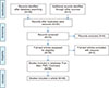
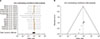
The literature was searched in the PubMed library and Cochrane Library (Fig. 1). The bibliographic information is available with the URL id: https://www.ncbi.nlm.nih.gov/sites/myncbi/1R55CXpfm9S5E/bibliography/52548262/public/?sort=date&direction=ascending. The reviewed references were the published articles (full text and abstract) involving human and animal experimental studies (flow diagram).
The details of the search strategy, and inclusion and exclusion criteria are available on Prospero ‘International prospective register of systematic reviews’ (CRD42018093657).
The website ‘Research gate’ was accessed for full length articles accessible at URL: https://www.researchgate.net/profile/Sankalp_Sharma.
The analyses of the present study-data were performed under the following parameters:
(a) Iron overload disease/conditions and their respective redox iron imbalances;
(b) An intervention in the form of iron therapy with iron containing drugs and respective mortality due to ‘redox iron elevation’ among the iron treatment recipients.
The summary-effect and assessment of the findings were performed in GRADE-pro GDT table and Revman 5.3 software, and recommendations were subsequently presented for further evaluation.
BLOOD TRANSFUSION MEDIATED REDOX IRON ELEVATION
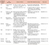
According to the U.S. Food and Drug Administration (USFDA) norms, the quality indicators for the red cell transfusion mandate at least 75% RBC survivability ‘in vivo’ within 24 hours of the blood transfusion [49]. A single unit RBC causes approximately 60 fold heme-iron (approximating 25% RBCs hemolysis) to be released in the intravascular compartment [4]. A release of NTBI/LPI after an ‘extravascular destruction of senescent RBCs’ and cell free hemoglobin (CFH) mediates reaction with nitric oxide and hydrogen peroxide (Fenton and peroxidase reaction) to generate hydroxyl and ferric radical of heme (oxo-ferryl Hb and ferric Hb), which further cause an oxidative lipid peroxidation mediated cellular damage, subsequently posing a risk of adverse outcomes among hospital admissions (Table 1) [13458910111213].
An increased post-transfusion NTBI, LPI and TS levels (human and animal studies) or elevated TS level (population studies) shows an increased in mortality (Table 1) [124578910111213]. The oxidative stress within post-operative patients (≥4 units transfusions) and near-expiry blood units is associated with reduced RBC deformability and raised redox iron levels respectively (Table 1) [314].
IRON OVERLOADING DISEASE CONDITIONS, ROLE OF HEPCIDIN
Iron overloading disease conditions such as thalassemia major (TM) and thalassemia intermedia (TI) cause ineffective erythropoiesis (IE) and decrease ‘serum hepcidin levels’, a metabolic state that promotes iron absorption and overload in the bone marrow and body organs [1516]. A high transfusion iron overloading rate (TILR) >0.2 mg/kg/day further mediate an increase in TS and redox NTBI [6161718].
An expansion of erythroid precursors in IE cause hemi-chrome formation due to extravascular hemolysis and lipid peroxidation-based reaction [151617].
TI manifests as a suppressed hepcidin level, and elevated TS, NTBI, and serum ferritin (erythroid expansion mediated iron overload) (Table 1) [151618]. TM in-contrast causes erythroid suppression, elevated hepcidin concentra-tion; secondary to multiple transfusions [high (TILR)] and decreased in iron absorption (RBC-transfusion mediated iron overload) [151617].
Hepcidin suppression is also manifested in chronic inflammatory states (such as Hepatitis C), due to reactive oxygen species mediated increase in histone deacetylase (HDAC), induction of hypoxia inducing factors (HIF), increased NTBI, iron accumulation in the liver, and oxidant damage in the form of liver fibrosis (Table 1) [1920].
IRON OVERLOADING DISEASES AND IRON INDICES - ROLE OF CHELATION THERAPY
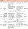
The NTBI and LPI (redox active) are the directly chelatable iron (DCI) forms [18]. These iron forms are indicators of high TS in patients with iron overload (Table 1) [161821].
In a comparative studies focusing on TS and subsequent increase in NTBI [1821]; a positive correlation of TS with NTBI has been demonstrated (Table 1) [61821]. In a comparison of hereditary hemochromatosis and thalassemia's a TS <85% in hemachromatosis display a NTBI 0.4 to 3.0 µM whereas in thalassemias TS of (80–100%) is accompanied with NTBI levels (0.4–10 µM) respectively [18].
An increased plasma level of DCI is an early indicator of iron overload and is targeted along with ferritin through the iron chelation regimen (Table 2) [6171820]. A significant loss in correlation between DCI and TS has been observed after the commencement of chelating therapy (Tables 1, 2, Supplementary Table 1) [6171820].
The analysis evaluating the association of TS with NTBI (r=0.77, P=0.0001) [176] showed a loss of correlation after commencement of chelation treatment (r=0.125, P=0.61) (Table 2) [6172223]. In the EPIC study (N=1,744) focusing on transfusion dependent patients, a dose-based decrease in the serum ferritin levels with deferasirox treatment was observed [24]. Stored blood however has a low TS (6.014±1.813–6.857±2.006 nM/mL; N=60) with a high NTBI (45% of total extracellular iron) with a steady ROS increase during the storage period (Table 2) [3].
A standardized chelation regime is more effective in early chelation of NTBI and LPI as compared to ferritin or RBC membrane iron (RBCM) (Table 2) [6172223]. The NTBI and ferritin iron showed correlation with inflammation and iron status. LPI (≥0.6 units) showed a significant correlation with high sensitive C-reactive protein (hsCRP). A significantly raised NTBI levels is observed with anemic (Hb<10; N=13) compared to non-anemic patients (P<0.05) and hepatitis C virus (HCV) infected patients (N=13) compared to non HCV patients (P<0.05) respectively (Table 2) [20].
IRON FORMS - ROLE OF ANTIOXIDANTS
Antioxidants mediate a significant reduction in the oxidative damage by decreasing the ROS in patients. An increased serum transferrin level, especially in thalassemia patients and immunocompromised infants, also mediates an anti-oxidative action (Table 2) [352526]. A reduction in biochemical LPI values in non-chelated TM children (3–13 yr) [25] was attributable to a high concentration of antioxidants in children; however, a progressive decrease in antioxidant protection was observed with an increasing age (Table 2) [525].
Total iron levels and superoxide free radicals correlate positively with oxidized form of ascorbate and negatively with reduced ascorbate (Table 2) [2126]. Ascorbate reduces NTBI to the ferrous state (Fe2+) and gets oxidized to ascorbate free radicle [26]. Ferrous (Fe2+) in-turn reduces oxygen to superoxide radical (hydrogen peroxide) (Table 1) [32526]. Stored RBC units show a negative correlation between total blood glutathione (antioxidant) and total extracellular iron (r=0.723) and NTBI (r=0.691); P<0.001 and a positive correlation of malondialdehyde [(MDA), a pro-oxidant] to extracellular Hb (r=0.736; P<0.001; N=60) (Table 1) [3]. A majority of the TM patients with cardiac complications showed an increased NTBI concentration [92536]. An extravascular RBC destruction releases NTBI; the resultant toxicity is subsequently reduced with chelation therapy. Experimental therapy directed towards reducing TS, such as apo-transferrin injections showed a reduced TS as well as reduced toxic hemichrome levels. The haptoglobulin therapy causes an attenuation in the ROS levels (Table 1) [91623].
IRON DRUGS AND REDOX ACTIVE IRON LEVELS
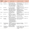
Oral iron preparations, iron ferrous salts, and iron-polymaltose display a linear increase in NTBI concentration after attaining a transport-maximum of paracellular enterocyte transportation into the blood stream [27]. Oral ferrous sulphate (100 mg) displayed significant NTBI levels in contrast to 100 mg of iron-polymaltose (IP) 0.7 µM (IP NTBI levels are comparable to placebo, water) in the iron adequate test subjects (Table 2) [28].
Intravenous high molecular weight iron salts (iron dextran, iron carboxymaltose) have a minimal reactivity with transferrin and show a significantly reduced increase of NTBI concentration even at high doses (Table 2) [272829303132].
A comparison of rise in NTBI after iron sucrose and iron dextran (86±42% vs. 45±45%; P<0.05) showed a significant difference of an increase in protein carbonylation (marker of oxidative stress) by iron sucrose as compared to iron dextran (P<0.05) and a higher NTBI levels of ferric gluconate and Iron sucrose as compared to iron dextran (P<0.001; 0.002 respectively) (Table 2) [2930].
Weak-labile iron drug-salts (iron sorbitol, iron citrate) cause rapid TS and increase in the NTBI levels, and renal elimination [27].
“A comparison of three ferrous salts showed the following: i) NTBI values of 6–12 µM/L within first eight hours post-dosage (100 mg); ii) TS levels below 100%, and iii) correlation of NTBI with transferrin saturation (r=0.76, P=0.001)” (Table 2) [3132]. Iron sucrose is implicated with the ROS based renal damage in hemodialysis patients, and a transient increase in TS (>80%) [33].
Low dose iron preparations (10 mg) showed a detectable LPI after iron intake (ferrous ascorbate, ferrous glycine sulphate), sometimes up to weeks after therapy (Table 2, Supplementary Table 2) [263233]. Transient renal tubular injuries have been reported attributable to the oxidative stress (iron sucrose 30 min post-infusion) [33].
MORTALITY COMPARISON OF IRON OVERLOADING CONDITIONS — META-ANALYSIS
The mortality indices in the population study [34] comparing TM (N=284; dead 40) and TI (N=95; dead 13) patients (N=379) showed a statistically different survival (P<0.0001) before and after an introduction of Blood chelation therapy [HR in TM compared to TI 6.8 (95% CI, 2.6–17.5) to 2.8 (95% CI, 0.8–9.2) before and after 1965 (era of iron chelation therapy) heart damage in the study emerged as a predominant cause of death in TM patients (Table 1) [34].
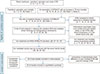
An analysis of the role of redox iron levels towards mortality was performed within the ‘PICOs selected studies’ by evaluating conditions with known redox iron overload (Fig. 2, Tables 1, 2).
The meta-analysis of ROS mediated mortality was evaluated under following parameters: Mortality within general population with high TS, NTBI/LPI levels within stored RBCs, post transfusion NTBI/LPI (after transfusion of old RBCs), correlation of iron chelation therapy with directly chelatable redox iron and serum ferritin, and oxidant damage on RBCs along with correlation of redox iron with pro-oxidants and antioxidant levels (Table 1).
The iron therapy intervention was evaluated as follows: NTBI elevation with iron drugs, correlation of iron therapy with serum LPI, an increase in serum iron concentration with various iron drugs, oxidative stress following iron therapy, prognostic indicators of patient on iron therapy, serum ferritin levels, and patient mortality (Table 2).
The findings and analysis of the studies are summarized in Table 3.
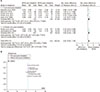
To evaluate an average increase of NTBI among patients on chronic transfusion therapy, a standard mean difference (SMD) for pre-chelated and post-chelated redox iron levels and post-iron therapy redox iron levels were compared using forest plot (Supplementary Table 1, Fig. 3) [31721222328293031353637]. One study (Pootrakul et al.) was excluded from the final analysis (Fig. 3) due to a high variation in the post-chelation ROS levels from the remaining studies under evaluation [18] (Supplementary Table 1). In a study (Collard et al.) (Fig. 4) a comparison of fresh Blood (3 days) and old Blood (35 days) Iron ROS levels was made to assess the difference between the NTBI of fresh blood and old blood units respectively. The cumulative standardized mean-difference for the pre-chelated and post-chelated is 0.97 (0.62, 1.32; N=384; chi2=0.01; I2=69%); test for overall effect: Z=5.42 (P<0.00001) (Fig. 3).
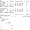
Patients on oral and intravenous (i.v) iron drugs were analyzed using forest plot for baseline level increase in the redox iron concentration (Fig. 4). The pre-therapy ROS levels were assumed to be 0.4 µM/L unless specified. Assessment for statistical heterogeneity or a pooled study analysis was not performed in the selected studies that aimed at evaluating the ROS levels within iron therapy and stored blood, respectively. The clinical heterogeneity identified included diverse kinetics and different reactivity profile for various iron drugs (Fig. 4).
The post-iron therapy ROS was 1.09 [0.73, 1.45; N=80; Tau2=0.00; chi2=3.68; df=4 (P=0.45); I2=0%] (Fig. 4). In subgroup of iron therapy and stored blood ROS level respectively, the standard mean difference is 5.44 [(4.00, 6.87); N=50; Tau2=1.41; chi2=8.88; df=4 (P=0.06); I2=55%]. The total standard mean difference is 2.89 (1.81, 3.98); I2=88%; Z=5.22 and P<0.00001 (Fig. 4A, B).
The clinical conditions were evaluated to discern the role of iron constituents in mortality among hospital admissions. The patients included were the blood transfusion recipients, post-operative patients, and patients on iron therapy.
An overall reduced mortality was observed among patients (N=572,046) (odds ratio, 0.89; 95% CI, 0.39, 2.02). An increased mortality was observed with elevated serum ferritin secondary to high iron dose theraphy (Fig. 5, Supplementary Table 3) [3839404142]. An increased mortality has also been shown among patients with serum ferritin >800 ng/mL [4344]. A serum ferritin <500 ng/mL with TS of <30% i.v iron therapy showed a reduced mortality (Table 3) [414344].
The assessment of mortality among the patient population (blood transfusion recipients and iron therapy) without including studies correlating high serum ferritin and mortality showed a odds ratio of 0.78 (0.24 to 2.46) indicating the secondary role of redox iron towards mortality (no separate chart shown) (Forest Plot 3; Supplementary Table 3).
A study (N=32,435) focused on iron therapy within the incident dialysis patients demonstrated a reduced all-cause mortality and well-tolerated serum ferritin concentration (600–800 ng/mL) [39]. Studies have also reported a reduced mortality due to ‘heart failure’ or ‘cardiovascular causes and sepsis’ or ‘post-operative cardiac valve replacement’ of patients on iron therapy as compared to placebo [384045].
OBSERVATIONS AND SUMMARY OF FINDINGS
Blood transfusions with older RBCs and ineffective erythropoiesis cause an increase in TS and redox-active NTBI (Quality of evidence high ; low risk of bias) (Table 4). NTBI showed a positive correlation with TS, which determines its pro-oxidant potential, tissue damage, and mortality (Quality of evidence moderate, selective reporting) (Tables 1, 4). Iron loading disease conditions increase NTBI, LPI (due to ineffective erythropoiesis or multiple transfusions), and redox active iron forms that are highly responsive to chelation therapy (Quality of evidence high ; low risk of bias) (Table 4). Iron ROS mediated tissue toxicity caused by the LPI is indirectly estimated by the antioxidant status and MDA status of an individual, with a reduced toxicity within patients having high antioxidant levels and low pro-oxidant level (Quality of evidence moderate; risk of bias) (Table 4).
Iron ROS mediated tissue toxicity caused by the LPI is indirectly estimated by the antioxidant status and MDA status of an individual, with a reduced toxicity within patients having high antioxidant levels and low pro-oxidant level (low risk of bias) (Fig. 2, Table 3). Iron formulations both oral and parenteral forms prescribed for various clinical in dications cause an increase (above normal range) in NTBI and LPI concentrations (Quality of evidence high; low risk of bias) (Table 4). Iron drug therapy has been proven to reduce mortality among hospital admissions despite an increase in the labile iron forms, under various clinical settings (low risk of bias) (Fig. 2, Table 3). Iron mediated toxicity may-be monitored with serum ferritin levels than that with the labile ROS iron forms, in terms of mortality indices in hospitalized patients (low risk of bias) (Fig. 2, Table 3).
DISCUSSIONS AND CONCLUSIONS
Transfusion dependent disease conditions such as thalassemia, myelodysplastic syndrome, and sickle cell disease cause ‘ineffective erythropoiesis’, along with the production of redox iron forms (NTBI or LPI) and tissue iron accumulation [151646]. These disease conditions showed a higher production of ROS and lipid hydroxyl-peroxides as well as an extensive formation of hydroxyl (OH)-free radicals through the Fenton reaction [39102646].
The oxidized ascorbate level present in the stored blood showed a strong correlation with the MDA, and was also associated with an uncontrolled release of iron from ferritin [14212633].
Ascorbic acid, glutathione, and transferrin (free-radical scavengers) determine the extent of superoxide-based lipid peroxidation and cellular damage (Table 1) [3172125]. The redox reactive iron showed a significant response to the chelation therapy with a quantitative reduction in the serum ROS iron and ferritin levels compared to the baseline (Table 1) [6171823]. The TS levels may not show any variation during chelation treatment even after a significant variation in the redox reactive LPI and storage ferritin forms of iron; however, several studies have reported a contradictory response to chelation on TS possibly due to different rate of decay of serum ferritin and serum transferrin saturation respectively (Table 1) [182123].
Iron drugs cause an increase in the redox iron variants in the blood; however, a reduction in the mortality secondary to the iron drugs has been observed across a range of iron formulations such as i.v. ferric carboxymaltose, ferric gluconate, and iron sucrose respectively (Table 2, Fig. 5).
The results of the PIVATOL study (proactive IV iron therapy in hemodialysis patients) for assessing the probability of all-cause-mortality in high dose iron recipient i.e. ‘hemodialysis patients’ [38], and multicenter trial on heart failure patients (CONFIRM-HF; N=304) showed the effect of ferric carboxymaltose injections (500–1,000 mg) [New-York Heart Association (NYHA) class II or III] with a reduced all-cause mortality as well as cardiovascular mortality (52 wk after the treatment) [38].
The TS, NTBI, LPI, and serum ferritin needs to be correlated with underlying diseases such as thalassemia major, and patients with hepatitis C infection, along with patient specific parameters such as anti-oxidant status and serum ferritin levels. Mortality secondary to the ROS iron however needs to be explored further.
 XML Download