

 PDF
PDF ePub
ePub Citation
 Print
Print


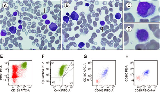
A 70-year-old Caucasian man was referred to our hospital because of pancytopenia. His peripheral blood count showed the following: hemoglobin level, 10.0 g/dL; white blood cells, 3×10³/µL; neutrophils, 1×10³/µL; lymphocytes, 1.9×10³/µL; monocytes, 0.1×10³/µL; and platelets, 80×103/µL. Blood chemistry analysis revealed 3 g/dL of monoclonal protein [immunoglobulin G (IgG) lambda]. Bone marrow aspirate showed infiltration by 20% plasma cells and 10% lymphocytes with hairy morphology (A–D, arrows). Immunophenotyping using a 6–8 color method showed the presence of two clones: 1) plasma cells with restriction for cytoplasmic immunoglobulin lambda light chains (E, F, green dots) and 2) B-lymphocytes [cluster of differentiation (CD)19+, CD20+] with immunophenotypic features of classic hairy cell leukemia (CD103+, CD25+, CD11c+, CD200+) (G, H, blue dots) with restriction for surface immunoglobulin lambda light chains (not shown). The BRAFV600E mutation was detected in the bone marrow aspirate sample, and bone marrow trephine biopsy was consistent with the above findings. Therefore, he was diagnosed with simultaneous IgG lambda plasma cell myeloma and classic hairy cell leukemia. The association of plasma cell myeloma and hairy cell leukemia, either synchronous or metachronous, has rarely been reported. In such cases, immunophenotyping with multiparameter flow cytometry is useful to detect hairy cells.
 XML Download