

 PDF
PDF ePub
ePub Citation
 Print
Print


INTRODUCTION
Erectile dysfunction (ED) is a chronic disease that lowers an individual's quality of life [1]. Phosphodiesterase 5 inhibitors (PDE5I) are one of the first-line drugs for ED treatment [2]. However, these medications merely improve ED symptoms rather than change the pathophysiologic derangements of ED, such as distal vessel disorder caused by diabetes mellitus (DM), corpus cavernosum damage due to trauma and nerve injury resulting from radical prostatectomy [3]. In recent years, low-intensity extracorporeal shock wave therapy (ESWT) has been widely used with exciting results in the medical domain as a novel and promising approach for a range of conditions, including, for example musculoskeletal disorders [4], myocardial infarction [5], and non-healing wounds [6].
In urology, ESWT has been used not only for Peyronie's disease and chronic pelvic pain but also for ED [7]. Vardi et al. [8] recorded detailed instructions about the ESWT approach as early as 2010. Over the next several years, many researchers published their studies using ESWT as an ED treatment, in which the major results showed that ESWT had a positive influence on ED [910]. In our previous study, we found ESWT could increase the expression of vascular endothelial growth factor (VEGF) in the corpus cavernosum, thereby promoting the repair of injured tissue [1112].
Peripheral vascular injury caused by chronic hyperglycemia in the corpus cavernosum is an important cause of DM induced ED [13]. Meanwhile, oxidative stress plays a key role in vascular injury induced ED [14]. In our previous study in an animal model, we found that modified Ojayeonjonghwan (Korean herbal formula, KH-204), an oriental herbal medicine decreased oxidative stress [1516]. Thus, we believe that KH-204 may, at least in part, promote the repair of injured penile tissue.
This study was designed to investigate the application of the combination of ESWT and KH-204 for ED. Our goal was to obtain authentic data to determine the efficacy of the combination ESWT and KH-204 in the treatment of ED.
Go to : 
MATERIALS AND METHODS
1. Experimental animals and study design
Eight-week-old male Sprague-Dawley rats weighing 270 to 300 g (Orient Bio Co., Seongnam, Korea) were used in this study. DM was induced by an intraperitoneal injection of streptozotocin (50 mg/kg in sodium citrate buffer, pH 4.5). After 72 hours, DM was confirmed by blood glucose levels greater than 300 mg/dL determined by Accu-Check Active blood glucose monitor (Hoffmann-La Roche Ltd, Basel, Switzerland), and blood samples were obtained from tail veins. The rats were randomly divided into 5 groups (n=10 per group): normal, DM, DM+ESWT, DM+KH-204, and DM+ESWT+KH-204. After 12 weeks, all rats underwent intracavernosal pressure (ICP) measurement and were sacrificed under anesthesia after treatments. Rats were anesthetized with an intraperitoneal injection of tiletamine and zolazepam (30 mg/kg) and xylazine (10 mg/kg).
2. Preparation of a Korean herbal formula (KH-204)
The major ingredients in KH-204 include 5 plants, as previously described: Cornus officinalis (32%), Lycium chinense (32%), Rubus coreanus (16%), Cuscuta chinensis (16%), and Schisandra chinensis (4%) [17]. It was developed by a company that makes oriental herbal medicines, KEMIMEDI Co., Ltd. (Seoul, Korea). The quality of each extract was confirmed using high-performance liquid chromatography (HPLC) as previously described [17]. The marker compounds of C. officinalis, L. chinense, R. coreanus, C. chinensis, and S. chinensis are loganin, betaine, ellagic acid, hyperoside, and schizandrin in the HPLC chromatogram, respectively. Each herb (20 kg) was extracted in 200 L of distilled 30% ethanol and refluxed at 98℃±2℃ for 3 hours. The extract was filtered, and the liquid from the filtrates was removed by a rotary evaporator and a spray dryer. KH-204 was dissolved in distilled water and administered orally through an 8 F red Rob-Nel catheter once a day for 4 weeks.
3. ESWT treatment
After 4 weeks of DM induction, rats in the DM+ESWT, and DM+ESWT+KH-204 groups underwent MT2000H shockwave treatment (Urontech, Hwaseong, Korea). Under anesthesia, each rat was placed in the supine position, and shaved, and the prepuce was degloved. Ultrasound gel was applied to the penis and a shockwave applicator was placed at the penis. A total of 300 shocks were delivered at an energy level of 0.1 mJ/mm2 and a frequency of 120 shocks/min. This procedure was performed 3 times a week for 2 weeks, which is similar to the treatment course used for patients with ED in clinical practice. In DM+ESWT+KH-204 group, ESWT and KH-204 treatment were started together.
4. ICP measurement
Erectile function was assessed 1 week after the treatment course for all 5 groups. After placing the rat in the supine position, the corpus cavernosum, crus of the penis and the pelvic ganglion were exposed through abdominal incision.
The carotid artery and cavernous nerve were exposed to detect the mean arterial pressure (MAP) and ICP. BD Intramedic PE-50 tubing (BD, Franklin Lakes, NJ, USA) was inserted into the carotid artery to measure the MAP. At the same time, a 23-gauge butterfly needle filled with heparin was inserted in the corpus cavernosum, and then connected to a pressure transducer (Grass model S48K, Grass Instrument Division; Astro-Med Inc., West Warwick, RI, USA) to measure the ICP. A bipolar electrical stimulator was used to stimulate the pelvic ganglion and the cavernosal nerve for 50 seconds at 10 V and 0.5 milliseconds at 2.4 mA, respectively. ICP and MAP were recorded by a pressure transducer (Grass Stimulator Recorder, Grass model S48K, Grass Instrument Division). The maximal ICP was calculated and recorded on a computer with a PowerLab commercial data acquisition system (AD Instruments, Dunedin, New Zealand). BD Intramedic PE-50 tubing was used to measure MAP. The ICP/MAP ratio and ratio of area under the curve to MAP was measured to assess erectile function. After the stimulation test, the penis was harvested from each rat. Samples were fixed in 4% paraformaldehyde for 24 hours at 4℃ before paraffin blocks were created [18].
5. Masson's trichrome staining
Masson's trichrome staining was performed on corporal tissue sections. The middle portions of the penile samples were fixed overnight in 4% formalin, washed, and stored in 70% alcohol at 4℃ until processing for paraffin-embedded tissue sectioning (4 µm). After staining, the color distribution in the tissue was measured using Image Pro Plus version 5.0 (Media Cybernetics, Silver Spring, MD, USA). After the entire color distribution of the image was calculated, the muscle tissue was expressed using the color blue. The mean ratio of collagen and muscle fiber was calculated.
6. Histochemistry
The following primary antibodies were used: neuron-specific β-III tubulin (diluted 1:200, Abcam, Cambridge, UK), neuronal nitric oxide synthase (nNOS, diluted 1:200; Santa Cruz Biotechnologies, Santa Cruz, CA, USA), α-smooth muscle actin (α-SMA, diluted 1:500; Abcam), VEGF (diluted 1:200; Santa Cruz Biotechnologies) and 6-diamidino-2-phenylindole (DAPI; Vector Laboratories Inc., Burlingame, CA, USA) was used to stain nuclei. Digital images were obtained using a Zeiss LSM 510 Meta confocal microscope (Zeiss, Oberkochen, Germany), and the mean intensity was calculated using ZEN 2012 (Zeiss).
7. Western blot analysis of endothelial NOS and platelet endothelial cell adhesion molecule-1 expression
Corpus cavernosum tissue was homogenized using RIPA buffer (Cell Signaling Technology, Danvers, MA, USA) containing an ethylenediaminetetra acetic acid-free protease inhibitor cocktail and a phosphatase inhibitor cocktail (Roche Diagnostics GmbH, Basel, Switzerland). The resultant lysates were clarified by centrifugation at 12,000×g for 10 minutes at 4℃ and their supernatants extracted. Proteins were electrophoresed on NuPAGE 4% to 12% Bis-Tris gels (Invitrogen, Carlsbad, CA, USA) and transferred onto nitrocellulose membranes. The membranes were incubated with primary antibodies against platelet endothelial cell adhesion molecule-1 (PECAM-1, diluted 1:500; Abcam), endothelial NOS (eNOS, ab5589, diluted 1:1,000; Abcam), or phosphorylated eNOS (P-eNOS, Ser1177, diluted 1:500; Cell Signaling Technology). Next, the membranes were incubated with secondary antibodies conjugated to horseradish peroxidase for 1 hour at room temperature. Enhanced chemiluminescence reagents (Amersham, Arlington Heights, IL, USA) were used for densitometric analysis of band intensity.
8. Statistical analysis
The number of animals planned by the researchers is a statistically minimal number when reference is made to papers on animal experimental results similar to this experiment. Fifty rats were divided into groups according to the failure rate of diabetic model induction (about 30%, the probability of no diabetes induced by streptozotocin administration). All data are presented as means±standard deviations (SDs). Data were analyzed using IBM SPSS Statistics ver. 22.0 (IBM Co., Armonk, NY, USA). Data are expressed as means±standard errors. Results from different groups were compared using the Mann–Whitney U test. A p-value less than 0.05 was considered to be statistically significant.
9. Ethics statement
All animal experiments in this study were approved by the Institutional Animal Care and Use Committee of The Catholic University of Korea (approval number: CUMC-2016-0310-01).
Go to :
RESULTS
1. ICP measurement for improvement of ED
The DM group showed statistically significantly lower ICP values than control group, but showed significant recovery in the ESWT and KH-204 groups (Fig. 1). The mean ICP/MAP ratios for the control, DM, DM+ESWT, DM+KH-204, and DM+ESWT+KH-204 groups were 0.88±0.13, 0.25±0.04, 0.47±0.16, 0.40±0.15, and 0.64±0.15 respectively. The mean ratio was significantly lower in the DM group than in the control group (p<0.001), but recovered in the ESWT+KH-204 treated group compared with the DM group significantly (p=0.003). There were no statistical differences between the ratios of the ESWT and KH-204 single treated groups. The ESWT+KH-204 group showed a higher ratio compared with the ESWT and KH-204 single treatment groups (p=0.042 and p=0.049 respectively).
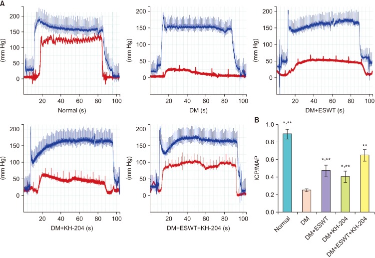 | Fig. 1Intracavernosal pressure (ICP), ICP/mean arterial pressure (MAP) of each groups. (A) Representative ICP recordings in response to cavernous nerve stimulation. (B) ICP/MAP ratios after nerve stimulation of the different groups. DM, diabetes mellitus; ESWT, extracorporeal shock wave therapy; KH, Korean herbal formula. ICP: red line, MAP: blue line. Data are expressed as means±standard deviations. *Statistical significance in comparison with the DM+ESWT+KH-204 group. **Statistical significance in comparison with the DM group.
|
2. ESWT+KH-204 effects on stimulation of smooth muscle/collagen ratio
The DM group showed statistically significantly less corporal smooth muscle tissue and higher collagen deposition than the control group (Fig. 2). The mean muscle/collagen ratios±SDs for the control, DM, DM+ESWT, DM+KH-204, and DM+ESWT+KH-204 groups were 0.21±0.04, 0.06±0.02, 0.10±0.02, 0.09±0.03, and 0.15±0.05 respectively. The mean ratio was significantly lower in the DM group than in the control group (p<0.001), but it recovered in the ESWT+KH-204 treated group compared with the DM group (p=0.003). The rats treated with ESWT+KH-204 showed a higher ratio than the single treatment groups (p=0.044 and p=0.048 respectively).
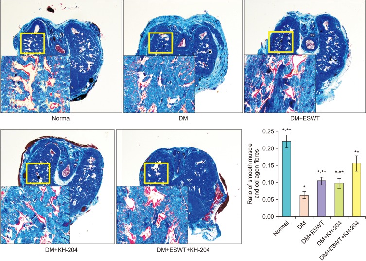 | Fig. 2Results of smooth muscle content evaluation by Masson's trichrome staining. Mean of the smooth muscle and collagen fiber ratios of the different groups (graph) (×40; inset, ×100). DM, diabetes mellitus; ESWT, extracorporeal shock wave therapy; KH, Korean herbal formula. Data are expressed as means±standard deviations. *Statistical significance in comparison with the DM+ESWT+KH-204 group. **Statistical significance in comparison with the DM group.
|
As shown in Fig. 3, α-SMA expression levels were elevated in the corpus cavernosum, indicating that smooth muscle contents in corpus cavernosal tissues. The mean intensities± SDs α-SMA of -positive areas for the control, DM, DM+ESWT, DM+KH-204, and DM+ESWT+KH-204 groups were 296.86±78.40, 80.55±30.77, 180.58±73.13, 147.92±48.34, and 249.61±75.01 respectively.
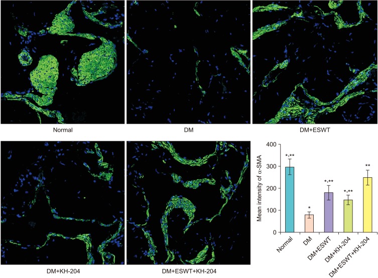 | Fig. 3Results of smooth muscle content evaluation by α-smooth muscle actin (α-SMA). Mean of the smooth muscle and collagen fiber ratios of the different groups (graph). DM, diabetes mellitus; ESWT, extracorporeal shock wave therapy; KH, Korean herbal formula. Data are expressed as means±standard deviations. *Statistical significance in comparison with the DM+ESWT+KH-204 group. **Statistical significance in comparison with the DM group.
|
3. ESWT+KH-204 results in more VEGF expression in corporal tissue
We performed immunohistochemical (IHC) staining to evaluate the expression of VEGF in corporal tissue (Fig. 4A). The mean intensities of VEGF staining for the control, DM, DM+ESWT, DM+KH-204, and DM+ESWT+KH-204 groups were 79.39±8.33, 30.29±9.43, 72.75±20.45, 65.82±11.61, and 89.22±14.35 respectively. It was decreased significantly in the DM group compared with the control group (p<0.001), while the ESWT+KH-204 group restored VEGF staining compared with the DM group significantly (p<0.001). The ESWT+KH-204 combined group showed higher intensities compared with the single-treatment groups (p=0.011 and p=0.031 respectively).
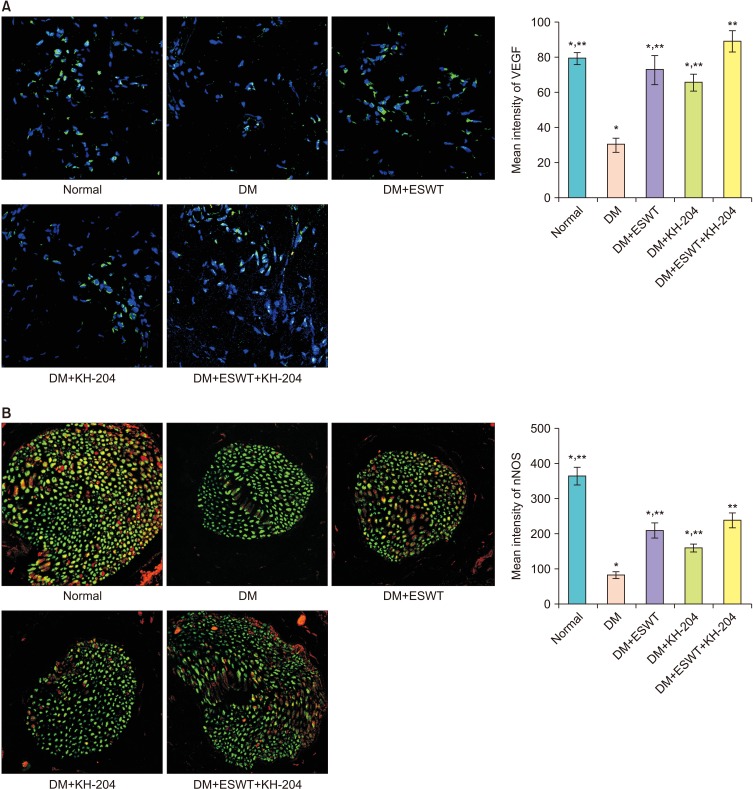 | Fig. 4(A) Immunohistochemical (IHC) staining of vascular endothelial growth factor (VEGF) expression in the corpus cavernosum of each group. Mean intensities of VEGF expression in the different groups are represented in the graphs. (B) IHC staining of neuronal nitric oxide synthase (nNOS) expression in the dorsal penile nerve of each group. Mean intensities of nNOS expression levels of the different groups are represented in the graphs. DM, diabetes mellitus; ESWT, extracorporeal shock wave therapy; KH, Korean herbal formula. Data are expressed as means±standard deviations. *Statistical significance in comparison with the DM+ESWT+KH-204 group. **Statistical significance in comparison with the DM group.
|
4. ESWT+KH-204 activates nNOS expression in the dorsal penile nerve
nNOS expression was assessed by IHC staining in dorsal nerve (Fig. 4B). In the DM group, rats showed a statistically significant decreased amount of nNOS-containing nerves. The mean intensities±SDs of nNOS-positive areas for the control, DM, DM+ESWT, DM+KH-204, and DM+ESWT+KH-204 groups were 366.16±60.95, 83.45±21.64, 210.82±54.16, 161.48±26.68, and 239.18±50.01 respectively (Fig. 4B). nNOS expression was significantly decreased in the DM group but it was significantly greater in the ESWT+KH-204 treatment group than in the DM group (p<0.001). The ESWT group recovered significantly higher nNOS levels as compared with the KH-204 group (p=0.045). The combined treatment group showed higher levels than the single treatment groups (p=0.039 and p=0.01 respectively).
5. ESWT+KH-204 promotes endothelial regeneration in corporal tissue
eNOS and PECAM-1 expression levels were assessed in corporal tissue by western blotting. P-eNOS expression was significantly lower in the DM group compared with the control group (p<0.001) and it was significantly restored with ESWT+KH-204 treatment (p=0.001). The mean densities of P-eNOS/eNOS for the control, DM, DM+ESWT, DM+KH-204, and DM+ESWT+KH-204 groups were 0.34±0.04, 0.14±0.03, 0.22±0.07, 0.23±0.06, and 0.30±0.05 respectively (Fig. 5A). The mean densities of the PECAM-1/β-actin ratio for the control, DM, DM+ESWT, DM+KH-204, and DM+ESWT+KH-204 groups were 0.34±0.05, 0.10±0.03, 0.20±0.06, 0.18±0.05, and 0.28±0.04 respectively (Fig. 5B). The ESWT+KH-204 treated group showed significantly restored PECAM-1 expression compared with DM rats (p<0.001). The combination treatment group showed higher densities and ratios compared with the single treatment groups (ESWT and KH-204; p=0.007 and p=0.02 respectively) in PECAM-1/β-actin ratio.
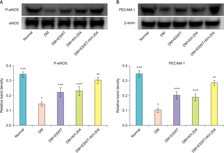 | Fig. 5(A) Comparison of the relative platelet endothelial cell adhesion molecule-1 (PECAM-1)/β-actin band densities of the different groups by Western blot. (B) Phosphorylated endothelial nitric oxide synthase (P-eNOS) expression in the corporal tissue of each group assessed by Western blotting. The relative band densities of P-eNOS/eNOS (graph). DM, diabetes mellitus; ESWT, extracorporeal shock wave therapy; KH, Korean herbal formula. Data are expressed as means±standard deviations. *Statistical significance in comparison with the DM+ESWT+KH-204 group. **Statistical significance in comparison with the DM group.
|
Go to :
DISCUSSION
The use of ESWT for the clinical treatment of ED has been gradually increasing in recent years. Even though most studies showed that ESWT had a positive effect on ED, Lu et al. [19] found that ESWT was more effective in mild ED. From a series of randomized sham-controlled trials, some researchers obtained positive results that the ESWT approach showed significantly good results in ED related questionnaire, even in PDE5I non-responders [20]. As a first-line drug for ED, PDE5I still has some limitations. In addition to non-responses, side-effects such as headache, dyspepsia, facial flushing, nasal congestion, and backache, cannot be ignored by patients and urologists and patients with heart disease have limited use [21]. Moreover PED5I have reduced efficacy in patients with diabetic ED, and is not a fundamental treatment and requires continuous use [22]. The mechanism of ESWT has not yet been fully elucidated, but there are several hypotheses. The shear stress of ESWT induces endothelial proliferation due to intra and extracellular responses. The increase in eNOS and VEGF due to ESWT is known to promote angiogenesis and restore ED [23]. In addition, ESWT is reported to help recruitment of stem cells, which is thought to restore the ED by helping the recovery of the nerve and blood vessels of erectile tissues [24].
Gerber et al. [25] believed that one main cause for DM-induced ED was vascular injury in the corpus cavernosum due to chronic hyperglycemia. And distal vessel spasm in the corpus cavernosum causes penis tissue ischemia. Jeon et al. [11] found that in the corpus cavernosum of rats with DM induced ED, the endothelial tissue apparently decreased. They hypothesized that the poor blood supply in the corpus cavernosum was the key reason for DM induced ED. Some researchers have found that ESWT increased VEGF in the injured tissue, and thereby improved regeneration of endothelial cells and vascular repair [26]. In addition, we found that the VEGF content in the corpus cavernosum of ED rats was lower in comparison with the control group.
In our previous study, we found KH-204 as an oriental herbal medicine could effectively decrease oxidative stress [17]. Thus, we considered that KH-204 could protect the corpus cavernosum from vascular injury by reducing oxidative stress. In this study, we found that the ICP was higher in the ESWT and KH-204 groups than in the DM group. The vascular endothelium content was also higher in the DM+ESWT+KH-204 group than in the single treatment group. The ICP result showed a similar trend. On the one hand, we thought ESWT could increase VEGF in the corpus cavernosum, improve regeneration of endothelial cells, promote vascular repair, and restore the blood supply. On the other hand, KH-204 could decrease oxidative stress in the corpus cavernosum of DM rats, reduce vascular damage, and protect injured tissue. Thus, we hypothesize that the protection mechanism and recovery mechanism of vascular and nerve regeneration coexist simultaneously, and improve ED.
Qiu et al. [24] and Li et al. [27] found that ESWT increased the expression of nNOS in the corpus cavernosum of DM rats, and they believed nNOS played an important role in regulating the dilation of blood vessels. With regard to this result, we found that, compared with the results of the ESWT group, nNOS had a higher expression in the DM+ESWT+KH-204 group, which also explained the ICP results. The combination of ESWT and KH-204 stimulated the nerve of the penis to produce a large amount of nNOS. That released nNOS combines with surrounding vascular smooth muscle cells, to expand the blood vessels and achieve a normal erection. Wang et al. [28] found that oxidative stress could increase cell injury. We also believe that it was the long-term oxidative stress in cavernous tissues that resulted in nerve injury and a reduction in the release of nNOS, which would induce ED. Oxidative stress was effectively inhibited by KH-204, which could reduce nerve injury in cavernous tissues. Thus, adequate nNOS produced by the nerve of the penis could play a role in the erectile response and might be one of the prime reasons that KH-204 improved ED.
In this study, administration of KH-204 and ESWT showed a better influence on ED treatment, but the further interaction between KH-204 and ESWT was not elucidated. The effect of angiogenesis of ESWT and the recovery of vascular injury obtained by the anti-oxidative stress effect of KH-204 are thought to help the recovery of vascular ED cooperatively. In addition, stimulation of endothelial cell by ESWT increase nNOS expression by increasing VEGF expression and anti-oxidative stress of KH-204 may also increase nNOS, which may play a role in synergic effect of combination therapy. The combination therapy of ESWT and KH-204 is expected to show better results in the clinical field than the single treatments because of its synergistic effect.
Go to :
 XML Download