

 PDF
PDF ePub
ePub Citation
 Print
Print


1. Development of tsutsugamushi disease in Korea
1) Before 1910
Although it is possible to identify disease epidemics in the general records including the Samguksaki describing the Three Kingdoms period (before 668) and the later records of the Goryeo (918–1392) and Chosun (1392–1897) dynasties, it is difficult to identify the specific diseases because of insufficient description of detailed symptoms [12]. These diseases were described as “Yeok,” such as “Yeok Jil,” “On Yeok,” or “Jang Yeok” and usually characterized by mass occurrence, acute manifestations, consistent symptoms, high mortality, and typically accompanied by fever and rash.
Statistics of notifiable diseases according to the modern system started from 1886–1911, and the diseases that were common in Korea at the time were smallpox, chickenpox, measles, scarlet fever, shigellosis, cholera, typhoid, diphtheria, pertussis, influenza, malaria, relapsing fever, Japanese encephalitis, epidemic meningitis (meningococcal infection), epidemic typhus, tuberculosis, leprosy, sexually transmitted diseases including syphilis, soil-mediated parasitic infections, and lung or liver fluke. With the exception of cholera and several newly introduced diseases, these diseases are indigenized diseases in Korea; thus, they may have been prevalent in the past. Thus, diseases recorded as “Yeok” can be assumed as one of them.
Among rickettsial diseases, epidemic typhus meets the above features of “Yeok” and may have been known long ago in Korea. The epidemiology of domestic epidemic typhus has drastically changed since the United States military used a large amount of dichlorodiphenyltrichloroethane, commonly known as DDT, to control body louse in 1945–1953. Thus, the epidemiologic pattern of epidemic typhus before 1945 is assumed to have been similar. As an example, of 4,668 patients (4,416 Koreans and 252 Japanese) who were admitted due to epidemic typhus at Kyungsung Municipal Sunhwa Hospital, the dedicated hospital for isolation and management of patients with tuberculosis or class I infectious diseases, in 1939–1945, 41% were manual workers, and 9.2% was transmitted by family members. Rash was usually observed in patients but was not hemorrhagic, and hearing loss was a complication in 25% of patients. The positive rate for OX19 was 54.3%, and the mortality rate was 9.0%. There were no patient in January, 7 in February, 15 in March, 54 in April, 80 in May, 45 in June, 15 in July, 4 in August, 2 in September, 5 in October, 14 in November, and 11 in December. Thus, the number of patients was the highest in spring (from April to June) [3]. April-June was the period of spring poverty in Korea when the nutritional status was the worst. It is presumed that Korean individuals were more affected than Japanese immigrants living in Korea. Other studies also showed that epidemic typhus most frequently developed in April-June and mainly in Korean individuals, manual workers, and men [4]. Among Japanese individuals, it developed in unemployed persons. If epidemic typhus recurs due to famine (Brill-Zinsser disease) and body louse is present, the spread of Rickettsia prowazekii makes epidemic typhus prevalent. Thus, the outbreak that occurred in spring from the Three Kingdoms period may have resulted from epidemic typhus. In particular, the pestilence in the early Chosun dynasty occurred mainly in spring. It originated in Pyeongan-do province, where prisoners were held in captivity. Since then, it occurred mainly in the northern and northeastern provinces, which are colder than the southern provinces. It was related to starvation and did not occur when grains were distributed for relief. It also did not occur in the capital city or royal family, which was consistent with epidemic typhus.
Before the introduction of Western medicine in Korea, the disease was termed as “Soo Dok” (water poisoning) in the book Dong Ui Bo Gam (Valuable encyclopedia on Oriental medicine) by Joon Heo, published in 1613, and is similar to “Shashidu” (sand louse intoxication) recorded in Chinese medical books. This term “Soo Dok” was also described in the book Hyang Yak Gu Geup Bang (Domestic medicine for first aid remedy), first published in 1232–1251, but the details of this illness were not described. Tsutsugamushi disease is described in Dong Ui Bo Gam as follows:
In the valley stream of the Gangnam area (※ the south side of the Yangtze River), there is an insect called Danho, Sagong, or Yeog. It does not have eyes but can hear well. When it hears the sound of people in the water and stings and injects its poison, which is why it is called Sagong (※ shooting expert) ... When a person is poisoned, he or she has a fever and feels suffocated and dizzy. The eyes and head ache ... There is another disease, Gyeon, which is similar to Sagong. However, skin ulceration occurs in Sagong, while not in Gyeon ... Another insect called Saseul, which is present under the scales of snake ... When a snake tries to dust off Saseul from its scales, the insect enters the sand. When a person is poisoned, a skin ulcer appears ... When the poisoned part of the body is cut off, the person immediately recovers, but if left untreated, the person can die within 2–3 days ... For treatment, garlic is put in boiling water, and the person is asked to bathe using this water. If there are rashes in the body, the skin has been already rotten by water poison.
It is a disease that causes fever, eschar or ulcer, and rash after being bitten by vermin at the river; therefore, it is similar to tsutsugamushi disease that was first reported in Japan (see “classical type tsutsugamushi disease”). It may be interpreted as schistosomiasis or tungiasis because of the presence of the terms “sand,” “water,” and “insect”; however, neither of these diseases cause fever and rash unlike tsutsugamushi disease. Additionally, common skin infections such as furunculosis are not the cause because these bacterial infections do not cause rash. However, this description is a quotation from the Chinese medical book Ishiyen Lumen (Introduction to medicine), published by Chen Lee in around 1580; thus, it is unclear whether this illness actually occurred in Korea. Further, “Shueidu” (water poisoning) in Ishiyen Lumen is a concise summary of at least three diseases. “Shashidu” (sand louse intoxication) and “Segongdu” (shooting insect intoxication) were first reported in Zhou Hou Bei Ji Fang (A handbook of prescriptions for emergencies) by Hong Ge, which was published in 313. There were also records of this disease as “Shashihu,” “Segonghu,” and “Shueiduhu” (water poisoning syndrome) in Zhu Bing Yuan Hou Lun by Uhan Phang Chao, which was published in 610.
2) From 1910 to 1945
At this period, the Empire of Japan had ruled Korea, and Western medicine was introduced in Korea. Tsutsugamushi disease is known to be endemic only in certain regions of Japan, namely, Niigata, Yamagata, and Akita Prefectures, and was severe and characterized by eschar and lymphadenopathy. Thus, the development of tsutsugamushi disease is easily recognizable. However, there was no report on patients with this disease in Korea, and therefore domestic physicians may not have been interested in it. Among the available tests at that time, namely animal inoculation and Weil-Felix tests, the OX19 antigen in the Weil-Felix test was used for the diagnosis of epidemic typhus. The OXK antigen was not widely used in Korea.
Japanese individuals migrated after they invaded Taiwan and Korea. Therefore, if these two countries had tsutsugamushi disease, they might have experienced and diagnosed the disease. In Taiwan, tsutsugamushi disease developed in Japanese officers and migrants working in newly constructed plantations, which was observed by Hatori since 1914 [5]. In contrast, there was no such report in Korea.
In Korea, Weir, a medical missionary, reported fever of unknown origin in 1915 [6]. The fever naturally subsided within 10–14 days and was accompanied by relative bradycardia, rash, conjunctival hyperemia, splenomegaly, and hearing loss. Thus, the author suspected rickettsial disease and used the term “paratyphus”. The main finding of the study was as follows. In 1913–1914, there were two cases in March, three cases in April, eight cases in May, and two cases in June. Thus, the disease only developed in spring and was considered mild because there was no death among 15 patients. Therefore, it was not a typical epidemic typhus. Patients were found in Jemulpo, Incheon, which was the treaty port area at the end of the Korean Empire; therefore, it was either epidemic typhus or murine typhus. In Korea, murine typhus develops throughout the year, and its incidence increases in the fall. Tsutsugamushi disease develops during the summer or fall, therefore, the season of development of “paratyphus” did not correspond to that of these diseases. In conclusion, the disease reported by Weir only developed in March–June, and therefore it was more likely epidemic typhus rather than tsutsugamushi disease. It was also mild, and therefore it may have been Brill-Zinsser disease or mild epidemic typhus, as was present in Mexico or Russia.
From 1910 to 1945, there were many studies on epidemic typhus and murine typhus in Korea. However, it is not easy to distinguish between these two diseases. At that time, considering the epidemiology and clinical features, mass outbreak was thought to be epidemic typhus, and sporadic typhus was classified as “endemic typhus.” The latter included several rickettsial diseases, but in the 1910s - 1920s, these diseases were not distinguished and were regarded as one disease. In 1932, the criteria for distinguishing epidemic typhus from murine typhus were presented in the Japanese academia [7], which summarized the results of animal experiments in addition to epidemiologic and clinical findings of the above two diseases. However, it is difficult to perform an animal test in clinical practice; hence, most studies on typhus were conducted based on clinical (presence of encephalopathy or hemorrhagic rash) and epidemiologic findings (outbreak or familial occurrence), due to which the diagnosis was not accurate. For example, although it was a familial occurrence, the father and mother were diagnosed with epidemic typhus, while the children with murine typhus. Moreover, often, the term “intermediate type” of murine typhus and epidemic typhus was used due to the ambiguous clinical features. Based on these criteria, murine typhus was diagnosed in several regions in Japan, Taiwan, and Manchuria. Diagnosis was also made in Korea based on these criteria. In particular, there were several studies on murine typhus in Pyongyang, the second largest city of Chosun at that time and currently the capital city of North Korea. The city was expanding rapidly, particularly in the east areas of the Daedong River [8]. For example, in Pyongyang, in terms of murine typhus that developed from 1932 to 1935, there was fever and rash, but no eschar and lymphadenopathy. Low positive OX19 titer values were also common in normal persons, so that the positive criterion was ≥1:80. There was no description of the OXK antigen test, making it unclear whether Orientia tsutsugamushi infection was excluded. Of 101 patients, 88 were Japanese migrants, and 13 were Korean individuals. Interestingly, there were 5 patients with murine typhus in January, 0 in February, 2 in March, 1 in April, 4 in May, 1 in June, 2 in July, 2 in August, 18 in September, 29 in October, 24 in November, and 13 in December [9]. Such an epidemic in fall is similar to the season in which tsutsugamushi disease and murine typhus develop nowadays. Some patients with low OX19 levels but high OXK titer levels may have been tsutsugamushi disease [10].
Other evidence suggesting the existence of tsutsugamushi disease in Korea includes the steady occurrence of arthropod-mediated disease. Thus, it is rather reasonable to assume that this disease had developed in Korea even before 1945. In 1917, mites attached to wild rats collected in Suwon were similar to Trombicula akamushi, which was isolated in Yagamata Prefecture [11]; thus, mite vectors were present in Korea. Furthermore, Japanese physicians who had a lot of experience in tsutsugamushi disease were only familiar with typical tsutsugamushi disease; hence, they might have overlooked atypical cases. In Japan, tsutsugamushi disease that occurred before 1945 is called “classical type tsutsugamushi disease” and characterized by lymphadenopathy, eschar, fever, and rash. It also had a high mortality rate of 15 – 60% and was therefore considered severe. It also developed only in summer in persons who worked inside the riverbank along the large rivers of the three northwestern regions. In contrast, “new type tsutsugamushi disease” first developed among the Allied Forces in the vicinity of Mount Fuji in 1948. Thereafter, it developed sporadically throughout the country. In 1951 – 1952, there was a large outbreak of tsutsugamushi disease in the Izu Shichito Islands (“Shichito fever”) [12]. Since then, Japanese physicians became familiar with the benign type of tsutsugamushi disease. The new type was so mild that individuals who were diagnosed with it frequently recovered without antibiotic therapy. Moreover, there were no deaths. The exposure to scrub area rather than river was a risk factor, and the disease developed in fall, winter, or spring. Therefore, before 1945, even Japanese physicians with extensive experience on tsutsugamushi disease would not have suspected mild tsutsugamushi disease [13]. Thus, tsutsugamushi disease in Korea before 1945 was presumed to have mild or atypical manifestation similar to fever of unknown origin or murine typhus, and therefore domestic physicians seemed to have overlooked this disease.
According to the book Global Epidemiology, which was published in 1944 and recorded the endemic diseases in each country, “three types of typhus occur in Korea, and they are epidemic typhus, murine typhus (known as “Honan fever” in Korea) (※ “Honan fever” is a misnomer of “Honam fever”, which was proposed by Suo and Ishihara in 1939), and tsutsugamushi disease. Murine typhus is the most common, and tsutsugamushi disease the least. The mortality rate of tsutsugamushi disease in Korea is unknown. Mites are present, and the most important species is Trombicula akamushi” [14]. However, it is impossible to confirm the literature it was based on, but it is presumed that a small number of Western physicians were in Korea, and thus the data may have been obtained from them.
3) After 1945
The history of tsutsugamushi disease after the liberation of Korea from the Japanese Imperial rule in 1945 and the study results immediately after 1986 are described in the book Tsutsugamushi Disease in Korea by ex-professor Woo Hyun Chang as well as a review article [1516]. There were descriptions on rickettsial infections in Korea in the reports Public Health and Welfare Technical Bulletin (1947) and Epidemiology of the Diseases of Naval Importance in Korea (1948) [17], although these reports seemed to be based on insufficient data.
Two British soldiers each showed fever and eschar while being stationed near the Imjin River in June and July, respectively, 1951 during the Korean War. OXK-positive reactions were reported in the Weil-Felix test [18]. In the US Army, one was infected in Masan and three in the central front in November 1951 [19]. A US soldier developed fever, eschar, rash, and lymphadenopathy on October 16, 1953. He showed the negative OXK antigen reaction on day 8, but it increased to 1:640 on day 19, and rickettsial blood culture was positive. An Australian solider who worked at the western front developed the disease on November 3 and showed rash and eschar. The Weil-Felix test result was negative, but the culture was positive. Both patients improved with chloramphenicol treatment, and the symptoms, epidemiology, examination, and response to treatment were in accordance with tsutsugamushi disease, revealing that tsutsugamushi disease was also present in Korea [2021]. O. tsutsugamushi was isolated from 12 wild rats collected in Cheolwon, Geumhwa, and Yonchon in 1952–1953. It was also isolated from Trombicula pallida larvae, demonstrating that this infection was widespread in wild rats in the central area. Complement fixation antibody in rat blood was positive in one rat [22]. In 1964, an examination was conducted in Korean individuals. As a result of performing Weil-Felix test on 83 healthy Korean individuals (residents and soldiers) residing near the ceasefire line (Cheolwon, Yanggu, Pocheon, etc.), 8 showed positive reaction in the 1:20 dilution. All 45 persons living in Seoul who had never traveled to these areas showed negative reaction. In the experiment in which 106 wild rat tissues were inoculated into experimental animals, rickettsia reaction was negative [23]. This study demonstrated that infection was common in Korean individuals living in the endemic regions but was not related to the symptoms. Due to the limitations of the Weil-Felix test, the results could not be further investigated. The failure to verify rickettsia in animal inoculation was also a weakness of this study. There was a report on Leptotrombidium mites including T. pallida, T. palpalis, and T. orientalis in 1954 [24]. Since then, there had been no report on rickettsia in Korea for 30 years. However, in 1969, febrile diseases developed in the Korean Army who participated in the Vietnam War, and 13 sera showed positive reaction for Weil-Felix OXK antigen [25].
In October 1985, a Japanese individual who was touring the Jeju Island and playing golf, suffered from the tsutsugamushi disease immediately after returning home, which was reported in 1986 but did not draw attention [26]. At this time, some Korean individuals exposed to grass in rural areas presented with fever similar to typhoid fever and rash larger than rose spots of typhoid fever. Moreover, some patients improved with chloramphenicol therapy, but the disease was misdiagnosed as culture-negative typhoid fever. Further, since the outbreak of leptospirosis was first noticed in Korea at that time, it was considered first. There were no facilities available to diagnose rickettsial infection in Korea. In 1986, serologically diagnosed cases of tsutsugamushi disease in Korean individuals living domestically were reported in Chinhae by Yi et al. and in Seoul by Lee et al. [2728]. In 1987, Chang and Kang isolated the bacteria from patients and demonstrated that tsutsugamushi disease developed in the domestic population [29]. Since then, many clinicians and researchers have studied the epidemiology, characteristics of bacteria, pathology, clinical features, and treatment of tsutsugamushi disease in Korea. In particular, leptospirosis has been identified as the cause of “pulmonary hemorrhagic syndrome” in the central region since 1975, and tsutsugamushi disease has also been confirmed as one of the cause since the mid-1980s. Thereafter, outbreaks of acute febrile illness that occurred in the fall were recognized as a new syndrome and called “autumn febrile syndrome”. Additionally, at that time, murine typhus, leptospirosis, and hemorrhagic fever with renal syndrome were the main causes of this syndrome, but their incidence decreased, and the incidence of tsutsugamushi disease relatively increased. In the 1980s and 1990s, clinical and epidemiologic features were reported in nearly all university hospitals in each province. However, since 2000, the number of patients has increased according to the statistics of the Korea Centers for Disease Control and Prevention (KCDC), but the number of clinical studies has decreased.
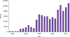
The number of reported patients has been steadily increasing; as tsutsugamushi disease was designated as a legal communicable disease in 1994, the reporting system improved, and physicians' awareness increased. The number of patients reported to the KCDC increased from 1,000 to 2,000 in the early 2000s to 4,698 in 2004. Subsequently, it increased sharply and reached 10,365 in 2013 (Fig. 1). The increase of outdoor activity and antibiotic use changes may contribute this change. Further, global warming has increased the density of mites, possibly leading to an increase in tsutsugamushi disease.
2. Disease feature in Korea
The most important feature of tsutsugamushi disease is being arthropod-mediated, maintaining the infection in nature through the relationship of warm-blooded animals, mites, and O. tsutsugamushi. Humans are warm-blooded animals and accidentally infected. Therefore, to understand tsutsugamushi disease, it is necessary to study these three factors and human reactions, which make it more complicated to study than other diseases. However, it shows excellent responses to antibiotics, and many aspects to be studied have been overlooked. The three factors above are briefly summarized below.
1) Bacteria
O. tsutsugamushi is a strictly intracellular bacterium that requires host cells to grow and was once classified a virus due to its small size. In 1995, it was reclassified from Rickettsia to Orientia. It was changed to O. tsutsugamushi based on the combination of Rickettsia orientalis and R. tsutsugamushi [30]. Its serotypes are very diverse. In Korea, the distribution of serotypes was different depending on the region. In research on blood culture isolates using monoclonal antibodies, Karp and Gilliam types were distributed in Gyeonggi-do and Gangwon-do, and the Boryong type (Kuroki type in Japan) was distributed mainly in Chungcheongnam-do [31]. There were also a few reports on the Yonchon type [32]. Since then, eschar or blood was amplified by polymerase chain reaction (PCR); then, the genotypes were investigated through sequencing. Boryong and Kawasaki types were the main genotypes [33]. Patients in Gangwon-do showed more severe symptoms than those in the southern area in the 1980s – 1990s. Considering the severity and regional serotype/genotype distributions of the patients, some researchers argued that it was similar to the relationship between the Japanese classical type (Gilliam, Karp, and Kato types, which are severe) and new type (Kuroki and Kawasaki types, which are mild). In 2007, the genome sequence of the Boryong strain was first decoded by Cho et al. [34], followed by the genome sequence of Ikeda and Karp strains.
In 2000, the threat of bioterrorism was emphasized, and laboratory safety was reinforced. As a result, research on tsutsugamushi disease required biosafety grade 3 facility. Therefore, the number of studies that required culture were drastically decreased due to installation and maintenance cost.
2) Mite
Leptotrombidium mites go through four stages: egg, larva (also called chigger), nymph, and adult. The larva only feeds on the body fluids of animals, and nymphs and adults live by eating insect eggs under the leaves or in the ground. The humidity of the soil they inhabit is important for their survival, and a place with tall grasses, such as Kunai grass in New Guinea or Susuki grass in Japan, to protect them from sunlight, is optimal. In areas inhabited by humans, the grass is cut off and soil is tread on, creating an inadequate environment for the mites to live. Therefore, within 1–2 years of residence on a deserted meadow, a person develops tsutsugamushi disease, but the risk decreases when the person continues to live there. The larva is reddish and 0.25 mm in length, and therefore it is difficult to identify it with the naked eyes of unskilled persons. When attached to an animal, it releases saliva containing proteolytic enzymes, dissolving the skin to make a stylostome and then feeding on the body fluid through this tube. This act is described as “bloodsucking,” but unlike mosquitoes, it does not suck blood. People do not feel any symptoms. The saliva contains bacteria, which enter simultaneously and cause tsutsugamushi disease. Only 0.2 - 2.1% of the larvae collected from epidemic regions had bacteria. Another study reported that the probability of a person developing tsutsugamushi disease after being bitten by a larva was about 2.5%. Usually, once attached, it sucks the body fluids for 3 - 4 days and falls off. Next, it metamorphoses into a nymph. Infected adults lay infected eggs via transovarian transmission, which develop into infectious larvae. They may be infected (transtadial) while developing from larvae to adults, maintaining infection in nature. Considering this life cycle, transmission by the larvae from an infected to an uninfected animal is impossible. However, some larvae (10%) attach to other animals to continue sucking when they fall off before engorged fully. The transmission of infection from an animal to another in this way has been demonstrated. Unlike louse in epidemic typhus, mites with this bacterium do not die.
The mite species is an important factor in determining the epidemiology of this disease. It may be also related to differences in clinical features. Thus, it has been studied continuously. In Southeast Asia, L. deliensis, which is similar to L. akamushi, is the main mediator. In Korea, L. pallidum and L. scutellare are dominant species [35]. In particular, in the southern part of the country, L. scutellare increases 1 - 2 months earlier in fall than the outbreak of the disease. In the northern and central regions, L. pallidum is abundant and increases in number in spring or fall.
3) Warm-blooded animal
Warm-blooded animals are considered as less important than causative bacteria or mites. However, wild rodents or other small animals must coexist with mites so that they can complete their life cycle. Among domestic wild rodents, Apodemus agrarius is the most common, and the positive rate of indirect immunofluorescent antibody for O. tsutsugamushi is 20–100%, and high in fall and winter [36].
4) Epidemiology
The disease develops throughout the country, especially in inland areas in Jeolla-do and Gyeongsang-do and Chungcheongnam-do (Fig. 2) and is epidemic from October to November. However, the number of patients during the non-epidemic period has increased recently. The disease is mainly reported in people living in the countryside or enjoying outdoor activities. Moreover, the cases related to mountaineering and parks in the city are also increasing. It is more prevalent in female residents in rural areas and male visitors in outdoor activities. In terms of age, it is common in the age of 50 - 70 years [37].
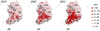 | Figure 2Incidence rates of tsutsugamushi disease in Korea, by area and year.SE, Seoul; GG, Gyeonggi-do; GW, Gangwon-do; CB, Chungcheongbuk-do; CN, Chungcheongnam-do; JB, Jeollabuk-do; JN, Jeollanam-do; GB, Gyeongsangbuk-do; GN, Gyeongsangnam-do; JJ, Jeju Special self-governing Province.
Cited from Lee HW, et al. Current situation of scrub typhus in South Korea from 2001-2013. Parasit Vectors 2015;8:238.
|
5) Clinical features
The clinical features of tsutsugamushi disease reported in Korea are similar to those of its classical type in Japan. The delayed treatment is associated with high complications and mortality [38], but the reported mortality in Korea is generally low (<1%), as antibiotics is used. Most Korean patients present the reinfection type rather than the primary antibody response types [27], as reported in Taiwan [39]. However, in the tropical region, it is difficult to diagnose it based only on symptoms because eschar or rash is not observed in many cases. For example, patients with O. tsutsugamushi infection, which was reported in Malaysia in 1976, did not show any rash or eschar, even though the diagnosis was confirmed [40].
The phenomenon in which O. tsutsugamushi causes persistent infection in animals has been well known previously [41], but there have been few studies on humans. In 1948, persistent infection was confirmed in one patient who was injected with O. tsutsugamushi for the treatment of neurosyphilis [42]. In 1952, one patient showed persistent infection in the lymph node [43]. After 60 years, in 2012, it was demonstrated that the bacteria persisted in the blood of six patients. In particular, the surface antigen coding gene sequences of the sequentially isolated bacteria in the acute and chronic phases were identified to be same. Thus, the possibility of reinfection was excluded, and persistent infection was confirmed [44]. In 2014, persons who did not have any risk factor of exposure to mites developed O. tsutsugamushi pneumonia, which may have resulted from the recurrence of latent infection [4546].
6) Diagnosis
In the 1980s, when tsutsugamushi disease re-emerged in Korean individuals, an indirect immunofluorescence method was already established, and was used in domestic universities, hospitals, and commercial laboratories. PCR was also performed. As the Korea National Institute of Health expanded into the KCDC, the number of samples that could be processed increased, and the test was free. Therefore, many hospitals stopped the test and outsourced the tests to the KCDC. Additionally, the National Health Insurance Service reimbursed the examination cost for only one of the two tests when the IgG and IgM antibodies were tested simultaneously or for one or two items if several antibody tests were performed for suspected organisms. Thus, it is difficult to diagnose the causative bacteria in patients showing atypical clinical features. Cell culture or animal inoculation was not performed in clinical medicine due to the difficulty in acquiring new technology licensing. Since 1993, several rapid diagnostic kits have been developed and used in developing countries as well as in Korea [47].
In Korea, the importance of the test is low because most Korean patients present typical features. However, the clinical features are atypical globally, and thus it is difficult to diagnose. As a result, the treatment is delayed, mortality is still high (5 – 17%), and pregnancy-related complications such as abortion are common in pregnant women.
7) Treatment
Currently, doxycycline is mainly used, and its effect is excellent. However, this antibiotic is generally not recommended for the following conditions: pregnancy, young children, inability to take oral medicine, and doxycycline resistance. The use of doxycycline for several days in pregnant women and children does not cause major problems, but macrolide antibiotics are safe even in this case, and therefore it can be a safe alternative [4849]. Moreover, azithromycin has an injectable form and thus can be used in people who have difficulty in oral administration. Tigecycline is another tetracycline antibiotic that shows a good in vitro efficacy and has an intravenous form [50]. The therapeutic effect of chloramphenicol has been proved for the first time, but it has a slower response than tetracycline and involves serious side effects. Thus, it is not currently used. Although rifampicin showed efficacy in comparative studies, there is no reason to use this drug for the treatment of tsutsugamushi disease. Although quinolone preparations have shown some therapeutic effects, quinolone resistance genes have been detected, and there have been cases of treatment failures. Thus, they have not been used as a first-line therapy.
The antibiotic effect seems to be lower than that in the past. In 1978, a study in Malaysia found that even a single dose of doxycycline led to full recovery [51]. However, according to a domestic research in 1995, even doxycycline treatment for 3 days did not alleviate fever in some patients [52]. For this reason, the treatment period for tsutsugamushi disease has not yet been established. Currently, it is being used for 1 week in Korea and longer in Japan.
Doxycycline resistance was reported in Thailand in 1996 [53]. It took 2 more days for patients infected in Chiang Rai to recover from fever after the start of doxycycline treatment than patients in Mae Sod region, which was the control group. When the bacteria isolated in Chiang Rai were inoculated into mice, the effect of antibiotics was low. However, no delayed response to antibiotics was demonstrated in a domestic study in 2008. No resistant strain was found in the antibiotic susceptibility test for the isolated strains [54]. There have also been no further reports on treatment response delay by other researchers in the same region of Thailand over 20 years [55]. In Australia in 2016, tsutsugamushi disease developed in soldiers taking prophylactic doxycycline. Susceptibility was detected in one patient when the causative bacteria were isolated and the antibiotic susceptibility test was performed [56]. A recent study evaluating doxycycline resistance using quantitative PCR did not detect resistant strain among 48 isolates [57]. Thus, this subject might be a matter of growth characteristics of the bacterial strains instead of antibiotic resistance [58].
3. Quarantine and management
It is a mite-mediated disease; therefore, reducing the mite bite can decrease the risk of tsutsugamushi disease. These are the following methods: getting rid of mite habitats (burning grass or bulldozing), sprinkling persistent pesticides on the grass around their habitats, and spraying insect repellents on the body or clothes, which showed a clear effect in soldiers during the World War II. However, it is questionable whether these methods can be used by endemic area residents in the non-war situation because these methods destroy the environment and make the land unproductive. It is also unclear whether it is more efficient than early diagnosis and treatment. Prophylactically using doxycycline has a preventive effect. It is used when the risk is temporarily high, as in a soldier training on the grass. However, even in such cases, it is common not to take the medicine, and thus the disease appears in some cases [56]. It is hard for residents to recognize the risk, and they have to take it every year, making it difficult for them to take prophylactic antibiotics. Thus, prophylactic antibiotics have not been formally used domestically.
Although prevention is the best, vaccines developed previously have not proved effective in clinical studies. One exception is the trial conducted by Kawamura using a live strain showing low virulence [59]. Moreover, these bacteria have varying serotypes, and therefore the antigens contained in the vaccines do not induce cross immunity [60]. As a result, no vaccines are currently in use or under development.
As described above, there are no established methods to control the outbreak of tsutsugamushi disease. Rapid diagnosis and treatment have a significant effect in reducing deaths, and therefore, Korea relies more on medical care than preventive care. Since tsutsugamushi disease was designated as a notifiable communicable disease in 1994, occurrence according to the period and region has been investigated nationwide. Based on these data, the KCDC and public health centers have promoted preventive education and early treatment for this disease to local residents during the risk period every year. However, the increase in the number of outbreaks since the 2000s shows that these measures were ineffective. Insect repellents are recommended, but it is not easy to apply them every time, and thus compliance is not good. Although not intended, the expansion of cultivated land or residential area will reduce forests, decreasing the exposure to mites. Moreover, the expansion of cultivated land will decrease the number of mice, reducing the occurrence of tsutsugamushi disease.
 XML Download