

 PDF
PDF ePub
ePub Citation
 Print
Print


INTRODUCTION
Tooth replantation has been proposed as an alternative technique to manage early tooth loss, especially in situations where dental implants and other prosthetic treatments are impossible [1]. In the field of pediatric dentistry, tooth replantation is indicated in situations such as tooth avulsion by traumatic dental injuries (TDIs). Avulsion of permanent teeth is commonly observed in children aged 7–9 years because the supporting tooth structures comprising loose periodontal ligament (PDL) and low-density alveolar bone, combined with an immature root structure, provide insufficient resistance to traumatic forces [23].
In avulsion injuries, severe damage to the PDL and loss of blood supply to the pulp tissue could lead to pulp necrosis and root resorption after replantation [45]. Extra-oral time prior to replantation is a critical factor for the survival of the replanted tooth [6]; therefore, immediate replantation is recommended for a better prognosis [25]. However, immediate replantation at the TDI site is difficult if there is a lack of knowledge regarding the replantation method and complex and/or complicated trauma affecting multiple body parts [7], which may lead to a delay in replantation. In recent years, abundant online information regarding the storage and transfer of avulsed teeth in a storage medium when immediate replantation is not feasible has become readily accessible. In cases of tooth avulsion, an appropriate storage medium is required to maintain the vitality of PDL cells until tooth replantation can be accomplished, and Hank's balanced salt solution (HBSS) has been recommended for this purpose [8].
The topical use of antibiotics, such as doxycycline or minocycline, prior to tooth replantation has been proposed [91011]. Antibiotics of the tetracycline class show broad-spectrum activity and easily bind to the root surface in a reversible manner without losing their antimicrobial action, even over a prolonged duration [12]. These antibiotics have been applied to the root surface in order to remove microorganisms and inhibit the development of an inflammatory response after replantation [1013].
Previous studies have shown consistent findings with respect to the effects of topical doxycycline treatment in patients undergoing tooth replantation [101114]. In fact, root surface treatment with doxycycline improved the likelihood of pulp revascularization after immediate replantation [1011]. Regarding delayed replantation, Jabarifar et al. [14] evaluated the effect of topical treatment with doxycycline prior to the replantation of canine teeth after 2 hours of storage. However, the authors provided limited evidence for the effectiveness of doxycycline because their study mainly focused on the effects of different types of storage media and did not include a control group of delayed replantation without doxycycline treatment.
Doxycycline has been evaluated for its efficacy in root surface treatment in both immediate and delayed replantation. Tooth replantation remains an important clinical issue, as it is frequently encountered by dental practitioners. Therefore, the present study aimed to evaluate the effects of root conditioning with doxycycline on periodontal and pulpal healing in replanted rat teeth preserved under different storage conditions.
MATERIALS AND METHODS
Animal preparation and definition of experimental groups
The experimental protocol of our study was reviewed and approved by the Ethics in Institutional Animal Care and Use Committee of Kyung Hee Medical Center, Kyung Hee University, Seoul, Korea (KHMC-IACUC-16-022). A total of 20 male Sprague-Dawley rats (Samtaco, Osan, Korea) aged 6–8 weeks and weighing approximately 150–200 g were used. This study used a split-mouth design, in which 40 maxillary first molars with complete root formation were allocated to the root surface treatment with doxycycline groups and control groups, under different storage conditions. Accordingly, the teeth were randomly divided into the following 4 groups (n=10 per group): i) Group I (control): replantation after 5 minutes of dry storage; ii) Group II (doxycycline treatment): replantation after 5 minutes of dry storage; iii) Group III (control): replantation after 60 minutes of storage in HBSS (Gibco Laboratories, Grand Island, NY, USA); iv) Group IV (doxycycline treatment): replantation after 60 minutes of storage in HBSS.
The teeth in the doxycycline treatment groups were soaked in doxycycline (1 mg/20 mL saline; Hana Pharm Co., Seoul, Korea) for 5 minutes prior to replantation [8].
Surgical procedures
For the surgical intervention, the animals were anesthetized by administering Zoletil 50 (Virbac Lab, Carros, France) intramuscularly at a dose of 100–150 mg/kg and underwent a 5-day treatment with 0.4% β–aminopropionitrile (β-APN; Sigma-Aldrich, St. Louis, MO, USA) to avoid traumatic extraction [15]. Forty bilateral maxillary first molars with complete root formation were extracted from 20 animals. Preoperatively, the oral cavity of the animals was cleaned with 2% chlorhexidine solution. Extraction of the maxillary first molars was performed gently using sterile extraction forceps. After excluding any roots that fractured during extraction, the allotment of 10 teeth per group was maintained by including additional experimental animals. The extracted teeth were exposed to a dry environment for 5 minutes at room temperature or preserved in HBSS solution for 60 minutes; subsequently, the teeth and sockets were cleaned with saline prior to replantation. After replantation, a single dose of 20,000 IU of penicillin G (Alvogen potassium penicillin G, Alvogen, Seoul, Korea) was administered intramuscularly in all animals, and they were fed a soft diet for 7 days [7]; euthanasia was performed 8 weeks after replantation (Figure 1).
Micro-computed tomography analysis
Micro-computed tomography (micro-CT) was performed at the Advanced Institutes of Convergence Technology (Genoss Co., Ltd., Suwon, Korea). Micro-CT data of the relevant maxilla were acquired on a SkyScan 1173 scanner (Bruker-microCT, Kontich, Belgium) at 14.91 μm resolution (130 kV and 60 μA). The acquired data were reconstructed with NRecon software (version 1.7.0.4, SkyScan, Kontich, Belgium). The micro-CT analysis was carried out by an experienced pediatric dentist (Nam OH), wherein the presence of periapical radiolucency feature indicated pulpal healing and the severity of root resorption indicated periodontal healing. The severity of root resorption was categorized as follows: i) Grade 0: no root resorption; ii) Grade 1 (mild): root resorption less than one-third of the total root length; iii) Grade 2 (moderate): root resorption less than two-thirds of the total root length; iv) Grade 3 (severe): root resorption more than two-thirds of the total root length.
Histological processing
The specimens were washed, dehydrated, embedded in paraffin, and sectioned at a thickness of 5–8 μm, using a microtome, in the transverse plane from the middle third of the mesiobuccal root. The slides were stained with hematoxylin and eosin and scanned (Panoramic 250 Flash III, 3DHISTECH, Budapest, Hungary), followed by fixation in 10% buffered formalin and decalcification with 10% formic acid.
Histomorphometric analysis
The scanned slides were observed using slide-viewing software (CaseViewer ver. 2.1, 3DHISTECH). The histomorphometric analysis was carried out by 3 experienced and trained examiners (Nam OH, Kim MS, and Lee HS), who were blinded to the group allocation. The analysis included the following parameters: i) Pulpal healing: area of pulpal inflammation; ii) Periodontal healing: area of surface root resorption, inflammatory root resorption, and replacement root resorption (ankylosis).
Both pulpal healing and periodontal healing were graded on a 4-point scale for each parameter as follows: grade 0, not observed; grade 1, <25% observed; grade 2, <50% observed; grade 3, <75% observed; and grade 4, ≥75% observed [16].
Statistical analysis
The histomorphometric results were represented as mean±standard deviation. Data were analyzed using SPSS version 15.0 (SPSS Inc., Chicago, IL, USA). The micro-CT results were analyzed using the χ2 test and Kruskal-Wallis test. Inter-examiner repeatability in the histomorphometric analysis was assessed based on the intra-class coefficient (ICC). The normality test for the histomorphometric analysis was performed using the Kolmogorov-Smirnov test, which revealed that the data were not normally distributed. The histomorphometric results were therefore analyzed using the Mann-Whitney test. P values <0.05 were considered to indicate statistical significance.
RESULTS
Micro–CT analysis
In teeth subjected to replantation after 5 minutes of dry storage, no statistically significant difference was observed between groups I and II (Table 1). In contrast, the teeth subjected to 60 minutes of storage in HBSS showed lower degree of root resorption in group IV than in group III (P<0.05), with no significant difference in the frequency of periapical radiolucency between these groups.
Table 1
Micro-computed tomography analysis

The chi-square test was used for periapical radiolucency and the Kruskal-Wallis test was used for root resorption.
Group I: replantation with no treatment after 5 minutes of dry storage, Group II: replantation with doxycycline treatment after 5 minutes of dry storage, Group III: replantation with no treatment after 60 minutes of storage in HBSS, Group IV: replantation with doxycycline treatment after 60 minutes of storage in HBSS, HBSS: Hank's balanced saline solution.
a)Statistically significant difference observed between groups III and IV (P<0.05).
![]()
Histomorphometric analyses
The ICC values indicated highly acceptable inter-examiner reliability in the histomorphometric analysis (ICC, 0.802–1.000). In the comparison of groups I and II, no significant differences were found in pulpal and periodontal healing (Table 2 and Figure 2). In group IV, the mean grade of surface root resorption was 1.37±0.77 and the mean grade of inflammatory root resorption was 1.33±0.71, whereas in group III, the grades were 2.03±0.96 and 2.00±0.95, respectively (Table 3 and Figure 3). Significantly lower grades of surface resorption and inflammatory resorption were observed in group IV than in group III (P=0.009 for surface resorption and P=0.011 for inflammatory resorption). The histomorphometric analysis revealed a mean grade of replacement resorption of 0±0 in group I, 0.10±0.31 in group II, 0.10±0.31 in group III, and 0.10±0.31 in group IV, with no significant differences (P>0.05). Moreover, no significant difference was found in pulpal healing between groups III and IV (P>0.05).
Table 2
Histomorphometric analysis of the groups that underwent replantation after 5 minutes of dry storage

The Mann-Whitney test was used for statistical analysis.
Group I: replantation with no treatment after 5 minutes of dry storage, Group II: replantation with doxycycline treatment after 5 minutes of dry storage.
![]()
 | Figure 2Sections of teeth replanted after 5 minutes of dry storage. (A) Gross histological characteristics of a representative root from group I reveals newly formed PDL-like tissues attached to most of the root surface, while some areas are without PDL-like tissue attachment, showing inflammatory resorption (asterisk). (B) Sagittal micro-CT image of the root in group I shows a visible CM in the canal space. (C) High magnification of the square region of the pulp in the control group shows disruption of the odontoblastic layer (arrows). (D) High magnification of the square region of the pulp proper in the control group shows inflammatory cells and fibroblasts in the pulp proper. (E) Gross histological characteristics of a representative root from group II reveals newly formed PDL-like tissues attached to most of the root surface, while some areas fail to exhibit PDL-like tissue attachment. (F) Sagittal micro-CT image of a root from group II. (G) High magnification of the square region of the pulp in the doxycycline group shows disruption of the odontoblastic layer (arrows). (H) High magnification of the square region of the pulp in the doxycycline group shows congestion of the blood vessels in the pulp proper. (I) Histologic grade of pulpal inflammation. (J) Histologic grade of surface root resorption. (K) Histologic grade of inflammatory resorption.PDL: periodontal ligament, CT: computed tomography, CM: calcified mass.
|
Table 3
Histomorphometric analysis in the groups that underwent replantation after 60 minutes of storage in HBSS

Group III: replantation with no treatment after 60 minutes of storage in HBSS, Group IV: replantation with doxycycline treatment after 60 minutes of storage in HBSS, HBSS: Hank's balanced saline solution.
a)Statistically significant difference between groups III and IV (P<0.05, Mann-Whitney test).
![]()
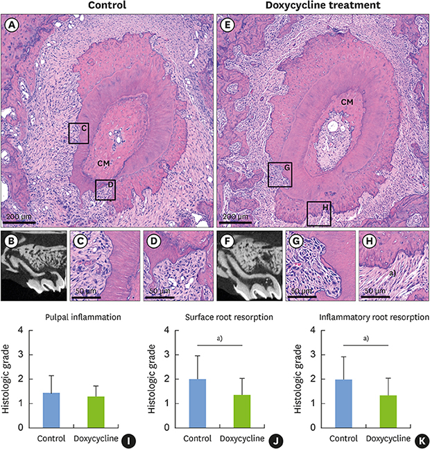
 | Figure 3Sections of teeth replanted after 60 minutes of storage in HBSS. (A) Gross histological characteristics of a representative root from group III reveals root resorption in almost half of the root surface area and a CM densely embedded in the pulp canal. (B) Sagittal micro-CT image of the root from group II shows severe root resorption. (C) High magnification of the square region of the root in the control group shows a moderate degree of inflammatory resorption. (D) High magnification of the square region of the root from the control group shows severe inflammatory resorption extending beyond the cementum. (E) Gross histological characteristics of a representative root from group IV shows some degree of root resorption and a CM embedded in the pulp canal. (F) Sagittal micro-CT image of a root from Group IV. (G) High magnification of the square region of the root in the doxycycline group shows severe inflammatory resorption extending beyond the cementum. (H) High magnification of the square region of the root in the doxycycline group shows PDL-like tissue attachment in the presence of surface root resorption. (I) Histologic grade of pulpal inflammation. (J) A significantly lower histologic grade of surface root resorption was achieved in the doxycycline group (P=0.009). (K) A significantly lower histologic grade of inflammatory resorption was achieved in the doxycycline group (P=0.007).HBSS: Hank's balanced saline solution, CM: calcified mass, CT: computed tomography, PDL: periodontal ligament.
a)Statistically significant difference between the control and doxycycline groups (P<0.05, Mann-Whitney test).
|
DISCUSSION
The healing process after replantation involves various types of pulpal and periodontal reactions. Delayed tooth replantation is associated with a high rate of unfavorable outcomes. To increase the survival rate of a replanted tooth in such a situation, the tooth should be stored in an appropriate storage medium to protect it from dehydration and preserve its vitality. Nevertheless, PDL cells cannot survive longer than 60 minutes under extra-oral conditions [8]. Based on these findings, studies focused on development of root-surface conditioning are needed.
In tooth replantation, prevention of inflammatory root resorption and reduction in the inflammatory response are important considerations for ensuring both pulpal and periodontal regeneration. Contaminated remnants around the root surface are responsible for increasing root resorption [1718], and their removal by mechanical or chemical root surface treatment prior to replantation is beneficial. Hence, root surface conditioning during tooth replantation with doxycycline is expected to have a positive effect due to the antimicrobial activity of doxycycline.
The results of this study showed that in the replantation groups where teeth were subjected to 5 minutes of dry storage, doxycycline enhanced neither pulpal nor periodontal healing compared with the control group. In contrast, recent studies have demonstrated that doxycycline promoted pulp revascularization after immediate replantation [91011], which could be attributed to a decrease in the number of micro-organisms in the pulpal canal due to the effect of doxycycline [911]. The discrepancies between the findings of this study and those of previous studies could be due to differences in the animal models and experimental conditions utilized. Furthermore, replantation after 5 minutes of dry storage simulates the ideal condition of immediate replantation, and the healing of teeth replanted under such conditions could be favorable, regardless of pretreatment with doxycycline.
Doxycycline downregulates the activity of matrix metalloproteinases (MMPs), which are the key enzymes in periodontal tissue destruction; hence, topical antimicrobial delivery of doxycycline has been considered as an adjunctive treatment approach in patients with periodontal disease [1920]. A previous rat study reported that increased MMP-1 and MMP-8 expression after replantation was associated with extra-oral time period of the rat tooth [7]. In this study, favorable periodontal healing was more likely to occur in the doxycycline groups, since doxycycline is known to cause more significant downregulation of MMP-8 expression than of MMP-1 expression.
Root resorption is a major complication following tooth replantation. The results of the replantation groups subjected to 60 minutes of storage in HBSS revealed that doxycycline inhibited both surface root resorption and inflammatory root resorption as compared to the control group, which is related to the negative effect of doxycycline on RANKL-induced osteoclastogenesis [21].
No significant differences were found in replacement root resorption (ankylosis) between the doxycycline and control groups. Replacement root resorption may occur after severe mechanical injury or dehydration of replanted teeth [6], and severe mechanical injuries of the root surface may occur due to accidental luxation of the teeth, such as through a fulcrum effect from the alveolar bone margin [9]. In the present study, atraumatic extractions of rat teeth were performed using β-APN, and the occurrence of replacement resorption was relatively low [15], which could explain the statistical results that were obtained.
In conclusion, the micro-CT and histomorphometric results obtained in the present study demonstrated that the use of doxycycline in root surface provided more favorable periodontal healing of replanted teeth stored for 60 minutes in HBSS. However, doxycycline did not improve periodontal healing of replanted teeth after 5 minutes of dry storage. Within the limitations of this study, root surface treatment with doxycycline can play an important role in delayed tooth replantation.
 XML Download