

 PDF
PDF ePub
ePub Citation
 Print
Print


INTRODUCTION
Choriocarcinoma is an aggressively growing and widely metastasizing tumor that originates from trophoblastic cells. Most of these tumors occur in the uterus and are related to a normal or abnormal pregnancy. They are often found in extragonadal sites, such as the mediastinum, retroperitoneum, pineal gland, gall bladder, urinary tract, and stomach. On the other hand, primary gastric choriocarcinoma (PGC) is quite rare.12 This paper reports a rare case of PGC coexisting with adenocarcinoma and discusses the characteristics of this neoplasm with a reference to the relevant literature.
CASE REPORT
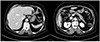
This study was approved by the Institutional Review Board of Wonkwang University Hospital (WKUH 2018-10-010), and informed consent was waived. A 76-year-old female patient visited the emergency department of Wonkwang University Hospital with abdominal discomfort and melena that had lasted for 2 months. She complained of mild, dull, and intermittent abdominal pain, but had no fever, diarrhea, or constipation. The patient's blood pressure was 110/70 mm/Hg; pulse rate, 68 beats/min; and body temperature, 36.8℃. The patient's laboratory test results revealed the following: hemoglobin, 5.4 g/dL; white blood cell count, 10.50×10³/mm3; CRP, 10.86 mg/L; CEA, 7.06 ng/mL; and the other blood chemistry parameters, within normal limits. Two packs of packed red blood cells were transfused. She also underwent esophagogastroduodenoscopy under the clinical impression of gastrointestinal bleeding. Esophagogastroduodenoscopy revealed a huge ulceroinfiltrative mass lesion with blood clots on the boundary between the greater curvature side and the posterior wall side of the stomach lower body (Fig. 1). CT revealed a 3-cm exophytic mass lesion with heterogeneous enhancement, an ulcer lesion at the posterior wall side of the stomach lower body, and multiple enlarged lymph nodes at the splenic artery and left gastric artery nodal stations (Fig. 2). PET/CT was performed to identify the other primary lesion or metastasis, but there was no abnormality of either ovaries or other organs. No distant metastasis to the other organs was found. The initial endoscopic biopsy result established a diagnosis of a well-differentiated adenocarcinoma. In June 2018, the patient underwent a radical subtotal gastrectomy with a D2 (1, 3, 4sb, 4d, 5, 6, 7, 8a, 9, 11p, and 12a) lymph node dissection and gastroduodenostomy. The surgical findings revealed a 7-cm mass lesion, diagnosed as Borrmann type II advanced gastric cancer in the posterior wall side of the stomach lower body with a 3-cm exophytic mass invading the mesocolon and multiple enlarged lymph nodes at the 6, 7, 8a, and 11p stations. Pathologically, the tumor had two components. The predominant portion of the tumor, which occupied more than 2/3 of the tumor, consisted of highly pleomorphic, bizarre cells with cytotrophoblastic and syncytiotrophoblastic differentiation. This findings strongly suggested a gastric choriocarcinoma. The remaining portion of the tumor was a well-differentiated tubular adenocarcinoma (Fig. 3A, B). The tumor had invaded the subserosa of the stomach and metastasized in the two lymph nodes (2/21). Lymphovascular invasion was noted but there was no perineural invasion. Immunohistochemical staining for β-human chorionic gonadotropin (hCG) was highly positive in the choriocarcinoma component (Fig. 3C). All resection margins were clear.
The final diagnosis was a choriocarcinoma coexisting with tubular adenocarcinoma of the stomach. After confirming the diagnosis based on the biopsy results, the serum β-hCG level was measured with the following value: 972.1 mIU/mL. With the absence of postoperative complications, the patient was discharged from hospital nine days after surgery. One week after discharge, she was scheduled to undergo chemotherapy, but the treatment was suspended temporarily because the patient demonstrated general weakness.
Unfortunately, 2 months after discharge, she revisited the emergency department of Wonkwang University Hospital with a complaint of general weakness, and CT revealed multiple hepatic metastases of the tumor. The serum β-hCG level was 443,560 mIU/mL. She died of hepatic failure five months after surgery.
DISCUSSION
Choriocarcinoma of the stomach was first described by Davidson3 in 1905, and accounts for approximately 0.08% of all gastric cancers.123 Because of rarity of this neoplasm, most studies have been case reports. Therefore, the clinicopathology, treatment modalities, and prognostic factors are not well established.
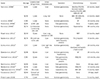
According to a large analysis done by Kobayashi et al.1 in 2005 to 53 patients who had PGC, the male to female ratio was 2.3:1, with a mean age of 62.4 years (median, 63 years; range, 32–84 years) in men and 54.8 years (median, 63 years; range, 26–74 years) in women, and most of the tumor was located in the lower third of the stomach (41%). These results were similar to gastric adenocarcinoma. In addition, nearly all tumors presented with hemorrhage or necrosis.1 The reported cases of primary gastric choriocarcinoma with a clear prognosis in the last 10 years were collected retrospectively from the English and Korean language literature. A total of 15 cases, including the present case, have been reported. The male to female ratio was 2:1, with a mean age of 65.7 years (median, 68.5 years; range, 44–85 years) in men and 65.4 years (median, 65 years; range, 57–76 years) in women. Most of the tumor was located in the lower third of the stomach (47%). This result was similar to previous studies (Table 1).2456789101112131415
Various theories for the pathogenesis of PGC have been suggested, which include the following: PGC originates from a displaced gonadal anlage, metastasizes from an unidentified primary lesion in the uterus, and develops from an underlying gastric teratoma. On the other hand, the most widely accepted hypothesis is the retro-differentiation of gastric adenocarcinoma. This hypothesis is that adenocarcinoma tissues retro-differentiate to the level of the embryonal ectoderm and retain their ability to produce trophoblasts. In addition, a pure choriocarcinoma can occur through the overgrowth and elimination of a pre-existing adenocarcinoma portion. The retro-differentiation theory is based on the fact that most PGC coexists with gastric adenocarcinoma and that there is a possible transition from an adenocarcinoma to choriocarcinoma. This is supported by the relatively similar clinical features of PGC to gastric adenocarcinoma, such as sex ratio, mean age, and location of the tumor, and about 70% of PGC was reported to be associated with gastric adenocarcinoma. In addition, a previous study on comparative genomic hybridization and fluorescence in situ hybridization reported that PGC genetically has the characteristics of both choriocarcinoma and adenocarcinoma.116171819
The clinical manifestations of PGC are also similar to those of gastric adenocarcinoma, accompanied by abdominal pain, weight loss, anorexia, nausea, and vomiting. Gastrointestinal bleeding is more frequent, and some effects of the hormone can lead to gynecomastia, precocious puberty, and vomiting, which are also observed during pregnancy.19 The present patient, however, had no manifestations of hormone-associated symptoms. PGC is difficult to diagnose in a preoperative endoscopic biopsy because most of the PGCs coexist with adenocarcinoma. Only 8–9.4% of patients are diagnosed correctly before laparotomy.117 In the present case, the initial diagnosis before surgery was a well-differentiated tubular adenocarcinoma.
The management of choriocarcinoma is not well established because of its rarity. Despite this, previous studies suggested curative surgery and adequate chemotherapy as the treatment of choice.111 The chemotherapy regimens used for gonadal choriocarcinoma are generally considered. Several investigators employed the regimen used for advanced gastric adenocarcinoma based on the retro-differentiation theory, but there is no established regimen.121020 Recently, there have been case reports of successful responses to multidisciplinary treatment. Takahashi et al.11 presented a case of gastric choriocarcinoma cured by multidisciplinary treatment, including gastrectomy, etoposide, methotrexate, actinomycin D, cyclophosphamide, and vincristine chemotherapy, and radio-frequency ablation. The patient had a long-term survival of 10 years. Nevertheless, the prognosis of gastric choriocarcinoma is still poor.
PGC easily metastasizes through the lymphatic and hematogenous routes. The common sites of metastasis are the lymph node, liver, peritoneum, and lung. Most patients die of hepatic failure due to tumor metastasis, hemorrhage of the tumor itself, and disseminated intravascular coagulation within one year after diagnosis.120 The prognostic factors of gastric choriocarcinoma are not well known. In 2005, Kobayashi et al.1 proposed synchronous liver metastasis, residual tumor after resection, and the absence of chemotherapy as the prognostic factors for gastric choriocarcinoma.
In conclusion, the authors a case of PGC coexisting with adenocarcinoma that was treated with a radical subtotal gastrectomy with a D2 lymph node dissection. PGC is a very rare disease, but the possibility of this disease must be considered during diagnosis.


 XML Download