

 PDF
PDF ePub
ePub Citation
 Print
Print


 , Hojun Yang, Jeong Hwan Kim, In-Kyung Sung, Hyung Seok Park
Abstract
Background/Aims
Methods
Results
Figures and Tables
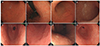
 | Fig. 1Two main subtypes of nodular gastritis. (A) Small-granular-type nodular gastritis consisting of multiple 1–2 mm subepithelial nodules in the antrum. (B) Large-nodular-type nodular gastritis consisting of multiple 3–4 mm subepithelial nodules in the antrum.
|
 | Fig. 2Follow-up endoscopic findings after nodule regression. (A) Small-granular-type nodular gastritis with whitish discoloration was observed in the antrum. With the progress of nodule regression, a salt-and-pepper appearance was observed in the antrum. (B) The salt-and-pepper appearance extended up to the lesser curvature side of the body, which is consistent with the atrophic border. (C) With the progression of the salt-and-pepper appearance, whitish discoloration with transparent submucosal vessels was visible in the antrum. An atrophic border was found at the greater curvature side of the proximal antrum. (D) Prominent submucosal vessels were observed from the antrum extending up to the lower body. The endoscopic diagnosis was consistent with chronic atrophic gastritis. (E) Large-nodular-type nodular gastritis was noticed in the distal antrum. (F) The nodules extended up to the proximal antrum. Some of the large nodules were closer to the diffuse irregular mucosal elevations observed in metaplastic gastritis than the nodules observed in nodular gastritis. (G) On a retroflexed view, diffuse irregular elevations were observed on the lesser curvature side of the body. A villous appearance was noted on the surface of whitish elevated lesions. (H) Diffuse irregular elevations were observed with whitish discoloration, indicating intestinal metaplasia. The endoscopic diagnosis was consistent with metaplastic gastritis.
|
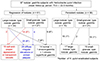 | Fig. 3Study flow of the 97 H. pylori-infected patients with nodular gastritis. The asterisks in parenthesis indicate the numbers of patients in whom H. pylori was eradicated. In total, 25 patients with large-nodular-type nodular gastritis (including six patients in whom H. pylori was eradicated) showed metaplastic gastritis on follow-up endoscopy, whereas 14 patients with small-granular-type nodular gastritis (including five patients in whom H. pylori was eradicated) showed chronic atrophic gastritis. Most of the patients with persistent nodules showed the same pattern on follow-up endoscopy; however, two patients with small-granular-type nodular gastritis progressed to large-nodular-type nodular gastritis. H. pylori, Helicobacter pylori.
|
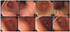 | Fig. 4Different prognosis of small-granular-type nodular gastritis according to the presence of a H. pylori infection. (A) Small-granular-type nodular gastritis was diagnosed along with a H. pylori infection in a 42-year-old woman. (B) Salt-and-pepper appearance was observed at the lesser curvature side of the lower body. (C) Four years after the H. pylori eradication, several linear hyperemic streaks were observed at the greater curvature side of the antrum. The endoscopic diagnosis was consistent with chronic superficial gastritis. (D) A salt-and-pepper appearance was no longer observed in the lower body. (E) Small-granular-type nodular gastritis was diagnosed along with a H. pylori infection in a 36-year-old man. (F) Small- and regular-sized nodules were extending up to the proximal antrum. (G) After eight years of persistent H. pylori infection, the nodules showed irregularity. (H) The size of the nodules increased with irregular changes. The endoscopic diagnosis was large-nodular-type nodular gastritis. H. pylori, Helicobacter pylori.
|
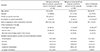
Table 1
Characteristics of 97 H. pylori-infected Patients with Nodular Gastritis
The differences between the two were analyzed using a t-test and chi-square test for the continuous and categorical variables, respectively. The continuous variables are presented as the mean±standard deviation, and the categorical variables are presented as the number of patients with the proportion (%).
H. pylori, Helicobacter pylori.
aTwo patients did not show H. pylori infiltration at the time of the biopsy because they were on medication for H. pylori eradication.
![]()
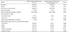
Table 2
Characteristics of 61 Patients with Nodular Gastritis according to the Findings of the Follow-up Endoscopy after Nodule Regression
Other endoscopic findings consisted of 14 normal findings, one of erosive gastritis, and one of chronic superficial gastritis. The differences among the three groups were analyzed by post-hoc analysis for the continuous variables, and a chi-square test with a Bonferroni correction for the categorical variables. Continuous variables are presented as the mean±standard deviation, and categorical variables are presented as the number of patients with proportion (%).
CAG, chronic atrophic gastritis; MG, metaplastic gastritis; H. pylori, Helicobacter pylori.
aSignificant difference (p<0.05) compared to the patients showing salt-and-pepper appearance and/or transparent vessels; bSignificant difference (p<0.05) compared to the patients showing diffuse irregular elevations and/or whitish plaques.
![]()
Table 3
Differences among 29 Patients Showing Diffuse Irregular Elevations after Nodule Regression
The differences between the two groups were analyzed using a t-test and chi-square test for the continuous and categorical variables, respectively.
The continuous variables are presented as the mean±standard deviation, and the categorical variables are presented as the number of patients with the proportion (%).
H. pylori, Helicobacter pylori.
![]()
 XML Download