

 PDF
PDF Citation
 Print
Print


Abbreviations
ASST
autologous serum skin test
ATA
anti-thyroid autoantibody
CSU
chronic spontaneous urticaria
CU
chronic urticaria
ECP
eosinophil cationic protein
FcεRIα
high affinity Fc epsilon receptor I alpha subunit
HLA
human leukocyte antigen
LAD-2
Laboratory of Allergic Disease 2
LAT
linker of activation of T cell
log
logarithm
NC
normal healthy control
TCTP
translationally controlled tumor protein
UAS
urticaria activity score
INTRODUCTION
Chronic spontaneous urticaria (CSU) is characterized by wheals and/or angioedema lasting for more than 6 wk, with no definite extrinsic cause (1). The prevalence of chronic urticaria (CU) has been found to range between 0.3%–11.3%, and the rate of hospitalization due to urticaria appears to be increasing (2). According to a 5-year nationwide epidemiological study in Korea, the overall ratio of CU among all urticaria patients was, on average, 23.5% (3).
Translationally controlled tumor protein (TCTP), also known as p23, fortilin, and histamine releasing factor, is a multifunctional protein widely expressed in almost all eukaryotic organisms (4). Previous reports have shown that intracellular TCTP expression is dependent on calcium concentrations and microtubule binding protein (56). In allergic disease, extracellular TCTP, which is observed in nasal and bronchoalveolar lavage and skin blister fluids, exhibits the capacity to induce histamine release from basophils (7). Indeed, recombinant TCTP has been found to enhance IL-4, IL-13, and histamine release from IgE sensitized human basophils (89). Also, other immune cells involved in allergic disease, such as eosinophils, B cells, bronchial epithelial cells, and T cells, appear to be at least partly regulated by TCTP (10111213). While some studies have suggested that the role of TCTP in basophil activation is dependent on IgE (14), Wantke et al. (15) demonstrated that TCTP exerts a stimulating effect via its specific receptor on the surface of human basophils and that it does not bind with IgE (914). Notwithstanding, a recent study reported that both serum TCTP levels and specific IgE Ab against TCTP were increased in CSU patients (16).
The activating mechanism of TCTP has not been fully elucidated in CSU. Kim et al. (13), however, demonstrated that dimerization of TCTP is essential for its cytokine-like activity in OVA-induced airway inflammed mice and atopic dermatitis-like NC/Nga mice in vivo (17). In the present study, we sought to compare TCTP levels in CSU patients and normal healthy controls (NCs) and to investigate whether monomeric and dimeric TCTP have different effects on basophil and mast cell activation.
MATERIALS AND METHODS
Clinical characteristics of the study subjects
We enrolled 70 NCs and 116 CSU patients who had urticaria symptoms for at least 6 wk (Table 1). Disease activity of CSU was assessed using urticaria activity score (UAS), which evaluates wheal characteristics, pruritus status, and symptom duration. Total scores ranged from 0–15, with higher scores indicating higher disease activity (18). The patients with UAS ≥13 has been classified into severe CSU (19). For assessing basophil activation, peripheral blood was collected from 24 CSU patients for whom antihistamine medication was withdrawn for at least 5 days before collecting their blood samples. The study was approved by the Institutional Review Board at the Ajou University Medical Center (AJIRB-BMR-MDB-17-221, AJIRB-GEN-SMP-13-108). Each patient provided written informed consent.
Table 1
Clinical characteristics of the study subjects
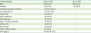
Values are presented as mean±standard deviation, number (%), or number (range).
H1RA, histamine receptor 1 antagonist.
![]()
Autologous serum skin test (ASST), atopy, total IgE, and other autoantibodies measurements
ASST was performed and interpreted according to appropriate guidelines (20). Atopy was defined as a positive result to at least one allergen on skin prick tests with common inhalant allergens (tree mixture, grass mixture, mugwort, ragweed, cat fur, dog fur, Dermatophagoides pteronyssinus, Dermatophagoides farinae, and Alternaria spp.) (Bencard, Bretford, UK). Total IgE and eosinophil cationic protein (ECP) levels were measured by the ImmunoCAP system (Thermo Fisher Scientific, Waltham, MA, USA) according to the manufacturer's instructions. Anti-thyroid autoantibodies (ATAs), including anti-thyroglobulin and thyroid microsomal antibodies, were detected by radioimmunoassay (BRAHMS Aktiengesellschaft, Hennigsdorf, Germany). To measure IgG to high affinity Fc epsilon receptor I alpha subunit (FcεRIα) in sera from CSU patients and NCs, we performed rapid dot-blot immunoassay as described previously (21).
Measurement of TCTP in serum
Serum TCTP concentrations were determined by ELISA (MyBioSource Ltd, San Diego, CA, USA) according to the manufacturer's instructions. Both intra-assay and inter-assay coefficients of variation (%) were less than 15%, and the sensitivity of the kit was 1 ng/ml.
Purification of recombinant TCTP protein
Recombinant monomeric and dimeric TCTP used for stimulation was purified in an Escherichia coli system, as described previously (13). Briefly, pRSET A/TCTP or Del-N11 TCTP was transformed to E. coli strain BL21(DE3) pLysS, and the cells were grown in Luria-Bertani medium containing 100 μg/ml of ampicillin and 34 μg/ml of chloramphenicol. The pre-culture medium was diluted and cultured until reaching an OD600 of 0.6–0.8. IPTG was added to induce recombinant protein, and then, the cell pellets were stored at −70°C or used directly. Cell pellets were disrupted by sonication, and supernatants containing soluble proteins were purified with HisPurTM Cobalt Resin (Thermo Fisher Scientific) according to the manufacturer's instructions. Subsequently, eluted proteins were loaded onto a HiTrap Q HP column (GE Healthcare, Princeton, NJ, USA), which was equilibrated with buffer A (20 mM Tris, 1 mM EDTA, 50 mM NaCl; pH 7.4). Proteins were separated by AKTA FPLC systems (GE Healthcare) and eluted with buffer B (20 mM Tris, 1 mM EDTA, 1 M NaCl; pH 7.4) at a constant flow rate of 1 ml/min. The eluted fractions were screened by SDS-PAGE and Coomassie blue staining. The purity of the proteins was 85%–98% and no other bands were observed visually except target protein (Supplementary Fig. 1). Samples containing monomeric or dimeric TCTP were collected, desalted with PBS, concentrated using Vivaspin 500 (Sartorius, Goettingen, Germany), and stored at −70°C until ready for use. To reduce lipopolysaccharide contamination, final proteins were additionally purified with EndoTrap (Cambrex, Walkersvile, MD, USA).
Western blotting for detecting dimerized TCTP in sera from CSU patients and NCs
Serum was diluted 2X Laemmli sample buffer and boiled for 5 min. After centrifugation, the supernatants were separated by 10% SDS-PAGE in non-reducing condition and electrophoretically transferred to nitrocellulose membranes (GE Healthcare). Membranes were blocked for 1 h at room temperature in 5% BSA/TBS-T. The primary Ab against TCTP (Santa Cruz Biotechnology, Dallas, TX, USA) was diluted 1:1,000 with blocking buffer and incubated overnight at 4°C with the membrane. After washing three times with TBS-T, the membranes were incubated with a goat anti-rabbit IgG-HRP conjugated secondary Ab (Bio-Rad Laboratories Inc., Hercules, CA, USA) for 1 h at room temperature. The membranes were washed again, visualized with ECL plus reagent (SurModics Inc., Eden Prairie, MN, USA), and detected by a LAS-3000 CCD camera (Fuji Film, Tokyo, Japan).
Basophil activation test
CD203c expression on the surface of basophils was measured by flow cytometry (22). Whole blood was collected in acid citrate dextrose tubes, and red blood cells were lysed with red blood cell lysis buffer (0.154 M NH4Cl, 10 mM KHCO3, 0.1 mM EDTA, pH 7.2–7.4). After washing with PBS, resuspended cells were stimulated by recombinant TCTP protein at different concentrations (4 to 32.5 ug/ml) for 2 h at 37ºC. After incubation, the cells were washed with PBS and stained by phycoerythrin-conjugated anti-human CD203c (Beckman Coulter, Marseille, France), FITC-conjugated anti-human CD123 (BD PharMingen, San Jose, CA, USA), and allophycocyanin-conjugated anti-human human leukocyte antigen (HLA)-DR (BD PharMingen) or isotype-matched controls on ice in the dark for 30 min. After washing once with PBS, the cells were analyzed on a FACS Canto II flow cytometer (Becton Dickinson, San Jose, CA, USA). Basophils were identified as CD123+ HLA-DR−populations. The percentage of cells expressing CD203c was evaluated by comparing TCTP stimulated basophils with non-stimulated (resting) basophils.
Measurement of ß-hexosaminidase release from Laboratory of Allergic Disease 2 (LAD-2) cells
LAD-2 mast cells were kindly provided by Dr. Arnold Kirshenbaum (National Institute of Allergy and Infectious Diseases, Bethesda, MD, USA). The release of β-hexosaminidase from LAD-2 cells was measured as previously described (23). Cells were treated by monomeric and dimeric TCTP (50 to 200 ug/ml) with or without simultaneous biotinylated-IgE (100 ng/ml; BioPorto Diagnostics, Hellerup, Denmark) overnight sensitization, followed by 30 min of stimulation with streptavidin peroxidase (100 ng/ml) or calcium ionophore A23187 (1 µM) in HEPES buffer containing 0.4% BSA at 37°C. Total β-hexosaminidase was obtained by lysing LAD-2 cells in 0.1% Triton X-100 in PBS. The supernatants were collected and incubated with an equal volume of p-nitrophenyl N-acetyl-β-D-glucosamide in citrate buffer (pH 4.5, 40 mM citric acid/20 mM Na2HPO4*7H20) for 30 min. The reactions were stopped by adding 0.4 M glycine buffer, and signals were read at 405 nM. The percentage of degranulation was calculated as 100×(ODstimulated−ODnon-stimulated)/ODtotal lysate.
Statistical analysis
Statistical analyses were performed using IBM SPSS version 20 for Windows (SPSS Inc., Chicago, IL, USA). Data are presented as a mean±standard deviation or as a median (interquartile range) value when variables were not normally distributed. A multiple regression model was applied to determine the variables most predictive of circulating TCTP concentrations as logarithm (log)-transformed values among those known to be associated with TCTP, including age, gender, atopy status, IgG to FcεRIα positivity, disease severity, and serum ECP level. Pearson correlation analysis identified associations among the continuous parameters. The p-values <0.05 were considered indicative of statistical significance.
RESULTS
Serum TCTP levels in CSU patients and NCs
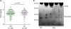
Median levels of serum TCTP in CSU patients (34.4 [2.93–242.9] ng/ml) were not different from those in NCs (37.5 [1.19–195.7] ng/ml, Fig. 1A). We attempted to differentiate dimeric from monomeric TCTP in the sera from CSU patients and NCs using non-reducing Western blot analysis. In doings so, we identified separate bands of higher and lower molecular weights. The bands located in a position of higher molecular weight were regarded as dimerized or oligomerized TCTP (Supplementary Fig. 2), which are conformationally modified. Relatively higher signal intensity for dimeric bands was noted for the sera from 4 patients with CSU, while the sera from NCs showed higher signal intensity for monomeric TCTP bands (Fig. 1B). However, we could not quantitatively analyze dimeric and monomeric TCTP amounts (Supplementary Fig. 3).
Correlation of serum TCTP levels with clinical characteristics of CSU
The median level of serum TCTP was higher in atopic CSU patients than in non-atopic patients (40.5 [25.5–65.0] ng/ml vs. 29.0 [12.0–61.0] ng/ml, respectively, p=0.019, Fig. 2A). However, no difference in TCTP levels was observed regarding to ASST positivity (38.8 [22.1–65.0] ng/ml vs. 30.4 [19.1–56.3] ng/ml, respectively, p=0.429). Patients with positive IgG to FcεRIα autoantibody had significantly higher serum TCTP levels than patients with negative IgG to FcεRIα autoantibody (50.1 [25.4–73.7] ng/ml vs. 31.4 [19.5–64.1] ng/ml, respectively, p=0.03, Fig. 2B). Serum TCTP levels did not differ according to the presence ofATA, anti-nuclear Ab, or angioedema among CSU patients. Regarding therapeutic responses to antihistamine, serum TCTP levels tended to be higher in refractory CSU patients than in patients responsive to antihistamines, although statistical significance was lacking (40.5 [22.2–77.1] ng/ml vs. 31.0 [19.4–56.0] ng/ml, p=0.141). However, a significant difference in serum TCTP levels was noted between severe and non-severe CSU patients (41.1 [28.0–70.4] ng/ml vs. 30.2 [19.4–54.3] ng/ml, p=0.049, Fig. 2C). Log-transformed serum TCTP levels were negatively correlated with age (r=−0.244; p=0.008), whereas they had a positive correlation with log-transformed ECP in CSU patients (r=0.346; p=0.002). No significant correlation was found between TCTP and total IgE levels in CSU patients (data not shown).
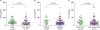 | Figure 2Association of serum TCTP levels according to atopy rate (A), IgG to FcεRIα positivity (B) and CSU severity (C).
|
With the results of univariate analyses, we performed multiple regression analysis to identify predictors of serum TCTP levels in patients with CSU. Significant positive correlations were noted between log-transformed TCTP in sera from CSU patients and both severe CSU (defined as a UAS ≥13; B=0.150, p=0.043) and log-transformed ECP levels (B=0.265, p=0.002) after adjustment for confounders, including age, gender, atopy, and IgG positivity to FcεRIα (Table 2).
Table 2
Predictors of log-transformed serum TCTP levels in patients with CSU

![]()
Basophil activation upon stimulation of monomeric and dimeric TCTPs
To investigate whether monomeric and dimeric TCTPs have the capability to activate basophils, we performed basophil activation tests in a total of 24 patients with CSU, measuring CD203c expression on the surface of basophils, since it had higher sensitivity than CD63 and was a predictor of severe CU in our previous report (22). The expression of CD203c on the surface of basophils was significantly increased upon stimulation with dimeric TCTP in a dose-dependent manner (p=0.01, Fig. 3A), while monomeric TCTP had no effect on CD203c expression.
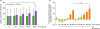 | Figure 3Effects of TCTP on basophil activation. (A) Basophils from 24 CSU subjects were treated simultaneously with monomeric and dimeric TCTP and with anti-IgE stimulation. Basophil activation was measured by surface CD203c expression using flow cytometry. (B) Degranulation of LAD-2 cells. Mast cell degranulation was investigated using the β-hexosaminidase release test. The positive control comprised LAD-2 cells sensitized with IgE and stimulated with streptavidin peroxidase or only stimulated with calcium ionophore. A statistically significant difference was defined as that with a two-tailed.ns, no significant.
*p-value<0.05; **p-value<0.01.
|
β-hexosaminidase release from LAD2 cells upon stimulation with TCTP
To confirm the effect of TCTP on human LAD2 cell degranulation, cells were treated with monomeric and dimeric TCTP with or without IgE sensitization. Dimeric TCTP induced a significantly higher level of β-hexosaminidase release in dose depedently when compared with untreated cells and cells treated with an equal concentration of monomeric TCTP (p<0.001, Fig. 3B). With or without IgE sensitization, there was no significant difference in beta-hexosaminidase release from LAD2 cells upon either monomeric or dimeric TCTP stimulation.
DISCUSSION
The present study demonstrated that dimeric TCTP can induce basophil activation and mast cell degranulation, while monomeric TCTP has no influence on basophils/mast cells. Even though the median levels of TCTP were not different between CSU patients and NCs, more intense bands of higher molecular weight reflective of the dimerized form of TCTP on Western blotting were noted in CSU patients than in NCs.
TCTP is ubiquitously expressed in all eukaryotic organisms and tissues; however, expression levels thereof vary among cell or tissue types. TCTP has been reported to be involved in the pathogenic mechanisms of asthma, immune responses to parasitic infections, atopic dermatitis, and CU (14161724). Kim et al. (13) revealed that TCTP conformationally changes via intermolecular disulphide bonding into its dimerized form in order to confer its cytokine-like activity. Thus, while TCTP might be present in normal conditions, sturctural modification may occur in an allergic enviroment rich in oxidants or endogenous and exogenous proteases that can induce dimerization of TCTP (13). Interestingly, mast cells release proteases, including tryptase and chymase, during degranulation, and studies have shown that serum tryptase levels are elevated in CU and associated with symptom occurrence and increased corticosteroid requirement (2526). In addition, an imbalance in reactive oxygen species and antioxidants has been found to worsen allergic inflammation including CSU (2728). We observed higher TCTP levels in CSU patients with IgG autoantibody against FcεRIα in the present study. Accordingly, we presummed that the autoimmunity represented by the positivity of IgG to FcεRIα could affect the dimerizing process of TCTP. Nevertheless, because the receptor for dimeric TCTP on basophils and mast cells has not been identified, the exact pathway of action for TCTP in the activation of basophils and mast cells remains to be elucidated. The crystal structure of human TCTP comprises a disulphide-linked dimer that confers an IgE binding site for degranulating mast cells (29). A recent study also detected higher levels of serum specific IgE Ab against TCTP in CSU patients (16), which supports our hypothesis that activation of TCTP may be associated with the autoimmune mechanism of CSU pathogenesis. TCTP has been found to play a role in immediate hypersensitivity by interacting with the Fab region of immunoglobulin and in mast cell activation in an Fc receptor-dependent manner (24). Moreover, prior studies demonstrated that different signal transduction events are initiated by TCTP on basophils in TCTP responders and non-responders (10). Therein, TCTP was found to prime basophils through FcεRIα or another adapter molecule that is distinct from IL-3 in TCTP non-responders, whereas TCTP induced secretion directly in TCTP responders (10). Based on our findings that dimeric, but not monomeric, TCTP could induce degranulation of peripheral basophils from CSU patients, we suspect that the dimerizing process might occur less in TCTP non-responders than in TCTP responders inducing greater degranulation, despite similar total TCTP levels.
Kashiwakura et al. (24) reported that active TCTP interacts with immunoglobilins by its NH2-terminal 19-residue peptide and H3 region with Fab region and crosslinking IgE to induce degranulation of mast cell via FcεRIα receptor. However, we found no significant difference in mase cell degranulation upon dimeric TCTP stimulation with or without IgE sensitization in the present study. To eliminate the controversy regarding whether TCTP dependently or independently activates mast cells via IgE (15), the exact activating pathway needs to be elucidated. A recent study demonstrated that dimeric TCTP induces IL-8 production in bronchial epithelial cells via the activation of adaptor protein 1 by phosphorylating MAPKs and the NF-κB pathway, which are also involved in mast cell activation, in a linker of activation of T cell (LAT)-dependent manner, thereby generating proinflammatory cytokines (3031). Meanwhile, FcεRIα aggregation has been found to lead to LAT phosphorylation through the phosphorylation of Syk, Akt, MEK, and ERK upon exposure to TCTP in TCTP-responders (1031). Consistent with these results, we speculate that dimeric TCTP may induce basophils/mast cells degranulation via the same signaling pathway. However, the lack of quantification of dimeric and monomeric TCTP levels in the present study is the major limitation to overcome. We just suggested the possible difference in the distribution of monomeric and dimeric TCTP from CSU patients to NCs visually and it is supported by results of ex vivo and in vitro experiments.
The release of proteases, leukotrienes, lipid mediators, and histamine from mast cells contributes not only to tissue inflammation but also to recruitment of eosinophils (32). Mast cells are also sources of IL-5, TNFα, and eotaxin, all of which elicit eosinophil recruitment and stimulation in urticarial lesions (3233). ECP, a typical marker of eosinophil activation, is known to be an indicator of disease severity in CU patients (34). Indeed, in the present study, we suggest that urticaria severity and serum ECP levels might be significant predictors of TCTP in patients with CSU. ECP has been shown to be concurrently expressed with VEGF in the lesional skin of CSU patients, and increases in VEGF have been observed in the plasma of CSU patients in association with urticaria severity (35). Accordingly, we presume that there may be a direct link between TCTP dimerization and ECP or basophil/mast cell-induced eosinophil activation that leads to more severe urticaria.
In conclusion, the current study demonstrated that dimerization of TCTP is important for activation of basophils and mast cells in CSU. Atopic status and the presence of autoantibody against FcεRIα were found to be associated with enhanced dimerization of TCTP. In light of these results, we deemed that dimeric TCTP induces basophil activation and mast cell degranulation leading to eosinophil activation and increased urticaria severity in CSU. Accordingly, inhibition of TCTP dimerization may be a potential therapeutic option for CSU patients.
 XML Download