

 PDF
PDF ePub
ePub Citation
 Print
Print


Introduction
Dental caries is the most frequent bacterial disease that affects the human oral environment.12 An accurate diagnosis of the presence or absence of this disease is a fundamental requirement of health care, although it may be a challenging task for clinicians, especially in the early stages of infection. A late diagnosis may allow the progression of the disease in enamel and dentin, consequently causing cavitation of the surface. While most occlusal lesions are visible in clinical examinations, proximal lesions are usually more difficult to detect, and their diagnosis often requires the combination of a careful visual inspection and an optimal radiographic exam. Studies have reported that the detection of caries lesions in radiographs is only possible after tissue demineralization reaches over 40%.34 Nonetheless, the interproximal radiograph is still the main auxiliary clinical tool used to detect proximal caries lesions in their early stages. However, this limitation has spurred researchers to explore ways of improving and comparing the diagnostic accuracy of various imaging systems and tools.5
Several digital systems for direct (charge-coupled device [CCD] and complementary metal oxide semiconductor [CMOS]) or indirect (photostimulable storage phosphor; PSP) image acquisition are available on the market. Many studies have shown that PSP has a number of advantages for proximal caries lesion detection.45 For example, the thickness and dimensions of PSP plates are similar to those of radiographic film, especially when compared with solid-state sensors, such as CCD and CMOS, and PSP systems do not have a cable, which helps ensure the patient's comfort during acquisition of the radiographic image.67
The spatial resolution of the PSP receptor is one of the parameters that determines the quality of the final image. The receptor's spatial resolution reflects the ability to discern details in radiographic images,8 varies according to the size of the picture element (pixel), and is often expressed as line pairs per millimeter (lp/mm). Since there is a direct relationship between the resolution selected before scanning and the scan time, it is worthwhile to study whether the resolution influences the diagnostic accuracy in the detection of caries lesions.
It has been suggested that radiographs with high spatial resolution allow better detection of radiographic details.9 Currently, some PSP plate systems offer the choice between high- and low-resolution settings during scanning, thereby allowing the acquisition of images with different resolutions, which facilitates an evaluation of the relationship between spatial resolution and diagnostic image quality. Moreover, at the present moment, the VistaScan Perio Plus (Dürr Dental, Bietigheim-Bissingen, Germany) is the only digital radiography system that allows 4 spatial resolutions. Therefore, this ex vivo study was conducted to evaluate the effect of different spatial resolutions of this PSP radiography system on the detection of proximal caries lesions.
Materials and Methods
Sample
Forty-five extracted human permanent premolars and molars were randomly divided and mounted into 9 plaster blocks. In each block, the teeth were positioned to provide contact between the proximal surfaces. The first tooth of the row was set as a non-test tooth, placed only to provide proximal contact with the first test tooth. A total of 72 proximal surfaces were analyzed; some of them were clinically cavitated, others had discolorations, and others were clinically sound. For ease of identification, metal markers were attached to the vestibular surface of each block with sticky wax.
Radiographic images
Radiographs were obtained using a GE 1000 X-ray machine (General Electric Company, Boston, MA, USA), operating at 65 kV, 10 mA, and 0.53 s of exposure time. The blocks were stabilized in a jig to create a 40-cm focus-tooth distance with a 1.5-cm tooth-receptor distance. In order to simulate the presence of soft tissues, a 12-mm-thick acrylic plate was placed between the tube and the tooth block.10 For all radiographs, the paralleling technique was employed. For each tooth, 4 images were recorded with a PSP system VistaScan Perio Plus, size 2 (3×4 cm). All 4 spatial resolutions available in the system (10 lp/mm, 20 lp/mm, 25 lp/mm, and 40 lp/mm) were used. Once a tooth was exposed, the plate was immediately scanned in 1 of the 4 resolutions. The scanning times and file sizes are shown in Table 1. The images were stored in 8 bits and saved in TIFF format in the DBSWIN® software (Dürr Dental, Bietigheim-Bissingen, Germany).
Image analysis
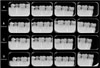
The images (Fig. 1) were exported to Adobe Photoshop® CS3 Extended (Adobe Systems Incorporated, San Jose, CA, USA). Three independent examiners (oral radiologists, previously trained with a minimum of 3 years of experience in digital imaging) masked, calibrated, and evaluated 288 images (72 proximal surfaces×4 different spatial resolutions) in a quiet, windowless room with dimmed lighting on an LG Flatron LCD monitor (21.5″, 1280×1024 pixels; LG Electronics Inc., Seoul, Republic of Korea). They could adjust the contrast, brightness, and magnification, and were instructed to rate the absence or presence of a carious lesion according to a 5-point confidence rating scale: 1, caries definitely absent; 2, caries probably absent; 3, unsure if present or absent; 4, caries probably present; and 5, caries definitely present. The metal pointer included in the image indicated which tooth was under evaluation (Fig. 1). Both proximal surfaces of the tooth were scored. For scores of 4 or 5, the examiners were also asked to identify whether the caries lesion was located only in the enamel or in both the enamel and dentin. A second evaluation of 20% of the images was performed at an interval of 15 days using the same parameters to assess the reproducibility of the scores.
Histological validation
After the teeth were radiographed, they were individually embedded in acrylic (Vipcril; Vipi, São Paulo, Brazil) and serially sectioned into 1000-µm-thick mesiodistal sections using a 300-µm diamond band. The tooth sections were cleaned of dust and mounted on microscope slides with transparent varnish. Two experienced observers (different from those who examined the radiographic images) examined the tooth sections using a Leica DMLP polarizing microscope (Leica Microsystems Inc., Wetzlar, Germany). They classified each tooth surface into the following categories: sound, lesion in enamel, and lesion in enamel and dentin. In case of disagreement between the observers, consensus was achieved through a joint assessment of the specimen under review. Both sides of each tooth section were examined. A caries lesion was defined as present when an opaque white or brown discoloration was observed in an area at risk of caries. When cavitated surfaces were present, an interruption of the outer tooth surface was seen.
Data analysis
Intra- and inter-examiner reproducibility was calculated using the weighted kappa test. The scores of each examiner were compared with the gold standard, and the area under the receiver operating characteristic (ROC) curve (AUROC) was used to calculate accuracy. To obtain binary sensitivity and specificity data, scores of 1 and 2 were combined as “no caries” and scores 4 and 5 were combined as “caries.” No examiner used a score of 3 for any surface. The accuracy, sensitivity, and specificity data were analyzed using the Shapiro-Wilk normality test, and mean values among examiners were compared using 1-way analysis of variance with the post hoc Tukey test. The null hypothesis was that there would be no differences among the spatial resolutions, with a significance level of 5%. Analyses were performed using MedCalc 15.8 (MedCalc Software, Ostend, Belgium).
Results
The histological examinations revealed that of the 72 proximal surfaces, 30 (41.7%) were sound, 18 (25%) showed enamel caries, and 24 (33.3%) had enamel-dentin caries.
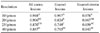
The sensitivity and specificity values did not differ significantly among the 4 spatial resolutions for any lesion depth (P>0.05) (Table 2). However, accuracy (as reflected by AUROC values) was affected by the spatial resolution (P<0.05). Accuracy values are shown in Table 3. For the detection of all caries lesions (enamel and enamel-dentin) the spatial resolution of 10 lp/mm showed the highest AUROC, with a significant difference between 10 lp/mm and all other resolutions (20 lp/mm [P=0.04], 25 lp/mm [P<0.001], and 40 lp/mm [P<0.001]). For detection of incipient caries lesions (enamel lesions) (Fig. 2), the spatial resolution of 10 lp/mm was also superior, with a significant difference compared to the other resolutions (20 lp/mm [P=0.04], 25 lp/mm [P<0.001], and 40 lp/mm [P<0.001]). However, the spatial resolution did not affect the detection of caries lesions in both the enamel and dentin (P>0.05).
The kappa values for intra- and inter-examiner agreement for the detection of caries ranged from 0.668 to 0.771 and from 0.731 to 0.766, respectively, which is considered substantial according to Landis and Koch.11
Discussion
Several studies have compared PSP with conventional radiographs for the detection of proximal caries lesions.412131415 All of them showed that digital images provided the same diagnostic information as conventional radiographs. Some PSP systems allow the selection of a higher or a lower spatial resolution during the scanning procedure, which may influence the diagnostic accuracy. The present study evaluated the 4 available spatial resolutions of the VistaScan Perio Plus device (10, 20, 25, and 40 lp/mm) in terms of the ability to detect proximal caries lesions.
In general, previous studies have revealed no influence of the spatial resolution of PSP on the detection of proximal caries lesions.5161718 In the present study, similar results were observed for the detection of caries lesions using resolutions of 20, 25, and 40 lp/mm. Wenzel et al.5 compared 2 spatial resolutions (20 lp/mm and 40 lp/mm) of the VistaScan Perio Plus system. The accuracy of these resolutions did not show a statistically significant difference in the detection of caries lesions, similarly to our study. Nikneshan et al.18 did not use the VistaScan device, so possible differences between the devices should be considered, as such differences may influence the obtained results. Berkhout et al.16 and Li et al.17 did not observe differences in caries lesion detection for the two resolutions studied (10 and 20 lp/mm); however, the authors did not present the accuracy values for the lesions at different locations (enamel and enamel-dentin).
Although we observed no difference for the resolutions of 20, 25, and 40 lp/mm in the present study, spatial resolution affected the detection of enamel caries lesions, but had no influence on the detection of carious dentin. The highest accuracy in our study was observed in images with low spatial resolution, for incipient (in enamel) caries lesions, but not for the detection of caries lesions in both enamel and dentin. This may be counterintuitive, as it seems logical that radiographic images with high spatial resolution offer more radiographic detail.9 However, this finding may be explained by the fact that images with a small pixel size (high resolution) are likely to show more noise than images with larger pixel sizes due to the lower level of dissipation of scattered X-rays among nearby pixels.41920 Caries lesions restricted to enamel and without cavitation are difficult to diagnose, meaning that small changes in the image may compromise their diagnosis. Image noise may compromise the accuracy of the diagnosis;19 thus, grainy images obtained with a higher spatial resolution may mask and/or simulate the presence of small caries lesions in enamel. Therefore, although they present fewer details, images with low spatial resolution also present less noise. It is worth noting that image noise only seems to affect the detection of incipient caries (caries lesions in enamel only), and not lesions involving dentin.
The results for sensitivity and specificity at the 4 resolutions did not show statistically significant differences. However, the sensitivity values were greater than those reported by Wenzel et al.5 This may indicate that the digital system used in this study was more effective for the detection of incipient caries lesions, particularly at the resolution of 10 lp/mm. However, the specificity values were similar in both studies. This indicates that both systems enabled the detection of sound surfaces, which constituted most of the sample. Li et al.17 compared the resolutions of 10 lp/mm and 20 lp/mm for the detection of caries lesions. Those authors did not find statistically significant differences between the resolutions, and the accuracy of the 10 lp/mm resolution was considered to be low. This seems to contradict our results, in which the 10 lp/mm resolution showed the highest accuracy. Nonetheless, it is important to keep in mind that the exposure parameters were not the same in both studies, which could result in different levels of noise in the images depending on the resolution used.
Other receptor characteristics, such as dynamic range and contrast resolution, may also influence the visibility of small details in digital images.17 The dynamic range refers to the exposure range in which a radiographic image can be produced without the occurrence of brightness and contrast changes in the image,21 and PSP plates have a wider dynamic range than most sensors and films.22 Contrast resolution is defined by the bit depth of the image, which consists of the amplitude of gray values in a radiographic image. The higher the bit depth of a system, the greater the grayscale and therefore the higher the contrast resolution of that system.23 In this study, we opted to use a smaller bit depth based on a previous study that found better sensitivity values with this depth in this unit.5
Another important factor affecting the detection of details in radiographic images is the human visual system. Findings should be interpreted in light of the limits of human vision, which is only capable of discerning up to a hundred shades of gray.24 Even though high-resolution image receptors are now capable of producing images with high detail and proper contrast, the human visual apparatus may be the main hindrance to improved radiographic caries detection. The detection of caries lesions using radiographic methods implies that the density difference between intact and demineralized hard tissues of the tooth is discernable, as a result of interactions between the X-rays and the minerals in the tooth structure. In proximal enamel lesions, the border between sound tissue and pathological regions can present low radiographic contrast, making it more difficult to perceive.14
The processing of high-resolution images requires increased scanning time. Accordingly, our results showed that for resolutions of 10 lp/mm, 20 lp/mm, 25 lp/mm, and 40 lp/mm, the scanning time was 7, 15, 24, and 31 seconds, respectively. In a busy dental practice where numerous radiographs are processed daily, high-resolution images imply a waiting time that is 4 times longer. A reduction in working time has been claimed to be one of the major advantages of digital radiography, and several studies have reported estimations of the time spent recording and scanning digital images with different receptors.22 Time is a sensitive issue, and to optimize clinical practice, unnecessary time consumption should be avoided whenever possible.
It seems possible to obtain good-quality images with medium or even low spatial resolution without a significant loss of information, in addition to the benefit of smaller file sizes. The high-resolution images in this study were almost 18 times larger than the low-resolution images. In a clinical setting, the required storage space and processor capacity may be factors worth considering. Since the low-resolution images showed a high accuracy for the detection of enamel caries lesions, it seems reasonable to consider the possibility that using high-resolution images for this purpose may not be justifiable as a first choice.
This study has the limitation inherent to ex vivo studies of not simulating real clinical situations. Factors such as object movement, metallic restorations, the tissues around teeth, and other parameters can complicate the detection of caries lesions. The detection of dental pathologies, such as root fracture, periapical lesions, and bone lesions, can be influenced by differences in resolution, equipment, and software. More studies should be conducted with other equipment and other diagnostic purposes.
Spatial resolution may influence the accuracy of the detection of incipient caries lesions in radiographs with PSP plates. Low-spatial-resolution images seem to be more appropriate for this purpose using the VistaScan device.


 XML Download