

 PDF
PDF ePub
ePub Citation
 Print
Print


INTRODUCTION
Women with breast cancer have > 2-fold higher risk of developing contralateral breast cancer (CBC) than women in the general population [1]. Because of the increasing rate of contralateral prophylactic mastectomy (CPM) and widespread use of adjuvant hormone therapies, the incidence of CBC has declined over the last decades [23]. However, approximately 5% of breast cancer patients develop CBC [4].
Several studies have reported the differences in characteristics and survival of unilateral breast cancer (UBC) and CBC patients, and CBC may be a distinct disease with unique biological behavior [56]. CBC is divided into synchronous contralateral breast cancer (sCBC), i.e., the first and second tumors develop simultaneously, and metachronous contralateral breast cancer (mCBC), wherein the second tumor develops later than the primary tumor [7]. A majority of studies reported various differences between sCBC and mCBC [67]. However, whether the development of sCBC or mCBC influenced patient prognosis remains unclear. Some studies suggested that sCBC patients had poor outcomes, whereas others indicated a similar survival in sCBC and mCBC patients [8910]. In addition, the cutoff value of the interval time to differentiate sCBC from mCBC remains inconsistent [71011].
In this study, we used the Surveillance, Epidemiology, and End Results (SEER) database to analyze the clinical, pathological, treatment-related characteristics, and outcomes of sCBC and mCBC and identify the prognostic factors for survival of these malignancies. The results will help in further understanding sCBC and mCBC and guide effective therapeutic strategies.
METHODS
Data source
We collected data from 18 SEER cancer registries using the MP-SIR of SEER*Stat software from the National Cancer Institute (http://www.seer.cancer.gov/seerstat; version 8.3.5), which currently covers 26% of the United States population.
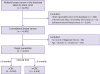
We collected information on female patients with invasive breast cancer from the SEER database from January 1, 2000 to December 31, 2010. Our cohort recruited patients aged 18–88 years [12], and we excluded patients with 1) a cancer documented only in death certificates or autopsy reports, 2) other reportable tumor in the database, 3) unrecorded time of diagnosis in the database, 4) ipsilateral second breast cancer, and 5) lesions with unknown laterality or no laterality information. CBC was considered synchronous if the second cancer was diagnosed within 6 months after the first tumor was diagnosed or metachronous if the tumor was diagnosed after ≥ 6 months (Figure 1) [1].
Figure 1
Identification of sCBC and mCBC in the SEER-18 registry.
sCBC = synchronous contralateral breast cancer; mCBC = metachronous contralateral breast cancer; SEER = Surveillance, Epidemiology, and End Results.
We also compared the following variables of sCBC and mCBC: age, race, tumor grade, tumor stage, estrogen receptor (ER), and progesterone receptor (PR) status, type of surgery, and type of radiotherapy. We analyzed age as a continuous or categorical variable (< 40, 40–49, 50–59, 60–69, 70–79, and ≥ 80) [13]. Race was classified as non-Hispanic white, non-Hispanic black, non-Hispanic Asian/Pacific Islander, Hispanic, and others. Tumor grade was categorized into well-differentiated (grade I), moderately differentiated (grade II), poorly differentiated (grade III), and undifferentiated (grade IV). Tumor stage was categorized according to the SEER historic stage A criteria as follows: localized, regional, and distant. Tumor histology was categorized into infiltrating ductal carcinoma (IDC), infiltrating lobular carcinoma (ILC), mixed IDC/ILC, and others according to the International Classification of Disease for Oncology. T grade was divided into 6 categories (Tis, T0, T1, T2, T3, and T4); axillary nodal involvement was categorized into N0, N1, N2, and N3; and distant metastasis was categorized into M0 and M1. ER and PR statuses were recoded as “positive,” “negative,” or “borderline.” Surgery was categorized into no surgery, lumpectomy, or mastectomy. Radiotherapy was divided into radiotherapy or no radiotherapy. Information on adjuvant hormone therapy and chemotherapy was not recorded in the SEER database.
Statistical analysis
Statistical analyses were performed using SPSS version 18.0 (SPSS, Chicago, USA). Pathologic, demographic, and therapeutic factors of sCBC and mCBC were compared using chi-square tests. The continuous variable (age) was compared using t-test. The Kaplan-Meier method was used to determine overall survival (OS) and breast cancer-specific survival (BCSS) curves of sCBC and mCBC, and the log-rank test was used to estimate differences in survival. In addition, we stratified patients by age at initial diagnosis and separately analyzed the BCSS of the different subgroups. OS was measured from the date of initial diagnosis of breast cancer to the date of death from any cause or the date of last follow-up (December 31, 2015). Furthermore, BCSS was calculated from the date of diagnosis to the date of death from breast cancer. Additionally, Cox proportional hazards regression analysis was performed to identify factors that predicted the outcome of CBC. The regression models included the patients' age and race, tumor grade, stage, histology, ER and PR status, radiation type, and surgery type of the 2 tumors. All statistical analyses were 2 sided, and p < 0.05 was considered statistically significant.
RESULTS
Patient characteristics
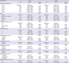
We identified 14,057 patients from the SEER 18 database. Overall, 8,139 (57.9%) patients had sCBC, and 5,918 (42.1%) patients had mCBC. The mean interval time of sCBC was 0.51 ± 0.91 months, and that of mCBCs was 45.62 ± 29.53 months. The characteristics of the first or second tumor of sCBC and mCBC were compared (Tables 1 and 2).
Table 1
Clinical characteristics of the first tumor of sCBC and mCBC
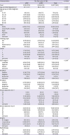
Data are shown as mean ± standard deviation or number (%).
The p < 0.05 was considered statistically significant.
sCBC = synchronous contralateral breast cancer; mCBC = metachronous contralateral breast cancer; CBC = contralateral breast cancer; IDC = infiltrating ductal carcinoma; ILC = infiltrating lobular carcinoma; SEER = Surveillance, Epidemiology, and End Results; AJCC = American Joint Committee on Cancer; ER = estrogen receptor; PR = progesterone receptor.
*Calculated after exclusion of patients with the unknown groups; †Calculated after exclusion of patients with the unknown or borderline ER/PR status.
Table 2
Clinical characteristics of the second tumor of sCBC and mCBC
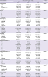
Values are presented as number (%).
The p < 0.05 was considered statistically significant.
sCBC = synchronous contralateral breast cancer; mCBC = metachronous contralateral breast cancer; CBC = contralateral breast cancer; IDC = infiltrating ductal carcinoma; ILC = infiltrating lobular carcinoma; SEER = Surveillance, Epidemiology, and End Results; AJCC = American Joint Committee on Cancer; ER = estrogen receptor; PR = progesterone receptor.
*Calculated after exclusion of patients with the unknown groups; †Calculated after exclusion of patients with the unknown or borderline ER/PR status.
The average ages of sCBC and mCBC patients were 61.7 ± 13.1 and 59.0 ± 13.6 years, respectively. The mCBC patients were younger than sCBC patients at initial diagnosis. The first tumors of mCBCs were more likely to be poorly differentiated and were more often IDC (70.5% vs. 64.8%) instead of ILC (8.3% vs. 13.9%) or mixed IDC/ILC (9.1% vs. 12.3%) compared with first tumors of sCBCs. The proportion of ER-negative first tumor was higher among mCBCs at 25.2% than among sCBCs at 12.5%. A similar pattern in PR status was also noted (negative PR: 33.1% in mCBC vs. 21.9% in sCBC). Meanwhile, mCBC patients tended to have less axillary nodal involvement (N0: 60.5% in mCBC vs. 52.4% in sCBC) and less distant metastasis (M0: 95.6% vs. 89.2%). The rates of lumpectomy and radiotherapy were higher in patients who subsequently developed mCBC at 54.0% (vs. 25.7% in sCBC) and 53.1% (vs. 32.2% in sCBC), respectively. We also compared the difference in pathological and treatment-related features of the second tumor of sCBC and mCBC. The results showed that the proportion of IDC (70.0% vs. 62.9%), grade III (34.5% vs. 17.9%) or IV (1.7% vs. 0.9%), ER negativity (26.9% vs. 9.4%), and PR negativity (41.7% vs. 18.7%) was higher in the second tumor of mCBC than in that of sCBC. We also found a considerably higher rate of lumpectomy (40.7% vs. 26.8%) and radiotherapy (31.7% vs. 26.1%) for the second tumor of mCBC than for that of sCBC (Table 2).
Survival outcomes of sCBC and mCBC
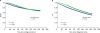
The median follow-up time was 86.2 months. Kaplan-Meier analysis demonstrated considerable difference in BCSS and OS rates between the 2 groups (Figure 2). Patients who developed sCBC had a worse OS than those who developed mCBC (5-year OS rates: 72% and 80%, respectively; 10-year OS rates: 55% and 62%, respectively). In addition, mCBC patients had a significantly favorable 5-year BCSS than sCBC patients, but they also had a worse long-term BCSS (5-year BCSS rates: 80% and 84% respectively; 10-year BCSS rates: 72% and 71%, respectively).
Figure 2
BCSS and OS curves of sCBC and mCBC. (A) Kaplan-Meier curves for BCSS of sCBC and mCBC. (B) Kaplan-Meier curves for OS of sCBC and mCBC.
The p < 0.05 was considered statistically significant.
BCSS = breast cancer-specific survival; OS = overall survival; sCBC = synchronous contralateral breast cancer; mCBC = metachronous contralateral breast cancer.
Subgroup analysis of BCSS based on different ages
The Kaplan-Meier method and log-rank test were used to analyze BCSS of patients with respect to age. When stratified by age at initial diagnosis, there was no substantial difference in BCSS rates between the two groups among patients aged 18–39, 40–49, 50–59, and 60–69 years (Figure 3); however, mCBC patients aged 70–88 years had better BCSS rates than sCBC patients (Figure 3). Furthermore, CBC patients who were younger than 40 years had poorer outcomes than those of other ages (Figure 4). Interestingly, patients aged 70–88 years who developed sCBC showed an adverse BCSS; however, such phenomenon was not detected in mCBC patients in the same age group (Figure 4). Because of the disparity in BCSS, we compared the characteristics of sCBC and mCBC patients who were older than 70 years and found that both first and second tumors of mCBC had advantages in clinicopathological characteristics (Supplementary Tables 1 and 2).
Figure 3
Kaplan-Meier survival curves of sCBC and mCBC patients grouped by age between 2000 and 2010. (A) 18–39 age group; (B) 40–49 age group; (C) 50–59 age group; (D) 60–69 age group; (E) 70–79 age group; and (F) 80–88 age group.
The p < 0.05 was considered statistically significant.
BCSS = breast cancer-specific survival; sCBC = synchronous contralateral breast cancer; mCBC = metachronous contralateral breast cancer.
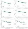
Figure 4
Survival curves of sCBC and mCBC patients of different age groups between 2000 and 2010. (A) Kaplan-Meier curves for BCSS of different age groups in sCBC patients. (B) Kaplan-Meier curves for BCSS of different age groups in mCBC patients.
The p < 0.05 was considered statistically significant.
BCSS = breast cancer-specific survival; sCBC = synchronous contralateral breast cancer; mCBC = metachronous contralateral breast cancer.

Predictive factors for poor outcomes of CBC
We further analyzed the predictive factors for outcomes of sCBC and mCBC patients (Table 3). For sCBC patients, the multivariate Cox proportional model identified the following independent prognostic factors for BCSS: age at diagnosis (p < 0.001), race (p < 0.001), grade (p < 0.001), and stage (p < 0.001) of first tumor, grade (p = 0.023) and stage (p < 0.001) of second tumor, ER status of second tumor (p = 0.005), and surgery for second tumor (p = 0.019). After adjusting for other characteristics, mCBC patients with older age (p = 0.001), first tumor classified as ILC (p = 0.019) or mixed IDC/ILC (p = 0.047), adverse grade (p < 0.001) and poorer differentiated stage (p < 0.001) of 2 tumors, surgery for 2 tumors (p < 0.001), mixed IDC/ILC (p = 0.006), ER negativity (p < 0.001) or PR negativity (p = 0.012) of the second tumor, and radiotherapy for the second tumor (p = 0.002) were significantly correlated with poor BCSS.
Table 3
Multivariable Cox proportional hazards analysis of BCSS of patients with CBC
The p < 0.05 was considered statistically significant.
BCSS = breast cancer-specific survival; CBC = contralateral breast cancer; sCBC = synchronous contralateral breast cancer; mCBC = metachronous contralateral breast cancer; IDC = infiltrating ductal carcinoma; ILC = infiltrating lobular carcinoma; SEER = Surveillance, Epidemiology, and End Results; HR = hazard ratio; CI = confidence interval; ER = estrogen receptor; PR = progesterone receptor.
DISCUSSION
Using the SEER database, we compared the clinical and pathological characteristics of sCBC and mCBC patients. We used 6 months as the interval time to divide CBC into sCBC or mCBC; this cutoff was previously used in other studies [710]. However, there is no consensus on the definite cutoff time for differentiating sCBC from mCBC. Several studies used a shorter cutoff time to distinguish between sCBC and mCBC [1114], and others chose longer cutoffs such as 12 months or 5 years [10].
Of 14,057 patients, 8,139 (57.9%) and 5,918 (42.1%) patients were diagnosed with sCBC and mCBC, respectively, between 2000 and 2010. A previous study reported that the incidence rate of mCBC was higher than that of sCBC [15]. However, in our study, the incidence of sCBC was higher than that of mCBC. There may be several explanations for this discrepancy. First, the cutoff interval time in our study was 6 months rather than 3 months, which may have influenced the proportion of patients classified as sCBC. Hartman et al. [13] indicated that the incidence of mCBC did not significantly decrease after ≥ 20 years. Another study conducted in Spain and including 120 cases of bilateral breast cancer revealed that approximately 40% of CBC patients were diagnosed with mCBC 10 years after the initial diagnosis [15]. Our study may not have included all mCBC patients owing to the short follow-up. Since the 1980s, the incidence rate of second primary tumor has decreased [2], possibly because of the increasing use of chemotherapy, hormonal therapy, and CPM [15]. Our results suggested that effective treatment substantially decreases the rate of mCBC but not of sCBC.
Pestalozzi et al. [16] reported incidences of ILC and IDC at 6.2% and 70.5%, respectively, among 13,220 patients with breast cancer from the IBCSG trial I between 1978 and 2002. Another study of 135,157 patients with invasive breast cancer from the SEER database from 1992 to 2001 reported rates of 76%, 8%, and 7% for IDC, ILC, and mixed IDC/ILC, respectively [17]. Our results suggested that the number of patients presenting with ILC was higher in CBC patients than in UBC patients, which is similar to that reported in several previous studies [1819] . Moreover, our study indicated that more first tumors of sCBC were ILC or mixed IDC/ILC, which was consistent with the results of Díaz et al. [15]. In summary, ILC patients should be assessed for symptoms of CBC more frequently within the first 6 months of diagnosis.
A study from the SEER database that enrolled 328,870 female patients with invasive breast cancer showed that 76.1% of these patients were ER positive and 65.6% were PR positive between 2001 and 2010 [20]. This was higher than the proportion of ER- or PR-positive first tumors of CBC in our study. We also found that the number of patients with an ER-negative first cancer was higher among mCBC patients than among sCBC patients. This may be because ER-positive/PR-positive tumors are more likely to be treated with endocrine therapy, which significantly reduces the risk of developing CBC [2122], particularly mCBC. In addition, our study indicated that the proportion of younger patients in the mCBC cohort was higher than that in the sCBC cohort, possibly resulting in a higher rate of patients who developed an ER-negative first tumor [23].
Several studies reported that sCBC was associated with poorer survival than mCBC [9242526]. However, Chen et al. [8] found that the time interval for differentiating sCBC from mCBC did not correlate with outcomes. In contrast, a study reported opposite results [27]. In our study, we found a significant early advantage in BCSS for the mCBC cohort; however, BCSS worsened starting from 8 years after initial diagnosis, possibly because of the adverse TNM stage of the first tumor of sCBC and worse characteristics of the second tumor of mCBCs. However, such patterns were not detected in terms of OS, which may be because of the higher number of elderly patients with sCBC who were more likely to develop geriatric conditions [13].
We compared BCSS among different age groups to investigate the association between age at initial diagnosis and prognosis. Interestingly, the subgroup analysis revealed that older patients (age >70 years) with mCBCs had longer BCSS than those with sCBCs. However, the same phenomenon was not observed in patients aged 18–69 years, and this may be because of the advantageous characteristics of both tumors of mCBCs among old patients. These results indicate that old age is not an unfavorable factor for survival of mCBC patients. We also found that young age (< 40 years) was associated with worse outcomes than other ages in both mCBC and sCBC patients. An alternative explanation is that young women diagnosed with CBC had more aggressive tumor characteristics, shorter interval time, and genetic predisposition [2628].
Different clinical features and outcomes were observed in mCBC and sCBC patients; however, only few studies focused on the poor prognostic indicators for survival of CBC patients. A study from Ireland that included 2,524 patients with breast cancer suggested that ER and PR positivity, lymph node negativity, and radiotherapy for the second primary tumor were important beneficial factors [24]. Another study showed that mCBC patients with lower stage, no lymph node involvement, ER positivity, and lower tumor grade had favorable OS [15].
Our results indicated that adverse grade and stage were poor prognostic indicators for BCSS of both sCBC and mCBC. In addition, age > 80 or < 50 years was associated with poor BCSS in mCBC patients. The first tumor classified as ILC or mixed IDC/ILC was an independent factor of poor prognosis for mCBC patients. To our knowledge, no study has reported a similar finding. Ibrahim et al. [9] found that hormone receptor-negative first tumor was a risk factor for poor prognosis. Our study showed that hormone receptor-negative second tumor was also an independent prognostic factor for BCSS. Unexpectedly, radiotherapy for the first tumor was not associated with favorable BCSS for CBC patients. A study from Japan indicated that CBC patients with short time interval (< 5 years) who were treated with adjuvant radiotherapy tended to have a more aggressive second cancer [29], which may explain why radiotherapy for the first tumor was not an independent favorable factor for sCBCs. In addition, mCBC patients who received radiotherapy for the second tumor had an advantage over those who did not receive radiotherapy in terms of BCSS; however, the percentage of patients who received radiotherapy for the second tumor was lower in the mCBC group than in the sCBC group. Therefore, secondary radiotherapy may be favorable for the survival of mCBC patients.
This large retrospective study with a relatively long follow-up period provides evidence on the differences in clinicopathological features and prognosis between sCBC and mCBC. Moreover, our study included the largest cohort to determine the prognostic factors for BCSS of sCBC and mCBC. Patients with ipsilateral second breast cancer were excluded because it was difficult to distinguish a new primary tumor from localized recurrent lesion. In addition, we excluded patients with other reported tumors in the database to avoid a survival bias. This study had several limitations. First, this was a retrospective study that may have included biases. Second, information about chemotherapy or endocrine therapy was not recorded in the SEER database, which may have influenced the outcomes. Third, the human epidermal growth factor receptor 2 status was not included in the SEER database until 2010.
In this study, the first tumors of sCBC tended to have higher stage and more lymph and distant metastases, whereas those of mCBC were more often poorly differentiated, ER-negative and PR-negative, and had less axillary nodal involvement. Moreover, second tumors of mCBC were more often IDC; grade III or IV; and had adverse stage, ER and PR negativity, and more axillary nodal involvement; in contrast, second tumors of sCBC were more likely to have lower grade, localized stage, ER/PR positivity, and no lymph and distant metastasis. BCSS curves showed better BCSS for mCBC in the early follow-up, but a worse outcome later. Furthermore, BCSS rates were not significantly different between sCBC and mCBC patients aged 18–69 years. The features of CBCs, such as adverse grade and late stage of two tumors, no surgery and radiotherapy, and ER or PR negativity of the second tumor, were associated with worse prognosis. Future research should focus on investigating biomarkers for predicting CBC and promoting more effective therapeutic modalities.
 XML Download