Journal List > J Korean Med Sci > v.34(Suppl 1) > 1127929

Kang, Cho, Hyun, Kim, Ko, Kang, Cheong, Kim, Moon, and Ha: A Pediatric Case of a D-Penicillamine Induced ANCA-associated Vasculitis Manifesting a Pulmonary-Renal Syndrome

Abstract

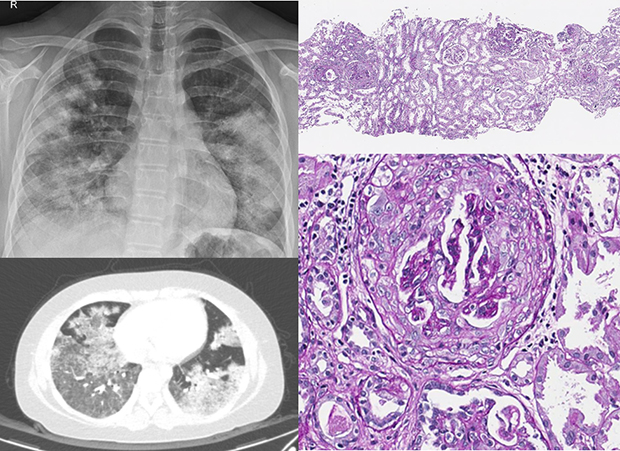
D-penicillamine has been reported to cause antineutrophil cytoplasmic antibody (ANCA)-associated vasculitis presenting as rapidly progressive glomerulonephritis or pulmonary-renal syndrome mostly in adults. We report a pediatric case of D-penicillamine induced ANCA-associated vasculitis that manifests as a pulmonary-renal syndrome with a mild renal manifestation. A 13-year-old girl who has been taking D-penicillamine for five years under the diagnosis of Wilson disease visited the emergency room because of hemoptysis and dyspnea. She had diffuse pulmonary hemorrhage, microscopic hematuria, and proteinuria. Myeloperoxidase ANCA was positive, and a renal biopsy revealed pauci-immune crescentic glomerulonephritis. Under the diagnosis of D-penicillamine-induced ANCA-associated vasculitis, D-penicillamine was switched to trientine, and the patient was treated with plasmapheresis, glucocorticoid, cyclophosphamide, and mycophenolate mofetil. Pulmonary hemorrhage improved rapidly followed by the disappearance of the hematuria and proteinuria five months later.

Graphical Abstract

jkms-34-e173-abf001

INTRODUCTION

Anti-neutrophil cytoplasmic antibody (ANCA)-associated vasculitis (AAV) is a small-vessel vasculitis characterized by the presence of ANCA and typical involvement of kidney and respiratory tract.1 D-penicillamine is one of the drugs that can induce drug-induced AAV.2 Rapidly progressive glomerulonephritis (RPGN) and pulmonary-renal syndrome (PRS) are primary manifestations of D-penicillamine-induced AAV.34567891011121314151617181920 Because most of them develop in adults, we could search only two cases of D-penicillamine-induced AAV diagnosed at age younger than 18 years,21 and both cases presented RPGN. Here, we report a case of D-penicillamine-induced AAV that developed in a 13-year-old girl and manifested a PRS without azotemia.

CASE DESCRIPTION

A 13-year-old female patient visited the emergency room for hemoptysis, fever, and dyspnea on September 8, 2017. She has been on D-penicillamine for five years under the diagnosis of Wilson disease with a well-controlled copper level. Urinalysis performed before the administration of D-penicillamine was normal. Since five months before admission, intermittent pain occurred at the ankle, foot, calf, and wrist. Two months prior to admission, urine test revealed microscopic hematuria (10-19 RBCs/HPF) and mild proteinuria (1+). Two weeks before admission, cough and fever developed followed by dyspnea and hemoptysis for a week before admission. She had no family history of autoimmune disease or Wilson disease.
At admission, blood pressure was 132/74 mmHg, the temperature was 36.5°C, pulse rate was 132/min, and respiratory rate was 24/min. Crackle sounds were heard at both lower lung fields, and heartbeats were regular without a murmur. Hemoglobin was 8.7 g/dL, reticulocyte count 2.5%, the white blood cell count was 9,670/µL, and platelet count was 344,000/µL. Serum hepatic enzymes were normal, and albumin level was 3.7 g/dL. Blood urea nitrogen and serum creatinine were 12 mg/dL and 0.62 mg/dL respectively and estimated GFR was 109.8 mL/min/1.73 m2. C-reactive protein level was 9.1 mg/dL, and erythrocyte sedimentation rate was 28 mm/hr. The patient had microscopic hematuria (5-9 RBCs/HPF) and proteinuria (urine protein creatinine ratio 1.05 mg/mg). Anti-myeloperoxidase (MPO) IgG titer was over 600 U/mL (reference < 20 U/mL) while anti-proteinase 3 (PR3) IgG titer was within normal limit. Complement levels were normal and antinuclear antibody, anti-double-stranded DNA antibody, rheumatoid factor, anti-cardiolipin antibody, anti-glomerular basement membrane antibody were all negative. There was no evidence of infection or hemolysis. Chest radiography and computed tomography suggested diffuse pulmonary hemorrhage (Fig. 1). The echocardiographic findings were not remarkable. Renal Doppler ultrasonography revealed normal size and shape of both kidneys and an equivocal increase in cortical echogenicity.
Fig. 1

Chest radiologic findings of the patient. (A) Bilateral extensive patchy consolidation at the plain film and (B) the multifocal patchy ground-glass opacity at chest computed tomography suggest diffuse pulmonary hemorrhage.

jkms-34-e173-g001
Under the tentative diagnosis of D-penicillamine-induced AAV, trientine was substituted for D-penicillamine. Because the patient's respiratory symptom aggravated, she was transferred to ICU and supported by a ventilator. Plasma exchange was performed for 3 days daily and methylprednisolone (1 g per day) was given intravenously for 3 days followed by the administration of IgG (0.7 g/kg) for 3 days.
A renal biopsy disclosed cellular and fibrocellular crescents in 39% (16/41) of the glomeruli. Glomerular size and mesangial cellularity were mildly increased. Immunofluorescent and electron microscopic findings were not noteworthy except focal effacement of the foot processes. These findings were compatible with the pauci-immune crescentic glomerulonephritis (Fig. 2).
Fig. 2

Photomicrograph of the kidney biopsy. (A) Crescent formation is observed in 39% of the total 41 glomeruli by the light microscopy (PAS, ×40). (B) A representative crescent (arrow) is shown at a high power magnification (PAS, ×400).

PAS = periodic acid–Schiff.
jkms-34-e173-g002
After confirming the renal pathology, intravenous cyclophosphamide (750 mg/m2) was administered, followed by administration of oral glucocorticoids (prednisolone 60 mg/day) and mycophenolate mofetil (1,500 mg/day). Acute phase reactants dropped a few days later with the rapid improvement of the respiratory failure. Proteinuria and microscopic hematuria disappeared five months later. MPO-ANCA titer decreased more gradually and still positive in a low titer (8.1 IU/mL) at 17 months after the withdrawal of D-penicillamine.

Ethics statement

This study was approved by the Institutional Review Boards (IRB) of the Seoul National University Hospital (H-1901-133-1005) and an informed consent was received from the patient and a caregiver. The images are published under agreement of the patient.

DISCUSSION

AAV is an autoimmune small-vessel vasculitis characterized by ANCA, an autoantibody specific for neutrophil granule components, MPO or protease 3 (PR3).1 Drugs such as propylthiouracil, hydralazine, penicillamine, allopurinol, sulfasalazine, and levamisole have been described to induce AAV, the majority with MPO-AAV.22 D-penicillamine-induced AAV is reported to affect kidneys with or without concurrent involvement of the respiratory tract, therefore, leads to either RPGN345678910112021 or PRS.1213141516171819
PRS describes a syndrome of respiratory failure in association with renal failure or a diffuse alveolar hemorrhage complicating acute glomerulonephritis.23 PRS often results in severe life-threatening systemic disease requiring urgent aggressive therapy and renal and lung biopsy reveals histological features of the glomerular crescents and pulmonary capillaritis.24 Serum creatinine is usually elevated in PRS, but it may not always be elevated.23 Among the 11 cases of penicillamine-associated PRS reported with the serum creatinine available, there was an adult woman who had normal serum creatinine.25 Our case also showed microscopic hematuria and proteinuria with normal renal function while renal biopsy revealed cellular or fibrocellular crescents in 39% of the glomeruli. McAdoo et al.26 analyzed 258 cases of necrotizing or crescentic glomerulonephritis and found that 14.7% of the patients represent preserved renal function. This silent crescentic glomerulonephritis with normal renal function is more likely to be identified in PRS because pulmonary manifestations facilitate earlier biopsy and diagnosis of the glomerulonephritis before renal function deteriorates. In our case, if the renal biopsy was performed later and the treatment was delayed, azotemia might have developed.
Most of the reported AAV patients were adults. Only two cases of D-penicillamine-induced AAV are reported at the age younger than 18 years, and both pediatric cases presented as RPGN.21 Therefore, to the best of our knowledge, this is the first pediatric case of D-penicillamine-induced AAV that manifests a PRS.
Loss of tolerance and development of autoimmunity to MPO and PR3 is the first step in the development of AAV. Mechanism of losing tolerance is unclear, though dysregulation of the adaptive immune response, neutrophil extracellular trap (NET), ANCA antigen on the surface of apoptotic neutrophil, and functional abnormality in regulatory T cell are implicated.1 Subsequently, T-cell helped activation of autoreactive B-cell produces ANCA. Finally, ANCA induces activation of circulating neutrophils that localize to vascular beds, degranulate and release NET resulting in endothelial injury.127
How individual drug triggers the AAV is not well understood. Because the molecular weight of most drugs is low, the formation of a bigger complex is required to provoke immune response.28 In animal experiments, penicillamine is reported to activate macrophages through binding to aldehyde-containing molecules and production of TNF-α, IL-6, and IL-23,2930 giving rise to induction of autoimmunity. Though mechanistic studies about penicillamine induced AAV is not sufficient, it can be compensated by investigations performed for other drug-induced AAVs, because the clinical manifestations are very similar.28 Propylthiouracil, another causative drug of AAV, is reported to be oxidized in the presence of activated neutrophils forming reactive drug metabolites that stimulate ANCA production.31 It was also reported that propylthiouracil induces abnormal conformation and impaired degradation of NET leading to the development of autoimmunity.32 In hydralazine-associated AAV, neutrophil apoptosis in response to hydralazine MPO binding is hypothesized to result in the production of autoantibodies.33
The prevalence of ANA in drug-associated AAV has been reported to vary from 17% to 80%. It was reported that the prevalence is higher in AAV induced by hydralazine and lower in the vasculitis associated with propylthiouracil.22 In authors review of the literature, ANA was present in 76% of the 17 reported cases of the D-penicillamine-induced AAV. Clinical significance of the ANA positivity in drug-induced AAV remains to be elucidated.
Because the diagnosis of drug-induced vasculitis is complicated by a variety of factors such as the dose and duration of medication, and frequency of diagnostic laboratory studies according to the physician's awareness of the disease, involved organs and clinical presentations may be diverse. Therefore, treatment should depend on the severity of clinical manifestations.28 The first step is to discontinue the offending agent. For those with active and significant organ involvement, additional immune suppression by glucocorticoid is necessary. For severe organ failure associated with alveolar hemorrhage or RPGN, cyclophosphamide or other immunosuppressants such as mycophenolate mofetil or rituximab may be added. Intravenous pulse methylprednisolone and plasmapheresis can be used in life-threatening cases. Supportive care would include ventilatory support and renal replacement for severe PRS.34
Delayed diagnosis of drug-induced AAV may cause serious results. Therefore, it is preferable to avoid long-term use of AAV inducible medications such as propylthiouracil, hydralazine, penicillamine, allopurinol, sulfasalazine, and levamisole. When long-term use of these drugs is inevitable, it is desirable to monitor respiratory symptoms and signs, and urinalysis closely, and when suspected, check ANCA titer and chest radiography. However, it should be emphasized that though positive seroconversion of ANCA is a clue to the diagnosis of drug-induced AAV, positive ANCA titer only is not sufficient to diagnose the drug-induced AAV and begin treatment, because only a small proportion of the patients with positive ANCA develop clinical vasculitis.28
In this case, crescentic glomerulonephritis was found in the renal biopsy despite the mild renal manifestations and completely normal renal function. Therefore, when urinary abnormalities are found in a patient with suspected AAV and pulmonary hemorrhage, even if the renal function is normal, renal biopsy is highly recommended.
Although anti-MPO antibody titer decreased steadily from over 600 to 8.1 IU/mL, the value was still above the normal range at 17 months after the withdrawal of D-penicillamine. This long-lasting ANCA activity is consistent with the finding in propylthiouracil (PTU)-induced AAV that anti-MPO titer was within the reference range only in six of the thirteen patients at a median follow-up of 38 months after discontinuation of PTU.35 Therefore, drug-induced ANCA seems to last for a substantial period after withdrawal of the triggering agent.

Notes

Disclosure The authors have no potential conflicts of interest to disclose

Author Contributions

  • Conceptualization: Kang HG, Ha IS.

  • Data curation: Kang S, Cho MH, Hyun H.

  • Formal analysis: Kang S, Cho MH, Hyun H.

  • Validation: Kim JH.

  • Writing - original draft: Kang S.

  • Writing - review & editing: Kim JH, Ko JS, Kang HG, Cheong HI, Kim WS, Moon KC.

References

1. Hutton HL, Holdsworth SR, Kitching AR. ANCA-associated vasculitis: pathogenesis, models, and preclinical testing. Semin Nephrol. 2017; 37(5):418–435.
crossref
2. Hogan JJ, Markowitz GS, Radhakrishnan J. Drug-induced glomerular disease: immune-mediated injury. Clin J Am Soc Nephrol. 2015; 10(7):1300–1310.
crossref
3. Banfi G, Imbasciati E, Guerra L, Mihatsch MJ, Ponticelli C. Extracapillary glomerulonephritis with necrotizing vasculitis in D-penicillamine-treated rheumatoid arthritis. Nephron. 1983; 33(1):56–60.
4. Sadjadi SA, Seelig MS, Berger AR, Milstoc M. Rapidly progressive glomerulonephritis in a patient with rheumatoid arthritis during treatment with high-dosage D-penicillamine. Am J Nephrol. 1985; 5(3):212–216.
5. Williams AJ, Fordham JN, Barnes CG, Goodwin FJ. Progressive proliferative glomerulonephritis in a patient with rheumatoid arthritis treated with D-penicillamine. Ann Rheum Dis. 1986; 45(1):82–84.
crossref
6. Devogelaer JP, Pirson Y, Vandenbroucke JM, Cosyns JP, Brichard S, Nagant de Deuxchaisnes C. D-penicillamine induced crescentic glomerulonephritis: report and review of the literature. J Rheumatol. 1987; 14(5):1036–1041.
7. Mathieson PW, Peat DS, Short A, Watts RA. Coexistent membranous nephropathy and ANCA-positive crescentic glomerulonephritis in association with penicillamine. Nephrol Dial Transplant. 1996; 11(5):863–866.
crossref
8. Hillis GS, Khan IH, Simpson JG, Rees AJ. Scleroderma, D-penicillamine treatment, and progressive renal failure associated with positive antimyeloperoxidase antineutrophil cytoplasmic antibodies. Am J Kidney Dis. 1997; 30(2):279–281.
crossref
9. Karpinski J, Jothy S, Radoux V, Levy M, Baran D. D-penicillamine-induced crescentic glomerulonephritis and antimyeloperoxidase antibodies in a patient with scleroderma. Am J Nephrol. 1997; 17(6):528–532.
10. Nanke Y, Akama H, Terai C, Kamatani N. Rapidly progressive glomerulonephritis with D-penicillamine. Am J Med Sci. 2000; 320(6):398–402.
crossref
11. Bienaimé F, Clerbaux G, Plaisier E, Mougenot B, Ronco P, Rougier JP. D-Penicillamine-induced ANCA-associated crescentic glomerulonephritis in Wilson disease. Am J Kidney Dis. 2007; 50(5):821–825.
crossref
12. Gibson T, Burry HC, Ogg C. Letter: Goodpasture syndrome and D-penicillamine. Ann Intern Med. 1976; 84(1):100.
13. Gavaghan TE, McNaught PJ, Ralston M, Hayes JM. Penicillamine-induced “Goodpasture's syndrome”: successful treatment of a fulminant case. Aust N Z J Med. 1981; 11(3):261–265.
14. Peces R, Riera JR, Arboleya LR, López-Larrea C, Alvarez J. Goodpasture's syndrome in a patient receiving penicillamine and carbimazole. Nephron. 1987; 45(4):316–320.
crossref
15. Macarrón P, García Díaz JE, Azofra JA, Martín de Francisco J, Gonzalez E, Fernandez G, et al. D-penicillamine therapy associated with rapidly progressive glomerulonephritis. Nephrol Dial Transplant. 1992; 7(2):161–164.
crossref
16. Endo H, Hosono T, Kondo H. Antineutrophil cytoplasmic autoantibodies in 6 patients with renal failure and systemic sclerosis. J Rheumatol. 1994; 21(5):864–870.
17. Gaskin G, Thompson EM, Pusey CD. Goodpasture-like syndrome associated with anti-myeloperoxidase antibodies following penicillamine treatment. Nephrol Dial Transplant. 1995; 10(10):1925–1928.
18. Derk CT, Jimenez SA. Goodpasture-like syndrome induced by D-penicillamine in a patient with systemic sclerosis: report and review of the literature. J Rheumatol. 2003; 30(7):1616–1620.
19. Sternlieb I, Bennett B, Scheinberg IH. D-penicillamine induced Goodpasture's syndrome in Wilson's disease. Ann Intern Med. 1975; 82(5):673–676.
crossref
20. Ntoso KA, Tomaszewski JE, Jimenez SA, Neilson EG. Penicillamine-induced rapidly progressive glomerulonephritis in patients with progressive systemic sclerosis: successful treatment of two patients and a review of the literature. Am J Kidney Dis. 1986; 8(3):159–163.
crossref
21. Lee Y, Lee ST, Cho H. D-penicillamine-induced ANA (+) ANCA (+) vasculitis in pediatric patients with Wilson's disease. Clin Nephrol. 2016; 85(5):296–300.
crossref
22. Choi HK, Merkel PA, Walker AM, Niles JL. Drug-associated antineutrophil cytoplasmic antibody-positive vasculitis: prevalence among patients with high titers of antimyeloperoxidase antibodies. Arthritis Rheum. 2000; 43(2):405–413.
crossref
23. West SC, Arulkumaran N, Ind PW, Pusey CD. Pulmonary-renal syndrome: a life threatening but treatable condition. Postgrad Med J. 2013; 89(1051):274–283.
crossref
24. Lee RW, D'Cruz DP. Pulmonary renal vasculitis syndromes. Autoimmun Rev. 2010; 9(10):657–660.
crossref
25. Matloff DS, Kaplan MM. D-Penicillamine-induced Goodpasture's-like syndrome in primary biliary cirrhosis--successful treatment with plasmapheresis and immunosuppressives. Gastroenterology. 1980; 78(5 Pt 1):1046–1049.
crossref
26. McAdoo SP, Tanna A, Randone O, Tam FW, Tarzi RM, Levy JB, et al. Necrotizing and crescentic glomerulonephritis presenting with preserved renal function in patients with underlying multisystem autoimmune disease: a retrospective case series. Rheumatology (Oxford). 2015; 54(6):1025–1032.
crossref
27. O'Sullivan KM, Lo CY, Summers SA, Elgass KD, McMillan PJ, Longano A, et al. Renal participation of myeloperoxidase in antineutrophil cytoplasmic antibody (ANCA)-associated glomerulonephritis. Kidney Int. 2015; 88(5):1030–1046.
28. Radić M, Martinović Kaliterna D, Radić J. Drug-induced vasculitis: a clinical and pathological review. Neth J Med. 2012; 70(1):12–17.
29. Li J, Uetrecht JP. D-penicillamine-induced autoimmunity: relationship to macrophage activation. Chem Res Toxicol. 2009; 22(9):1526–1533.
crossref
30. Li J, Mannargudi B, Uetrecht JP. Covalent binding of penicillamine to macrophages: implications for penicillamine-induced autoimmunity. Chem Res Toxicol. 2009; 22(7):1277–1284.
crossref
31. Jiang X, Khursigara G, Rubin RL. Transformation of lupus-inducing drugs to cytotoxic products by activated neutrophils. Science. 1994; 266(5186):810–813.
crossref
32. Nakazawa D, Tomaru U, Suzuki A, Masuda S, Hasegawa R, Kobayashi T, et al. Abnormal conformation and impaired degradation of propylthiouracil-induced neutrophil extracellular traps: implications of disordered neutrophil extracellular traps in a rat model of myeloperoxidase antineutrophil cytoplasmic antibody-associated vasculitis. Arthritis Rheum. 2012; 64(11):3779–3787.
33. Yokogawa N, Vivino FB. Hydralazine-induced autoimmune disease: comparison to idiopathic lupus and ANCA-positive vasculitis. Mod Rheumatol. 2009; 19(3):338–347.
crossref
34. Grau RG. Drug-induced vasculitis: new insights and a changing lineup of suspects. Curr Rheumatol Rep. 2015; 17(12):71.
crossref
35. Yang J, Yao LP, Dong MJ, Xu Q, Zhang J, Weng WW, et al. Clinical characteristics and outcomes of propylthiouracil-induced antineutrophil cytoplasmic antibody-associated vasculitis in patients with Graves' disease: a median 38-month retrospective cohort study from a single institution in China. Thyroid. 2017; 27(12):1469–1474.
crossref
TOOLS
ORCID iDs

Sena Kang
https://orcid.org/0000-0002-5891-6674

Myung Hyun Cho
https://orcid.org/0000-0002-3237-3173

Hyesun Hyun
https://orcid.org/0000-0001-8525-1471

Ji Hyun Kim
https://orcid.org/0000-0001-8477-0157

Jae Sung Ko
https://orcid.org/0000-0002-3064-2974

Hee Gyung Kang
https://orcid.org/0000-0001-8323-5320

Hae Il Cheong
https://orcid.org/0000-0001-7556-1265

Woo Sun Kim
https://orcid.org/0000-0003-2184-1311

Kyung Chul Moon
https://orcid.org/0000-0002-1969-8360

Il-Soo Ha
https://orcid.org/0000-0001-5428-6209

Similar articles