Abstract
Although acute traumatic fractures of the clavicle are relatively common, stress fractures of the proximal clavicle are extremely rare. Stress fractures of the clavicle have often been reported after a radical neck dissection or radiation but rarely occur during excessive repetitive exercise in professional athletes. The authors report a case of a stress fracture of the proximal clavicle during exercise in a young man with no specific preceding factors, which has not been reported in the Korean literature.
Go to : 
References
1. Shahane SM, Samant A, Pathak AC, Reddy R, Oswal C. Idiopathic non-traumatic or stress fracture of clavicle. J Case Rep Pract. 2014. 2:37–9.
2. Abbot AE, Hannafin JA. Stress fracture of the clavicle in a female lightweight rower: a case report and review of the literature. Am J Sports Med. 2001. 29:370–2.
3. Cummings CW, First R. Stress fracture of the clavicle after a radical neck dissection: case report. Plast Reconstr Surg. 1975. 55:366–7.
4. Choi EC, Kim DY, Koh YW, Kim HJ. Fracture of the clavicle after radical neck dissection, pectoralis major myocutaneous flapand postoperative radiotherapy. Korean J Otolaryngol. 1999. 42:1060–5.
5. Yamada K, Sugiura H, Suzuki Y. Stress fracture of the medial clavicle secondary to nervous tic. Skeletal Radiol. 2004. 33:534–6.

6. Wu CD, Chen YC. Stress fracture of the clavicle in a professional baseball player. J Shoulder Elbow Surg. 1998. 7:164–7.

7. Fallon KE, Fricker PA. Stress fracture of the clavicle in a young female gymnast. Br J Sports Med. 2001. 35:448–9.

8. Akasbi N, Elidrissi M, Tahiri L, Elmrini A, Harzy T. An unusual cause of shoulder pain in an elderly woman: a case report. J Med Case Rep. 2013. 7:271.

Go to : 
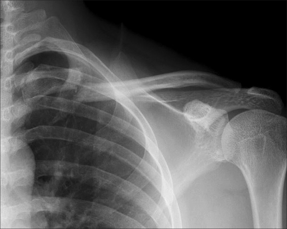
 | Figure 1.Initial left clavicle anteroposterior (A), caudal tilt (B), and axial lateral (C) radiographs showing no specific findings. |
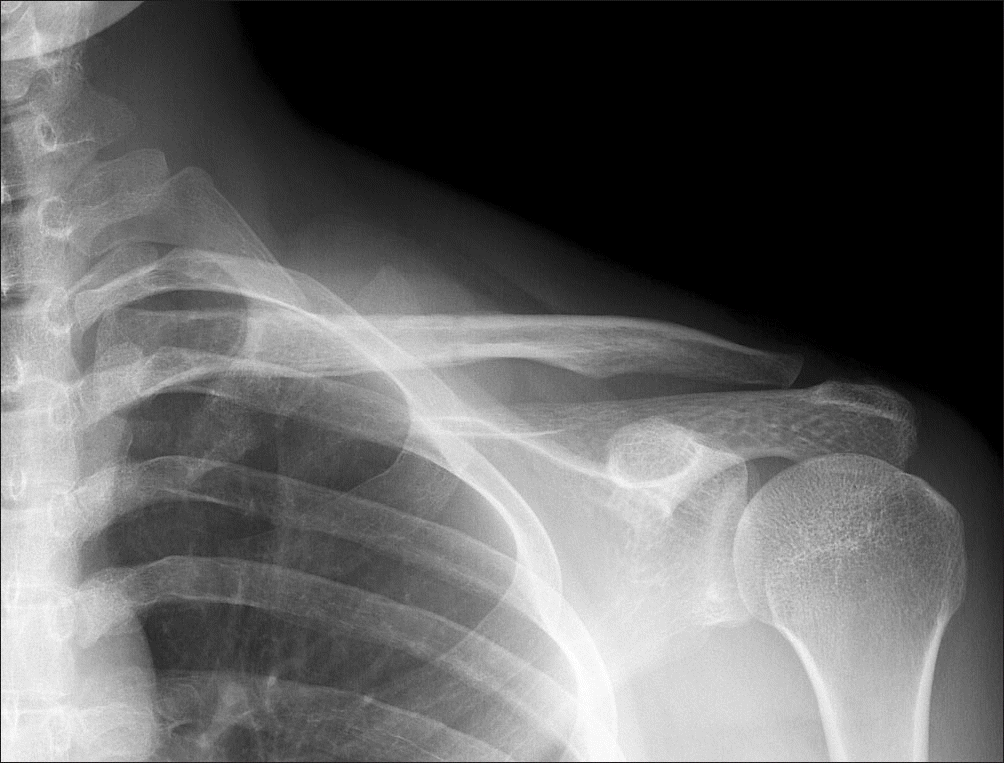
 | Figure 2.Follow-up radiograph showing oblique fracture lines with displacement at proximal portion of the left clavicle. |
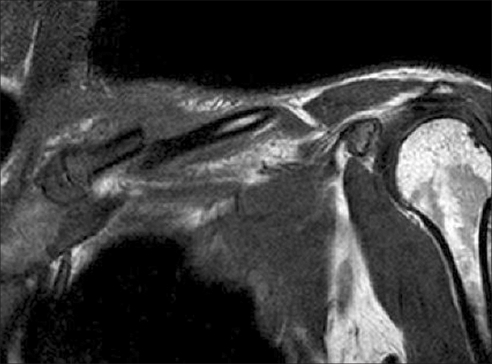
 | Figure 3.Coronal T1 weighted magnetic resonance imaging showing no other pathology, such as tumor or infection, other than the fracture associated with displacement in the left proximal clavicle. |


 PDF
PDF ePub
ePub Citation
 Print
Print


 XML Download