

 PDF
PDF ePub
ePub Citation
 Print
Print


Abstract
Avascular necrosis (AVN) is defined as the cellular death of bone and bone marrow components due to the loss of blood supply, and associated with post-traumatic or non-traumatic events. AVN usually involves the epiphysis of a long bone, such as the femoral and humeral heads, which are susceptible to osteonecrosis. Many studies have been conducted but they were restricted to investigations of femoral head avascular necrosis. The presence of osteonecrosis in the proximal femur may impair biological fixation after total hip arthroplasty. We report a 56-year-old male patient with avascular necrosis located not only at the femoral head, but also in the entire femur, including the medullary cavity, who underwent total hip arthroplasty 2 years earlier along with a review of the relevant literature.
Go to : 
References
1. Ficat RP, Arlet J. Ischémie et necrose osseuses. Paris;Masson: 1977. 1–224.
2. ARCO (Association Reserch Circularion Osseous) committee on terminology and classification. ARCO News. 1992. 4:41–6.
3. Sakai T, Sugano N, Nishii T, Haraguchi K, Yoshikawa H, Ohzono K. Bone scintigraphy for osteonecrosis of the knee in patients with non-traumatic osteonecrosis of the femoral head: comparison with magnetic resonance imaging. Ann Rheum Dis. 2001. 60:14–20.

4. Lafforgue P. Pathophysiology and natural history of avascular necrosis of bone. Joint Bone Spine. 2006. 73:500–7.
5. Kim YH, Kim JS. Histologic analysis of acetabular and proximal femoral bone in patients with osteonecrosis of the femoral head. J Bone Joint Surg Am. 2004. 86:2471–4.
6. Calder JD, Pearse MF, Revell PA. The extent of osteocyte death in the proximal femur of patients with osteonecrosis of the femoral head. J Bone Joint Surg Br. 2001. 83:419–22.
7. Sinha RK, Dungy DS, Yeon HB. Primary total hip arthroplasty with a proximally porous-coated femoral stem. J Bone Joint Surg Am. 2004. 86:1254–61.
8. Geesink RG, de Groot K, Klein CP. Bonding of bone to apatite-coated implants. J Bone Joint Surg Br. 1988. 70:17–22.
9. Archibeck MJ, Berger RA, Jacobs JJ. . Second-generation cementless total hip arthroplasty. Eight to eleven-year results. J Bone Joint Surg Am. 2001. 83:1666–73.
10. Rothman RH, Cohn JC. Cemented versus cementless total hip arthroplasty. A critical review. Clin Orthop Relat Res. 1990. 254:153–69.
Go to :
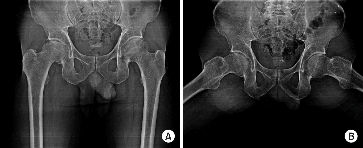 | Figure 1.Anterioposterior (A) and frog leg lateral (B) radiographs show collapse of the right femur head and subchondral fracture. |
 | Figure 2.Coronal (A) and axial (B) T2-weighted fat suppression magnetic resonance imaging show extended necrosis extended to the femur diaphysis. |
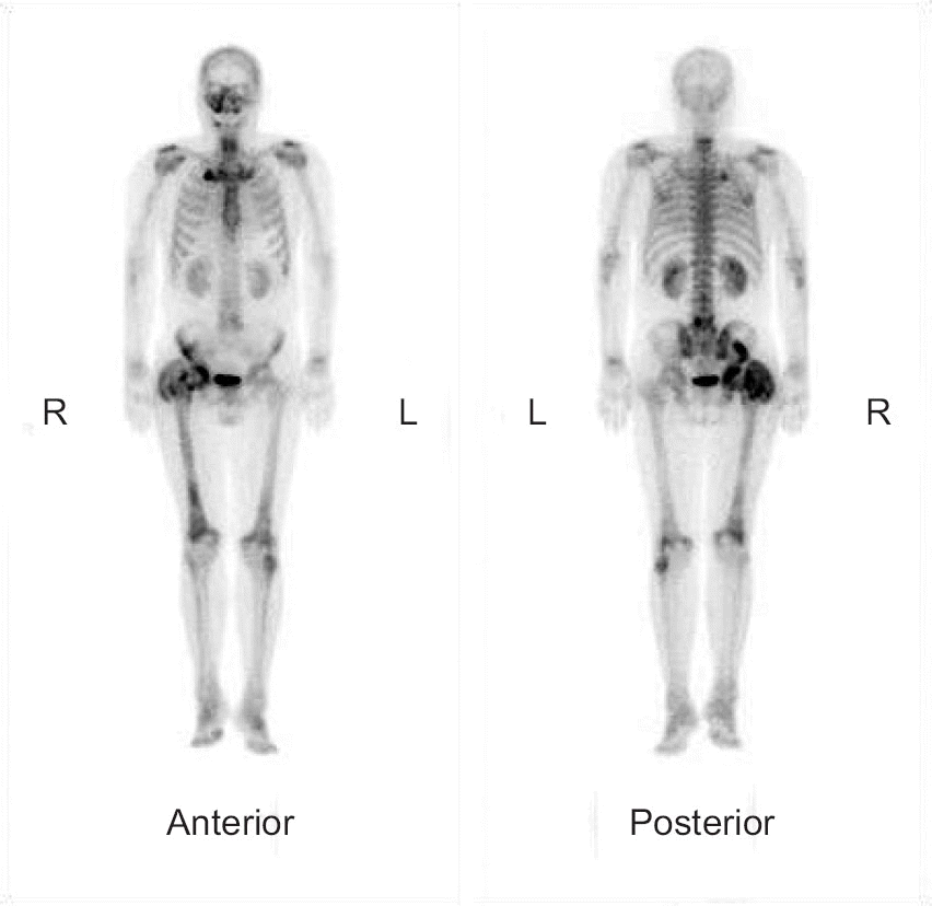 | Figure 3.Anterior and posterior view of technetium-99m bone scintigraphs showing diffuse uptake in the right femur suggesting entire femur avascular necrosis. |
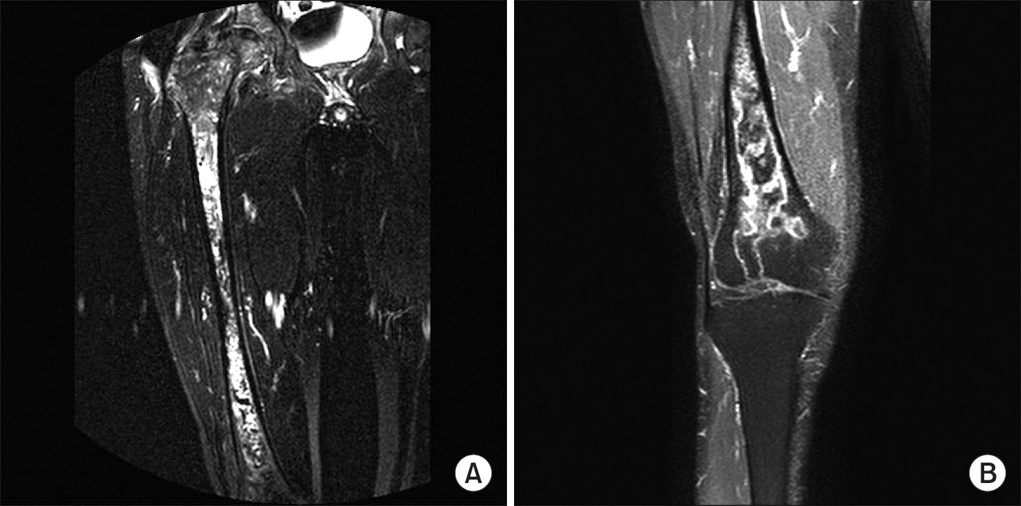 | Figure 4.Right femur (A) and knee (B) T2-weighted fat suppression magnetic resonance imaging show avascular necrosis extended to the lateral femoral condyle. |
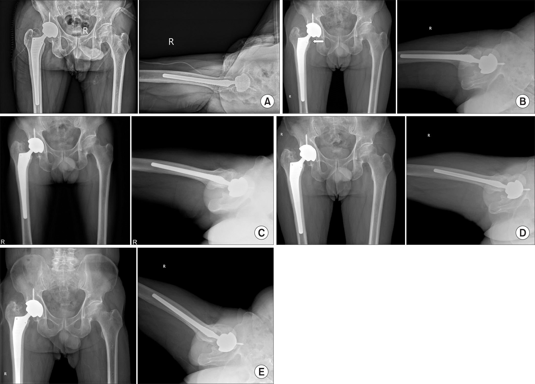 | Figure 5.(A) Postoperative anteroposterior and lateral radiographs show press-fitted femoral stem and acetabular component. (B) Four-month follow-up radiographs show calcar resorption and about 6-mm-sized subsidence is observed (white arrow). (C) Twelve-month follow-up radiographs show progress subsidence to approximately 10 mm but no further progression of subsidence is observed in the 18 months (D) and 24 months (E) follow-up radiographs. |
 XML Download