

 PDF
PDF ePub
ePub Citation
 Print
Print


Abstract
Purpose:
To analyze the risk factors for posterior migration of a single cage after transforminal lumbar interbody fusion (TLIF).
Materials and Methods:
This study was conducted retrospectively on 48 patients (60 discs) who were followed-up for 1 year after TLIF from January 2015 to January 2017. The patients were divided into two groups: group 1 containing 16 patients (17 discs) with cage migration and group 2 containing 32 patients (43 discs) without it. Information related to cage migration, such as the demographic factors, shape of disc, level and location of the cage inserted, and disc height change, was acquired from the medical records and radiologic images, and the possibility for generating posterior migration of cage was evaluated statistically.
Results:
The demographic factors and cage-inserted level were similar in the two groups (16 patients in group 1, 32 patients in group 2). In the migration group, number of patients with a pear-type disc, 9 patients, was significantly larger; the disc height change, 1.8 mm, was significantly smaller; and the cage was located frequently on non-center in the anteriorposterior view and center in the lateral view in 9 and 15 out of 16 patients, respectively.
Go to : 
References
1. Kimura H, Shikata J, Odate S, Soeda T, Yamamura S. Risk factors for cage retropulsion after posterior lumbar interbody fusion: analysis of 1070 cases. Spine (Phila Pa 1976). 2012. 37:1164–9.
2. Lee DY, Park YJ, Song SY, Jeong ST, Kim DH. Risk factors for posterior cage migration after lumbar interbody fusion surgery. Asian Spine J. 2018. 12:59–68.

3. Aoki Y, Yamagata M, Nakajima F. . Examining risk factors for posterior migration of fusion cages following transforaminal lumbar interbody fusion: a possible limitation of unilateral pedicle screw fixation. J Neurosurg Spine. 2010. 13:381–7.
4. Abbushi A, Cabraja M, Thomale UW, Woiciechowsky C, Kroppenstedt SN. The influence of cage positioning and cage type on cage migration and fusion rates in patients with monosegmental posterior lumbar interbody fusion and posterior fixation. Eur Spine J. 2009. 18:1621–8.
5. Fischgrund JS, Mackay M, Herkowitz HN, Brower R, Montgomery DM, Kurz LT. 1997 Volvo Award winner in clinical studies. Degenerative lumbar spondylolisthesis with spinal stenosis: a prospective, randomized study comparing decompressive laminectomy and arthrodesis with and without spinal instrumentation. Spine (Phila Pa 1976). 1997. 22:2807–12.
6. Kornblum MB, Fischgrund JS, Herkowitz HN, Abraham DA, Berkower DL, Ditkoff JS. Degenerative lumbar spondylolisthesis with spinal stenosis: a prospective long-term study comparing fusion and pseudarthrosis. Spine (Phila Pa 1976). 2004. 29:726–33.
7. Pan FM, Wang SJ, Yong ZY, Liu XM, Huang YF, Wu DS. Risk factors for cage retropulsion after lumbar interbody fusion surgery: series of cases and literature review. Int J Surg. 2016. 30:56–62.
8. Herkowitz HN, Garfin SR, Eismont FJ, Bell GR, Balderston RA. Rothman-simeone the spine 6th ed. Philadelphia;Saunders: 2011. 948.
9. Faundez AA, Mehbod AA, Wu C, Wu W, Ploumis A, Trans-feldt EE. Position of interbody spacer in transforaminal lumbar interbody fusion: effect on 3-dimensional stability and sagittal lumbar contour. J Spinal Disord Tech. 2008. 21:175–80.
10. Lowe TG, Hashim S, Wilson LA. . A biomechanical study of regional endplate strength and cage morphology as it relates to structural interbody support. Spine (Phila Pa 1976). 2004. 29:2389–94.
Go to :
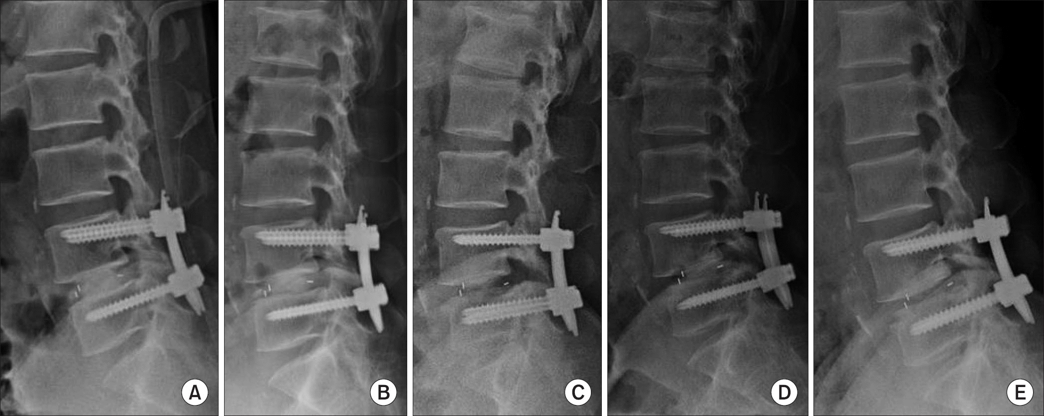 | Figure 1.Posterior migration of the cage. Postoperation (A), 3 months after operation (B), 6 months after operation (C), 9 months after operation (D), 12 months after operation (E). |
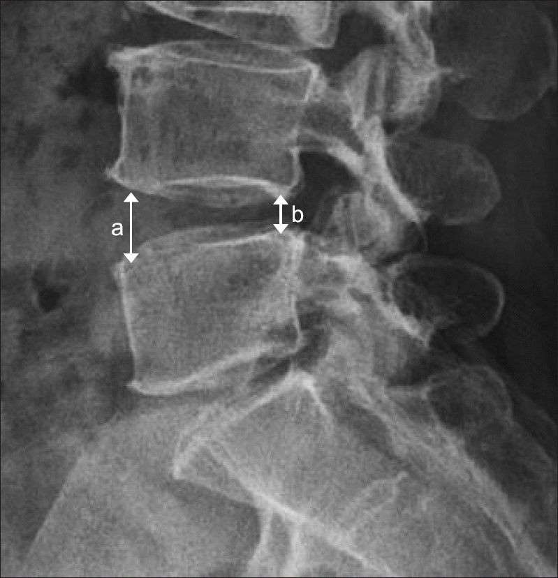 | Figure 3.Mean disc height. Mean disc height is (a+b)/2. a, anterior disc height; b, posterior disc height. |
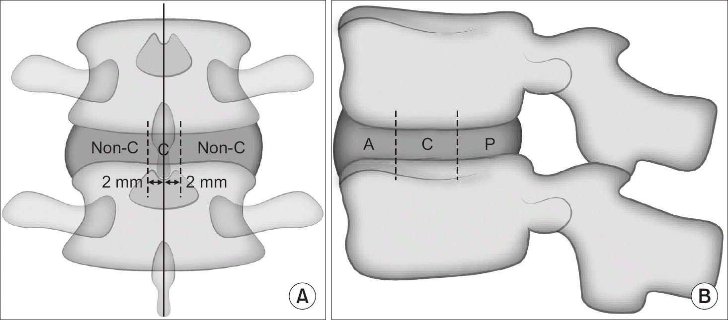 | Figure 4.Location of the cage inserted in the disc space. (A) Anterior posterior view. (B) Lateral view. C, center; A, anterior; P, posterior. |
Table 1.
Demographic Factors (Mean)
Table 2.
Radiology Evaluation (Mean)
Table 3.
Intraclass Correlation Coefficient (ICC)
| Observer | Variable | ICC |
|---|---|---|
| A | Posterior migration of cage | 0.996 |
| Disc height change | 0.991 | |
| B | Posterior migration of cage | 0.998 |
| Disc height change | 0.992 |
Table 4.
Interclass correlation coefficient (Icc)
| Variable | Observer∗ | Icc |
|---|---|---|
| Posterior migration of cage | A1–B1 | 0.991 |
| A2–B2 | 0.992 | |
| Disc height change | A1–B1 | 0.992 |
| A2–B2 | 0.993 |
 XML Download