

 PDF
PDF ePub
ePub Citation
 Print
Print


Abstract
Purpose:
To evaluate the results of tibial lateral plateau fractures using arthroscopic-assisted reduction and internal fixation without cortical window or bone grafts.
Materials and Methods:
From March 2009 to March 2017, 27 patients with Schatzker type II tibial plateau fractures with articular depression and displacement over 5 mm on a computed tomography (CT) scan, who were treated with arthroscopic reduction and internal fixation and followed-up for at least 18 months, were enrolled in this study. Under arthroscopic guidance, the depressed fracture fragment was reduced using a freer and fixed with 5.0 or 6.5 mm cannulated screws through the inframeniscal portal without a cortical window or bone graft. The clinical and radiological results were evaluated using a Rasmussen system. Second look arthroscopy was performed in thirteen patients during the implant removal operation.
Results:
All fractures healed completely with a mean union time of 8.7 weeks (range from 8 to 12 weeks). Twenty four patients had good to excellent clinical results and 25 patients had good to excellent radiological results according to the Rasmussen classification. A well-healed articular surface with fibrocartilage was also found in 13 cases with second look arthroscopy. The 8 cases on CT scan at outpatient department follow-up showed bone union without bone grafting.
Go to : 
References
1. Bennett WF, Browner B. Tibial plateau fractures: a study of associated soft tissue injuries. J Orthop Trauma. 1994. 8:183–8.
2. Tscherne H, Lobenhoffer P. Tibial plateau fractures. Management and expected results. Clin Orthop Relat Res. 1993. 292:87–100.

3. Chan YS, Chiu CH, Lo YP. . Arthroscopy-assisted surgery for tibial plateau fractures: 2-to 10-year follow-up results. Arthroscopy. 2008. 24:760–8.
4. Fowble CD, Zimmer JW, Schepsis AA. The role of arthrosco-py in the assessment and treatment of tibial plateau fractures. Arthroscopy. 1993. 9:584–90.
5. Guanche CA, Markman AW. Arthroscopic management of tibial plateau fractures. Arthroscopy. 1993. 9:467–71.
6. Holzach P, Matter P, Minter J. Arthroscopically assisted treatment of lateral tibial plateau fractures in skiers: use of a cannulated reduction system. J Orthop Trauma. 1994. 8:273–81.
7. Schatzker J, McBroom R, Bruce D. The tibial plateau fracture. The toronto experience 1968-1975. Clin Orthop Relat Res. 1979. 138:94–104.
8. Levy BA, Herrera DA, Macdonald P, Cole PA. The medial approach for arthroscopic-assisted fixation of lateral tibial plateau fractures: patient selection and mid- to long-term results. J Orthop Trauma. 2008. 22:201–5.
9. Nork SE, Schwartz AK, Agel J, Holt SK, Schrick JL, Winquist RA. Intramedullary nailing of distal metaphyseal tibial fractures. J Bone Joint Surg Am. 2005. 87:1213–21.
10. Rasmussen PS. Tibial condylar fractures. Impairment of knee joint stability as an indication for surgical treatment. J Bone Joint Surg Am. 1973. 55:1331–50.
11. Asik M, Cetik O, Talu U, Sozen YV. Arthroscopy-assisted operative management of tibial plateau fractures. Knee Surg Sports Traumatol Arthrosc. 2002. 10:364–70.
12. Suh JT, Ahn JM, Kim TW, Cho HM. Arthroscopically assisted reduction and internal fixation of intra-articular fractures of tibial plateau. J Korean Orthop Assoc. 2012. 47:96–103.
13. Lubowitz JH, Elson WS, Guttmann D. Part I: arthroscop-ic management of tibial plateau fractures. Arthroscopy. 2004. 20:1063–70.
14. Park IH, Lee KB, Park MR, Lee JY, Rhee DY. Arthroscopic management of the tibial condylar fractures. J Korean Or-thop Assoc. 1990. 25:1323–32.
15. Abdel-Hamid MZ, Chang CH, Chan YS. . Arthroscopic evaluation of soft tissue injuries in tibial plateau fractures: retrospective analysis of 98 cases. Arthroscopy. 2006. 22:669–75.
16. Marsh JL, Smith ST, Do TT. External fixation and limited internal fixation for complex fractures of the tibial plateau. J Bone Joint Surg Am. 1995. 77:661–73.
17. Watson JT. High-energy fractures of the tibial plateau. Or-thop Clin North Am. 1994. 25:723–52.
18. Young MJ, Barrack RL. Complications of internal fixation of tibial plateau fractures. Orthop Rev. 1994. 23:149–54.
19. Perry CR, Evans LG, Rice S, Fogarty J, Burdge RE. A new surgical approach to fractures of the lateral tibial plateau. J Bone Joint Surg Am. 1984. 66:1236–40.
20. McGlynn FJ, Caspari RB, Whipple TL, Meyers JF, Hutton PMJ. The role of arthroscopy in the treatment of tibial plateau fractures. Iowa Orthop J. 1986. 6:107–13.
21. Jennings JE. Arthroscopic management of tibial plateau fractures. Arthroscopy. 1985. 1:160–8.
22. Cho SK, Oh JD, Lee YS, Choi JT, Lim GH. Surgical treatment of tibial plateau fracture: validity of arthroscopy. J Korean Fract Soc. 1997. 10:832–42.
23. Honkonen SE. Indications for surgical treatment of tibial condyle fractures. Clin Orthop Relat Res. 1994. 302:199–205.
24. Perez Carro L. Arthroscopic management of tibial plateau fractures: special techniques. Arthroscopy. 1997. 13:265–7.
Go to :
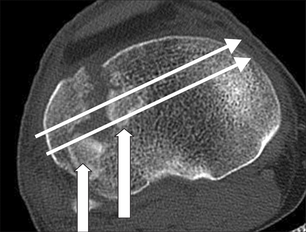 | Figure 1.Fragment interfering with the reduction is called a key fragment (thick and short arrows). The landmark of the screw insertion site was the fibular head and Gerdy’s tubercle. The thin and long arrows indicate the direction of screw insertion. |
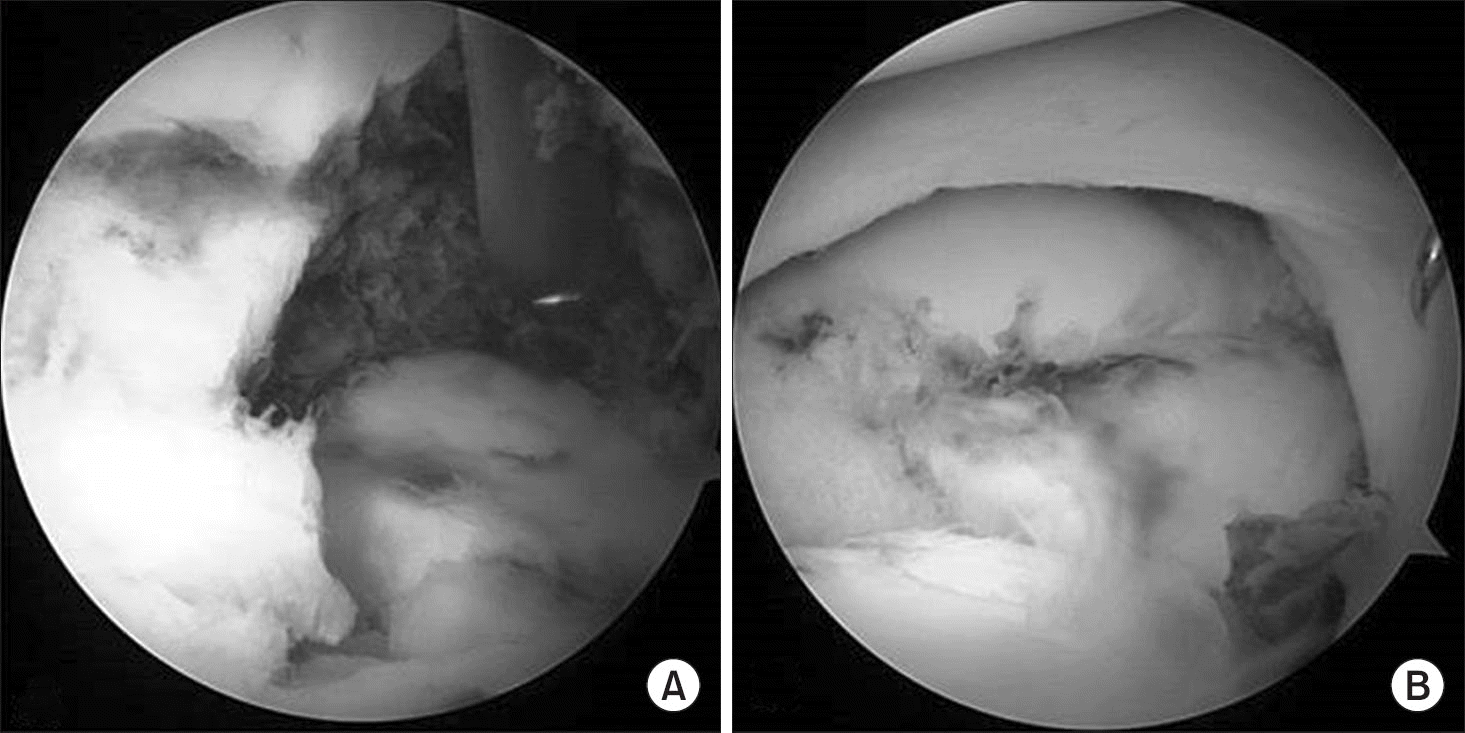 | Figure 2.Arthroscopic findings showed the depressed fracture fragment (A) and depressed fragment reduced using the freer (B). Three arthroscopic portals were used. The anteromedial portal was used for the viewing portal and 2 working (conventional anterolateral, anterolateral infra-meniscal portal) portals were used as the conventional anterolateral portal for inserting the skin hook to retract the anterior horn of lateral meniscus and anterolateral infra-meniscal portal was used to reduce the depressed fragment by the freer and small osteotome. |
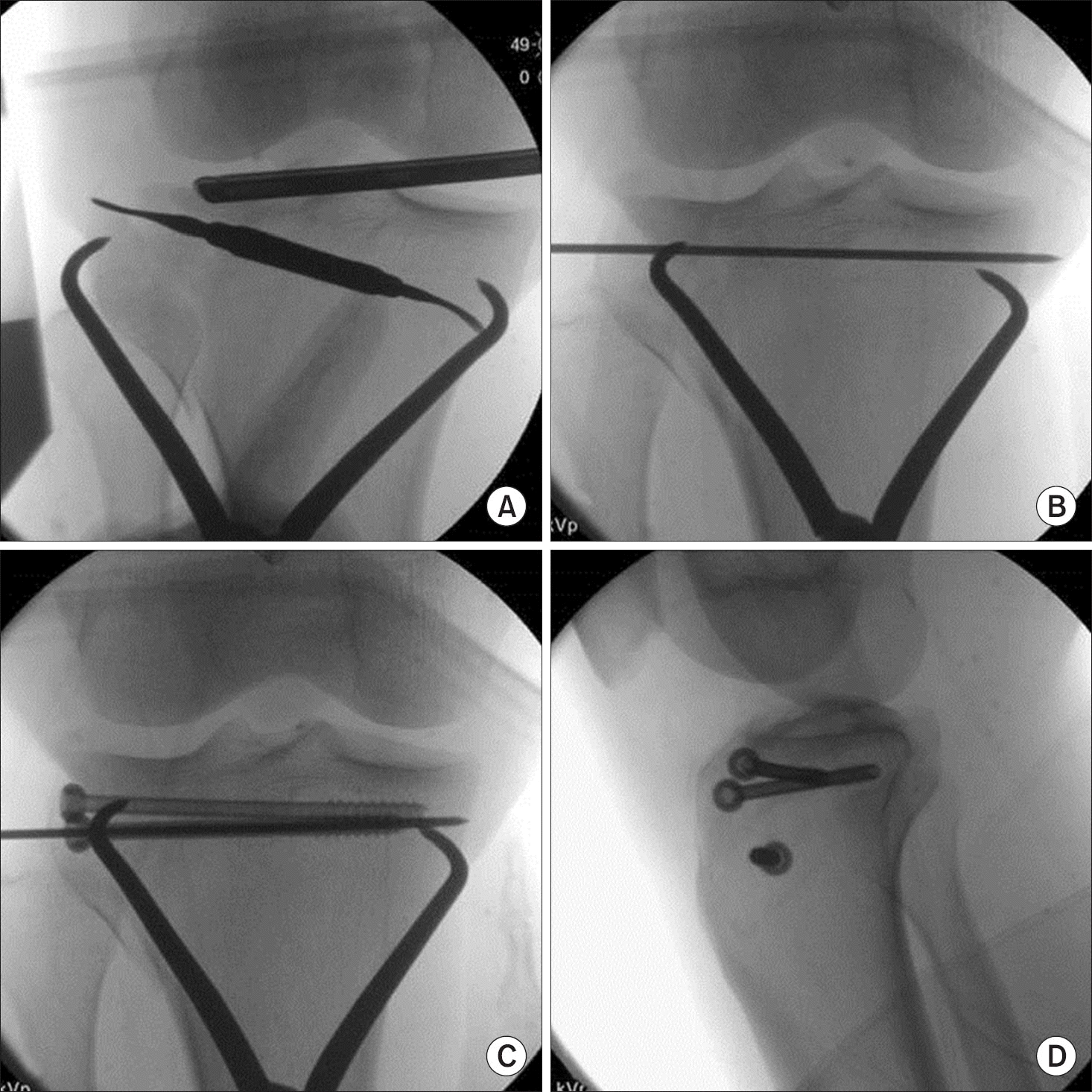 | Figure 3.(A) Intraoperative fluoroscopic photograph shows a reduction of a tibia plateau fracture using a freer. (B) A guide wire was inserted for fixation of a 5.0 cannulated screw. (C, D) The 5.0 mm cannulated screw with a washer is fixed. |
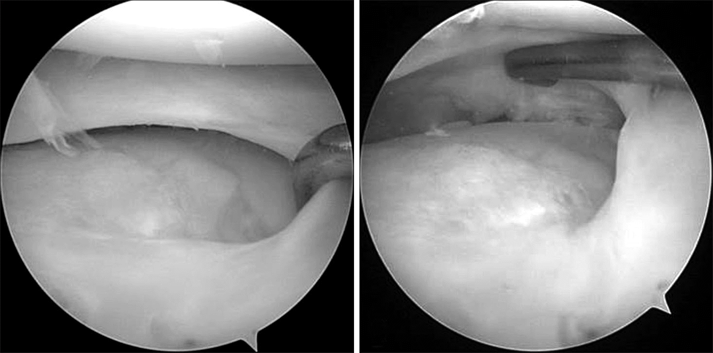 | Figure 4.Second look arthroscopy by the anteromedial portal shows a healed articular surface with fibrocartilage. |
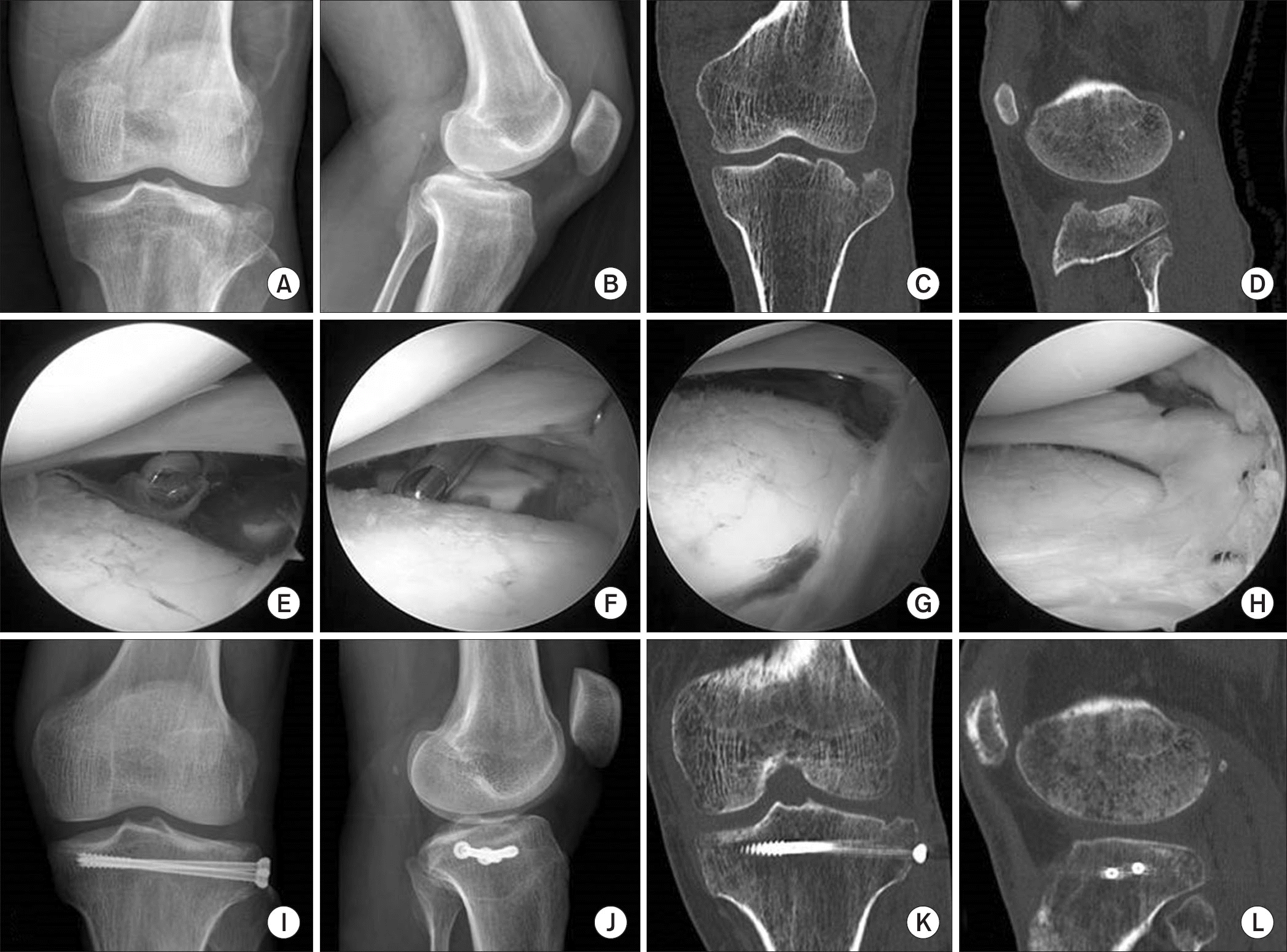 | Figure 5.A 47-year-old male with lateral plateau fracture. (A, B) Anteroposterior and lateral radiographs show a lateral tibial plateau fracture. (C, D) Preoperative computed tomography (CT) shows a Schatzker type II plateau fracture. (E, F) Arthroscopy by the anteromedial portal shows a depressed fracture fragment. (G) Arthroscopy-assisted reduction was done. (H) Lateral meniscal injury was found and repaired with absorbable sutures. (I, J) Internal fixation with 5.0 cannulated screws was done. (K, L) Postoperative CT shows an anatomically reduced articular surface. |
Table 1.
Patients’ Demographics (n=27)
Table 2.
Clinical and Radiological Results
 XML Download