

 PDF
PDF ePub
ePub Citation
 Print
Print


INTRODUCTION
Gastric cancer is a worldwide health problem; it is the third leading cause of cancer-related deaths and the fourth most common cancer worldwide [1]. Despite a progressive decrease in the incidence of gastric cancer, the overall survival of patients with gastric cancer has improved mainly because of early detection by health screening programs and adjuvant chemotherapy [2345]. Nevertheless, the prognosis of advanced gastric cancer with peritoneal seeding or distant metastasis remains dismal, despite recent advances in multidisciplinary approaches and the combination of systemic chemotherapy with various targeted therapies.
Positive peritoneal washing cytology (PWC) is a poor prognostic factor for early peritoneal recurrence and decreased disease-specific survival, regardless of macroscopic peritoneal carcinomatosis. Positive PWC in gastric cancer is classified as a M1 disease according to the current TNM staging system of the American Joint Committee on Cancer and the Japanese Gastric Cancer Treatment guidelines [67]. However, a recent meta-analysis suggested that the change in cytological status from positive to negative following neoadjuvant chemotherapy is associated with significantly improved overall survival [8]. Local therapeutic strategies such as intraperitoneal chemotherapy (IP CTX) or pressured intraperitoneal aerosol chemotherapy (PIPAC) are emerging as alternative solutions for stage IV gastric cancer; therefore, there is great interest in the design of intraoperative diagnostic methods for detecting peritoneal dissemination, especially in the absence of overt peritoneal seeding [910]. However, there is a limit to the practical application of intraoperative PWC for staging, mostly because of a lack of consensus in methodology, discrepancy between pathologists' experience, low sensitivity of the cytology examination, and long turnaround times for results.
From a methodological point of view, a fast and reliable staining method of intraoperative PWC is necessary to allow the referring surgeon to proceed to the next step promptly after the examination. To improve the usefulness of intraoperative PWC, we tested the Shorr staining protocol in gastric cancer [11]. The objective of the present study was to compare the pathologic results and survival outcomes of the Shorr staining method with those of the conventional Papanicolaou (Pap) staining with carcinoembryonic antigen (CEA) immunohistochemistry (IHC) method.
MATERIALS AND METHODS
This study was a retrospective study and approved by the Institutional Review Board for research using human subjects at Seoul National University Bundang Hospital (B-1711-432-113). Between November 2012 and December 2014, intraoperative PWC with Shorr staining was performed in 77 patients with clinical T3 or higher gastric cancer to validate peritoneal carcinomatosis pathologically which was confirmed by Pap with CEA IHC examination.
Processing of intraoperative PWC
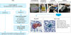
The schematic flow of intraoperative PWC is shown in Fig. 1. PWC was performed after laparotomy or the establishment of a pneumoperitoneum in laparoscopic surgery. If ascites was present in the peritoneal cavity, it was directly aspirated without saline injection. In the absence of ascites, 200 mL of saline was injected into the left subphrenic area, the subhepatic area, Douglas' pouch, and the intestinal mesentery [12]. Then, the patient's body was shaken gently during the position change from reverse Trendelenburg to Trendelenburg, or vice versa (Supplementary Video 1). After 3–5 minutes, fluid from the left subphrenic area, the subhepatic area, and Douglas' pouch was aspirated using a syringe with a long irrigation extension tubing tip. The aspirated ascites or fluid was immediately delivered to the department of pathology.
Fig. 1
Schematic of the process of intraoperative peritoneal washing cytology and Shorr staining. In papanicolaou staining, carcinoma cells are easily distinguished from mesothelial cells and inflammatory cells, which show large cell size and nuclear hyperchromasia. It is also possible to easily differentiate between carcinoma cells and other cells by using Shorr staining without drying or any other technical artifacts.
PWC = peritoneal washing cytology; Pap = Papanicolaou.
Shorr staining procedure for on-site cytology examination
The preparation steps prior to staining were as follows: aspirated fluid was centrifuged at 2,500 rpm for 3–5 minutes. The sediment was separated and subjected to cytospin at 2,000 rpm for 5 minutes. Then, the specimen was divided into 2 samples for Shorr staining and conventional Pap staining with CEA IHC. Fixation for Pap and Shorr staining was performed with 95% ethanol for 3 minutes, and the cytology slides for CEA immunostaining were fixed with 10% formalin. For Shorr staining, the slides were dipped in hematoxylin (90 seconds), 1% HCl (2 times, each 1 minute), and Shorr solution (Merck KGaA, Darmstadt, Germany) for 2 minutes. After staining, the slides were rinsed in 95% ethanol, 100% ethanol, and xylene. Shorr-stained smears were immediately reviewed by a certified cytotechnologist (Choi HY) and a designated pathologist (Lee HS), who were blinded to the clinical details of individual patients. Ordinary optic microscopy was used at 100× and 400× magnifications.
In order to clearly differentiate carcinoma cells from mesothelial cells, we routinely performed CEA IHC when there were a few atypical cells or atypism was not definite in the Pap alone-stained slides. Formalin-fixed cytology slides were used for CEA IHC using anti-CEA antibody (1:350, Dako, Glostrup, Denmark) with a Ventana Benchmark automatic immunostaining system (BenchMark XT; Ventana Medical System, Inc., Tucson, AZ, USA) according to the manufacturer's instructions. Antigen retrieval was performed with Cell Conditioning 1 (pH 8.4) for 30 minutes at 100°C, and immunoreactivity was detected using diaminobenzidine substrate.
The nuclear and cytoplasmic features in the Shorr-stained slides were almost equivalent to those in the Pap-stained slides. Therefore, the same diagnostic criteria were applied to the interpretation of both Shorr- and Pap-stained slides. Cytological results were classified as benign, atypical, suspicious for malignancy, or malignant, and compared with the results of conventional Pap staining with CEA IHC. The case-by-case final consensus result was discussed and determined in a common session.
Statistical analysis
Peritoneal carcinomatosis was diagnosed based on operative findings and pathologic confirmation. The sensitivity, specificity, positive predictive value, and negative predictive value for peritoneal carcinomatosis were calculated according to Shorr staining and conventional Pap staining with CEA IHC. All statistical analyses except concordance statistics (C-index) were performed using SPSS version 18.0 (SPSS Inc., Chicago, IL, USA). In order to compare agreement or disagreement between 2 methods, weighted Cohen's kappa was tested using the difference of both scores (Class x Shorr
− Class x Pap
). Kaplan-Meier curves were used to analyze overall survival and C-indices of Shorr staining and conventional Pap staining with CEA IHC were calculated and compared using R program version 3.4.3 with compareC package to evaluate whether there was discrimination in survival analysis according to the staining method. A P-value of <0.05 was considered statistically significant.
RESULTS
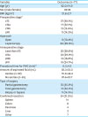
The patient characteristics are shown in Table 1. The mean age of the patients was 62.0 years, and the male to female ratio was 1.75:1. Preoperative clinical stages were T3 in 48%, T4a in 22.1%, T4b in 15.6%, and M1 in 14.2% of patients. Intraoperative PWC was performed under laparoscopy in 69 patients (89.6%). The average time of the procedure for intraoperative PWC (from saline injection to completion time of fluid aspiration) was 8.3 minutes, and the average amount of aspirated fluids was 83.3 ml. The average turnaround time from Shorr staining to pathologic review was 21.0 minutes (range, 17–28 minutes).
Table 1
Patient demographics and operative outcomes
Comparisons of sensitivity, specificity, and predictive value
The results of intraoperative PWC according to staining methods are shown in Table 2. In the conventional Pap method with CEA IHC, 16 (20.7%) patients were diagnosed with positive cytology and 6 (7.8%) patients showed atypical findings. Considering the atypical findings as negative cytology, the sensitivity, specificity, and positive predictive value for peritoneal carcinomatosis were 78.9%, 98.2%, and 93.7%, respectively. In the Shorr method, 15 (19.5%) patients were diagnosed with positive cytology and 7 (9.1%) patients showed atypical findings. Considering the atypical findings as negative cytology, the sensitivity, specificity, and positive predictive value for peritoneal carcinomatosis were 73.6%, 98.2%, and 93.3%, respectively.

Concordance between the Shorr method and the Pap method with CEA IHC
Diagnostic concordance between the 2 methods was observed in 90.9% (70/77) of patients (Table 3) and reproducibility of scoring was judged as excellent (weighted κ statistic=0.875). Diagnostic discordance was present in 7 patients and was mostly observed in association with atypical findings: 3 patients with atypical findings in the Shorr method were identified as having negative cytology by the Pap method with CEA IHC, and 3 patients showed the opposite pattern. One patient with atypical finding in the Shorr method was confirmed as having positive cytology by CEA IHC.
Table 3
Concordance between the Shorr stain and conventional Papanicolaou stain with CEA immunohistochemistry

| Shorr stain | Total | ||||
|---|---|---|---|---|---|
| Negative | Atypical | Positive | |||
| Pap with CEA IHC | |||||
| Negative | 52 | 3 | 0 | 55 | |
| Atypical | 3 | 3 | 0 | 6 | |
| Positive | 0 | 1 | 15 | 16 | |
| Total | 55 | 7 | 15 | 77 | |
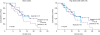
Overall survival rates according to the staining method are shown in Fig. 2. There was no significant difference in C-index between the 2 methods (0.459 in the Shorr vs. 0.458 in the Pap with CEA IHC, P=0.987). Because most disagreements in diagnoses occurred in patients with atypical findings, the survivals of patients with each finding were compared (Supplementary Fig. 1). The 2-year survivals in patients with negative finding were 30.8% in the Shorr staining and 33.0% in the Pap with CEA IHC (P=0.945). The 1-year survivals in patients with positive finding were 22.9% in the Shorr staining and 35.6% in the Pap with CEA IHC (P=0.798). And the survival of patients with atypical findings was compared and the results showed no significant difference between the 2 staining methods (P=0.676)
DISCUSSION
Despite recent advances in multidisciplinary approaches and the advent of targeted cancer therapy, the treatment of patients with stage IV gastric cancer remains challenging. A recent clinical trial demonstrated that palliative gastrectomy was not beneficial to improve survival even in gastric cancer patients with a single non-curable factor [13]. Therefore, systemic chemotherapy remains the standard of care, and surgical treatment is limited to incurable gastric cancer.
Various treatment strategies have been proposed as alternative solutions, especially for gastric cancer with positive peritoneal cytology findings (CY1) or overt peritoneal carcinomatosis. Kuramoto et al. [14] demonstrated the efficacy of extensive intraoperative peritoneal lavage (EIPL) followed by IP CTX in gastric cancer patients with P0CY1. In their study, the 5-year survival rate of patients with EIPL and IPC is 43.8%, and EIPL was the most significant prognostic factor in multivariate analysis. Recently, Ishigami et al. [15] showed that surgery after response to intraperitoneal and systemic chemotherapy significantly prolonged the survival of P1 and CY1 gastric cancer patients. The median survival time was 30.5 months, and the 1-year overall survival was 73.3%. The median survival time of patients with P0CY1 or P1 was significantly longer than that of patients with P2 or P3 disease. A novel treatment termed PIPAC is emerging as a salvage therapy for peritoneal carcinomatosis. PIPAC shows superior pharmacological properties with high local concentration and low systemic exposure, which could induce regression of peritoneal carcinomatosis using 10% of the usual systemic dose [16].
The shift in the treatment paradigm for gastric cancer with CY1 from systemic therapy alone to a combination of local and systemic therapies resulted in the adoption of intraperitoneal free cancer cell detection as the standard procedure for staging and for the selection of candidates for local therapies. Based on a study suggesting that the presence of free cancer cells in peritoneal washing fluid is responsible for the formation of micro-metastases and the subsequent extensive dissemination, cytologic examination was incorporated into the Japanese staging system for gastric cancer [1718]. However, the sensitivity of this assay is relatively low, ranging from 19% to 30% in gastric cancer invading the serosa, and only 54% of patients with peritoneal carcinomatosis are positive for cytology [1920212223]. Furthermore, the conventional Pap staining method, which is widely utilized in the practice of cytopathology, requires numerous procedures and approximately 30 minutes for wet fixation before staining. For these reasons, the conventional Pap staining method is not appropriate for intraoperative PWC, and there is an urgent need to develop rapid and reliable staining methods for intraoperative PWC.
Various staining protocols can be used in rapid cytologic examinations; however, staining methods other than the conventional Pap staining are rarely reported in gastric cancer. In a preliminary study, we tested 3 rapid staining methods for cytology: Hemacolor, Cytocolor, and Shorr staining. The Hemacolor stain was similar to the Wright and Giemsa smear and required a preparation time of <5 minutes; however, it was associated with nuclear hyperchromasia. The results of the Cytocolor stain were similar to those of the modified Pap stain; however, the nucleus appeared dark violet, and visualization of chromatin was poor. In addition, the preparation time was approximately 15 minutes. The Shorr stain produced a light brown-colored nucleus and well-visualized chromatin, which were similar to those produced by the conventional Pap stain, and it required approximately 10 minutes for preparation.
The Shorr stain is a rapid, mono-reagent staining method similar to the original protocol of the Pap stain [24]. Therefore, there is extensive agreement in the morphology results between the Pap- and Shorr-stained smear, and the Shorr-stained smear can be de-stained and re-stained with the Pap stain [2526]. However, samples that are deeply stained with the Shorr method, such as rich and bloody smears, show crowding and overlapping of cells, preventing accurate differentiation [24]. This method is widely used for rapid cytologic diagnoses such as imprint cytology of surgical frozen sections, which is common in malignant melanoma, breast tumors, and vaginal cytology [262728].
The present study demonstrated that the Shorr staining method is rapid, reliable, and useful for intraoperative PWC in gastric cancer. The sensitivity, specificity, and positive predictive value for peritoneal carcinomatosis were similar between the Shorr method and conventional Pap with CEA IHC (73.6%, 98.2%, 93.2% vs. 78.9%, 98.2%, and 93.7%). In addition, the concordance rate between the Shorr and Pap with CEA IHC staining methods was relatively high at 90.9% and reproducibility of scoring between 2 methods was excellent (weighted κ statistic=0.875), and most disagreements were related to negative or atypical findings. Moreover, the survival rates according to each staining method were similar and there was no significant difference in C-index between the 2 methods (0.459 in the Shorr vs. 0.458 in the Pap with CEA IHC, P=0.987). These findings suggest that the Shorr staining method can be used as an alternative method for intraoperative PWC, as its reliability for detecting free cancer cells was comparable to that of other methods regardless of the cytology examinations' low sensitivity. The main advantage of this method is its turnaround time of about 20 minutes. The average time for Shorr staining and pathologic review was 21.0 minutes. This allows the surgeon to obtain results from the pathologist within 30 minutes from specimen delivery, which facilitates the rapid determination of the intraoperative treatment strategy. The poor differentiation associated with the Shorr staining method in rich and bloody smears does not affect the pathological diagnosis, because most specimens are collected by laparoscopy before surgery and bleeding during intraoperative PWC is rare.
The present pilot study had several limitations. First, the cohort was small and the study was a single institution study; therefore, validation in a multicenter study or with a larger cohort is necessary. Second, the Shorr staining method was not compared with other rapid cytologic examinations such as rapid Pap staining, ultrafast Pap stain, and modified ultrafast Pap by Gill. Additional studies are needed to compare the Shorr staining method with rapid modified Pap tests. Third, there were several atypical findings that may have affected the diagnostic results. Additional examinations such as real-time reverse transcription polymerase chain reaction need to be performed to decrease the rate of atypical findings. Lastly, the disagreements in negative or atypical findings might be caused because a whole specimen was divided into 2 pieces each for Shorr- or Pap staining methods. Therefore, a randomized study using whole specimens would be required.
In conclusion, we found that Shorr staining is a rapid, reliable, and valuable method for intraoperative PWC in gastric cancer. This novel staining method may be of value to determine local therapeutic strategies such as IP CTX, especially in serosa-invading gastric cancer.

 XML Download