

 PDF
PDF ePub
ePub Citation
 Print
Print


Abstract
Purpose
To clarify the relationship between various factors (sociodemographic factors, health behavioral risk factors and health status, and diabetic factors) related to diabetic retinopathy and to suggest improvements regarding the associated medical examination.
Methods
The subjects were 1,444 diabetic patients diagnosed in the 5th Korean National Health and Nutrition Examination Survey (KNHANES), aged 19 years or older, who underwent non-mydriatic fundus photography. The criteria for diagnosing diabetes were a fasting glucose level ≥ 126 mg/dL and a previous diagnosis of diabetes or currently undergoing treatment. The diagnosis of diabetic retinopathy followed the modified Airlie House classification. Univariate and multivariate analyses of diabetic retinopathy were performed.
Results
Among the 1,444 patients who were diagnosed with diabetes, 277 had diabetic retinopathy; the prevalence rate was 19.18%. The higher the body mass index, the lower the risk of diabetic retinopathy by 0.924 times (p = 0.001; 95% confidence interval [CI], 0.883–0.966). The longer the duration of diabetes, the greater the risk of diabetic retinopathy; the prevalence period group of more than 11 years had a 26.025-fold higher risk than the newly diagnosed group (p < 0.001; 95% CI, 10.840–62.482). The risk of diabetic retinopathy increased with the hemoglobin A1c (HbA1c) level; the risk was 5.973-fold higher in the group with HbA1c above 11.0% (p < 0.001; 95% CI, 2.984–11.956) compared with the group with HbA1c < 6.0%. The risk of diabetic retinopathy was 2.050-fold greater with insulin injections (p = 0.003; 95% CI, 1.284–3.275).
Figures and Tables
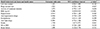
Table 1
Characteristics and comparison between without diabetic retinopathy and with diabetic retinopathy (sociodemographic factors)
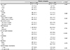
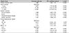
Table 2
Characteristics and comparison between without diabetic retinopathy and with diabetic retinopathy (health behavioral risk factors and health status)
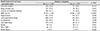
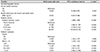
Table 3
Characteristics and comparison between without diabetic retinopathy and with diabetic retinopathy (diabetic factors)
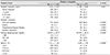
Table 4
Analysis of sociodemographic factors associated with diagnosis of diabetic retinopathy using logistic regression analysis (univariate analysis)
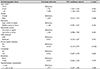
Table 5
Analysis of health behavioral risk factors and health status associated with diagnosis of diabetic retinopathy using logistic regression analysis (univariate analysis)
Notes
References
1. Kim YJ, Kim JG, Lee JY, et al. Development and progression of diabetic retinopathy and associated risk factors in korean patients with type 2 diabetes: the experience of a tertiary center. J Korean Med Sci. 2014; 29:1699–1705.

2. Zimmet P, Alberti KG, Shaw J. Global and societal implications of the diabetes epidemic. Nature. 2001; 414:782–787.
3. Cho NH. Prevalence of diabetes and management status in Korean population. Korean J Med. 2005; 68:1–3.
4. Kim SG, Choi DS. The present state of diabetes mellitus in Korea. J Korean Med Assoc. 2008; 51:791–798.
5. Resnikoff S, Pascolini D, Etya'ale D, et al. Global data on visual impairment in the year 2002. Bull World Health Organ. 2004; 82:844–851.
6. Jee D, Lee WK, Kang S. Prevalence and risk factors for diabetic retinopathy: the Korea National Health and Nutrition Examination Survey 2008-2011. Invest Ophthalmol Vis Sci. 2013; 54:6827–6833.
7. Lee DW, Bae JH, Song SJ. Efficacy of single-field non-mydriatic digital fundus photography for screening diabetic retinopathy. J Korean Ophthalmol Soc. 2011; 52:531–536.
8. Younis N, Broadbent DM, James M, et al. Current status of screening for diabetic retinopathy in the UK. Diabet Med. 2002; 19:Suppl 4. 44–49.
9. Maberley D, Cruess AF, Barile G, Slakter J. Digital photographic screening for diabetic retinopathy in the James Bay Cree. Ophthalmic Epidemiol. 2002; 9:169–178.
10. Gye HJ, Song SJ. New modalities for the diagnosis and treatment of diabetic retinopathy. Korean J Med. 2015; 89:271–276.
11. Gambill HD, Ogle KN, Kearns TP. Mydriatic effect of four drugs determined with pupillograph. Arch Ophthalmol. 1967; 77:740–746.
12. Herbert HM, Jordan K, Flanagan DW. Is screening with digital imaging using one retinal view adequate? Eye (Lond). 2003; 17:497–500.
13. Bae SH, Cho Belong, Son KY, et al. Interpretation of ophthalmoscopy. Korean J Fam Pract. 2013; 3:415–421.
14. Edwards AL. Funduscopic examination of patients with diabetes who are admitted to hospital. CMAJ. 1986; 134:1263–1265.
15. Lee KP, Kang JY, Lee JM, Jeong MK. The significance of fundus photographs during health mass screening. J Korean Acad Fam Med. 1999; 20:933–939.
16. Lim HT, Choi KS. Factors associated with screening for diabetic retinopathy in diabetic patients aged ≥40 years using the KNHANES IV. J Korean Ophthalmol Soc. 2012; 53:516–521.
17. Kim HK, Oh TS, Lee SM, Lee JB. The initial fundus examination and severity of diabetic retinopathy at a primary eye clinic. J Korean Ophthalmol Soc. 2005; 46:982–988.
18. Centers for Disease Control and Prevention (CDC). Cigarette smoking among adults-United States, 1992, and changes in the definition of current cigarette smoking. MMWR Morb Mortal Wkly Rep. 1994; 43:342–346.
19. Ainsworth BE, Haskell WL, Whitt MC, et al. Compendium of physical activities: an update of activity codes and MET intensities. Med Sci Sports Exerc. 2000; 32:9 Suppl. S498–S504.
20. Kim KS, Choi CH, Lee DY, Kim EJ. Epidemiological study on diabetes mellitus among rural Korean. J Korean Diabetes Assoc. 1972; 1:17–24.
21. Lee WK. Diabetic retinopathy. J Korean Med Assoc. 2005; 48:616–627.
22. Harris MI, Klein R, Welborn TA, Knuiman MW. Onset of NIDDM occurs at least 4–7 yr before clinical diagnosis. Diabetes Care. 1992; 15:815–819.
23. Klein R, Klein BE, Moss SE. The Wisconsin epidemiological study of diabetic retinopathy: a review. Diabetes Metab Rev. 1989; 5:559–570.
24. UK Prospective Diabetes Study (UKPDS) Group. Intensive blood-glucose control with sulphonylureas or insulin compared with conventional treatment and risk of complications in patients with type 2 diabetes (UKPDS 33). Lancet. 1998; 352:837–853.
25. Park CY, Park SE, Bae JC, et al. Prevalence of and risk factors for diabetic retinopathy in Koreans with type II diabetes: baseline characteristics of Seoul Metropolitan City-Diabetes Prevention Program (SMC-DPP) participants. Br J Ophthalmol. 2012; 96:151–155.
26. Kim JH, Kwon HS, Park YM, et al. Prevalence and associated factors of diabetic retinopathy in rural Korea: The Chungju metabolic disease cohort study. J Korean Med Sci. 2011; 26:1068–1073.
27. Yang JY, Kim NK, Lee YJ, et al. Prevalence and factors associated with diabetic retinopathy in a Korean adult population: The 2008-2009 Korea National Health and Nutrition Examination Survey. Diabetes Res Clin Pract. 2013; 102:218–224.
28. Hogan P, Dall T, Nikolov P. American Diabetes Association. Economic costs of diabetes in the US in 2002. Diabetes Care. 2003; 26:917–932.
29. Baek IR, Park HS, Byun SS. The determinants and medical care utilization behavior of private health insurance. The Journal of the Korea Contents Association. 2012; 12:295–305.
30. Kim YJ, Cho DY, Yi YH. Comparison of the demographic characteristics in private health insurance. Korean J Health Serv Manag. 2013; 7:143–151.
31. Yoon KH, Lee JH, Kim JW, et al. Epidemic obesity and type 2 diabetes in Asia. Lancet. 2006; 368:1681–1688.
32. Rema M, Premkumar S, Anitha B, et al. Prevalence of diabetic retinopathy in urban India: the Chennai Urban Rural Epidemiology Study (CURES) eye study, I. Invest Ophthalmol Vis Sci. 2005; 46:2328–2333.
33. Suzuki K, Watanabe K, Motegi T, Kajinuma H. High prevalence of proliferative retinopathy in diabetic patients with low pancreatic B-cell capacity. Diabetes Res Clin Pract. 1989; 6:45–52.
34. Liu JH, Tung TH, Tsai ST, et al. A community-based epidemiologic study of gender differences in the relationship between insulin resistance/beta-cell dysfunction and diabetic retinopathy among type 2 diabetic patients in Kinmen, Taiwan. Ophthalmologica. 2006; 220:252–258.
35. Mohamed Q, Gillies MC, Wong TY. Management of diabetic retinopathy: a systemic review. JAMA. 2007; 298:902–916.
36. Kohner EM, Oakley NW. Diabetic retinopathy. Metabolism. 1975; 24:1085–1102.
37. Aiello LM, Rand LI, Briones JC, et al. Diabetic retinopathy in Joslin Clinic Patients with Adult-Onset Diabetes. Ophthalmology. 1981; 88:619–623.
38. Park JY, Lee TY, Jang KS, Oh HY. A study on blood glucose level and self management among community dwelling type II diabetes patients. J Korean Acad Adult Nurs. 2010; 22:271–280.
39. Shin KH, Chi MJ. Fundus examination rate in diabetic and public health factors associated with fundus examination rate. J Korean Ophthalmol Soc. 2009; 50:1319–1325.
40. Ghasemi Falavarjani K, Wang K, Khadamy J, Sadda SR. Ultra-wide-field imaging in diabetic retinopathy; an overview. J Curr Ophthalmol. 2016; 28:57–60.
41. Kwak HW, Joo MJ, Yoo JH. The significance of fundus photography without mydriasis during health mass screening. J Korean Ophthalmol Soc. 1997; 38:1585–1589.
 XML Download