

 PDF
PDF ePub
ePub Citation
 Print
Print


Abstract
Purpose
To evaluate the clinical presentations of focal choroidal excavation and to report long-term outcomes of cases without retinal disorders at the initial presentation.
Methods
A retrospective review of medical records was performed for patients diagnosed with focal choroidal excavation. Concomitant retinal disorders at the initial presentation were identified. In cases without retinal disorders, the development of retinal disorders during follow-up was also evaluated.
Results
Forty-five eyes in 45 patients were examined in this study. Focal choroidal excavation was accompanied with retinal disorders in 16 eyes (35.6%). In the remaining 29 eyes, only focal choroidal excavation was noted without any accompanying retinal disorders. The accompanying retinal disorders included choroidal neovascularization (n = 8), central serous chorioretinopathy (n = 4), epiretinal membrane (n = 1), macular hole (n = 1), branch retinal vein occlusion (n = 1), and uveitis (n = 1). Of the 29 eyes without retinal disorders, 22 were followed up for a mean period of 33.5 ± 18.2 months. Consequently, choroidal neovascularization was found to have developed in one eye at 59 months, and subretinal fluid had developed in two eyes at 17 and 28 months, respectively.
Conclusions
Focal choroidal excavation was accompanied by retinal disorders in 35.6% of the included patients. In patients without retinal disorders, the development of a retinal disorder was noted in some eyes, suggesting the need for long-term regular follow-up in patients diagnosed with focal choroidal excavation.
Figures and Tables
Figure 1
A representative figure showing focal choroidal excavation. Note thick choroid with pachyvessels (asterisks).
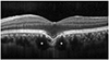
Figure 2
Clinical course of a focal choroidal excavation found in 29-year-old patient. At the diagnosis, there was no retinal disorder (A). The patients were regularly followed-up. At 59 months (B-D), subretinal fluid and hemorrhage was noted on fundus examination (B). Development of choroidal neovascularization was confirmed on fluorescein angiography (C) and optical coherence tomography (D). The patient was referred to the other hospital.
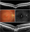
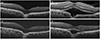
Figure 3
Clinical course of a focal choroidal excavation found in 37-year-old patient. At the diagnosis, there was no retinal disorder (A). The patients were regularly followed-up. Development of subretinal fluid was noted at 17 months (B), but spontaneously resolved after 2 months (C). At 28 months (D), recurrence of subretinal fluid was noted.
References
1. Jampol LM, Shankle J, Schroeder R, et al. Diagnostic and therapeutic challenges. Retina. 2006; 26:1072–1076.

2. Lee CS, Woo SJ, Kim YK, et al. Clinical and spectral-domain optical coherence tomography findings in patients with focal choroidal excavation. Ophthalmology. 2014; 121:1029–1035.
3. Say EA, Jani PD, Appenzeller MF, Houghton OM. Focal choroidal excavation associated with polypoidal choroidal vasculopathy. Ophthalmic Surg Lasers Imaging Retina. 2013; 44:409–411.
4. Xu H, Zeng F, Shi D, et al. Focal choroidal excavation complicated by choroidal neovascularization. Ophthalmology. 2014; 121:246–250.
5. Lee JH, Lee WK. Choroidal neovascularization associated with focal choroidal excavation. Am J Ophthalmol. 2014; 157:710–718.
6. Ellabban AA, Tsujikawa A, Ooto S, et al. Focal choroidal excavation in eyes with central serous chorioretinopathy. Am J Ophthalmol. 2013; 156:673–683.
7. Suzuki M, Gomi F, Hara C, et al. Characteristics of central serous chorioretinopathy complicated by focal choroidal excavation. Retina. 2014; 34:1216–1222.
8. Kim H, Woo SJ, Kim YK, et al. Focal choroidal excavation in multifocal choroiditis and punctate inner choroidopathy. Ophthalmology. 2015; 122:1534–1535.
9. Roy R, Saurabh K, Chandrasekharan DP, et al. Bilateral focal choroidal excavation in cone dystrophy. Clin Exp Optom. 2016; 99:198–199.
10. Kim WJ, Cho NC, Kweon EY. A case of focal choroidal excavation associated with chronic central serous chorioretinopathy. J Korean Ophthalmol Soc. 2015; 56:627–631.
11. Chung CY, Li SH, Li KKW. Focal choroidal excavation-morphological features and clinical correlation. Eye (Lond). 2017; 31:1373–1379.
12. Park JH, Sagong M, Chang WH. Three cases of focal choroidal excavation in the macula detected by spectral-domain optical coherence tomography. J Korean Ophthalmol Soc. 2014; 55:941–946.
13. Cheung CMG, Lee WK, Koizumi H, et al. Pachychoroid disease. Eye (Lond). 2019; 33:14–33.
14. Margolis R, Mukkamala SK, Jampol LM, et al. The expanded spectrum of focal choroidal excavation. Arch Ophthalmol. 2011; 129:1320–1325.
15. Park KA, Oh SY. The absence of focal choroidal excavation in children and adolescents without retinal or choroidal disorders or ocular trauma. Eye (Lond). 2015; 29:841–842.
16. Chung H, Byeon SH, Freund KB. Focal choroidal excavation and its association with pachychoroid spectrum disorders: a review of the literature and multimodal imaging findings. Retina. 2017; 37:199–221.
17. Obata R, Takahashi H, Ueta T, et al. Tomographic and angiographic characteristics of eyes with macular focal choroidal excavation. Retina. 2013; 33:1201–1210.
 XML Download