

 PDF
PDF ePub
ePub Citation
 Print
Print


Abstract
Purpose
The purpose of this study was to evaluate whether eye dominance changes after conventional pseudophakic monovision, and to identify factors that affect changes in eye dominance.
Methods
This retrospective study included 70 patients who underwent bilateral conventional monovision cataract surgery. Patients were divided into two groups based on whether they experienced a change in the dominant eye. We compared patients' uncorrected distance visual acuity (UCDVA), uncorrected near visual acuity (UCNVA), best-corrected visual acuity (BCVA), spherical equivalent, stereopsis, and time interval between cataract surgeries.
Results
The mean age was 71.26 ± 10.84 (range, 25–90) years, mean interval between surgery in each eye was 118.46 ±183.50 (range, 17–1,018) days, and mean postoperative diopter difference was 1.16 ± 0.53 (range, 0.00–2.75) diopters. After bilateral cataract surgery, 22 patients (31.43%) experienced a change in eye dominance, whereas 48 patients (68.57%) experienced no change. There were no differences in the time interval between cataract surgeries, preoperative UCDVA and UCNVA, pre- and postoperative BCVA, or stereopsis in either group. Patients who experienced a change in eye dominance showed smaller differences between preoperative and postoperative spherical equivalent, compared with patients who experienced no change in eye dominance (t-test, p < 0.05).
Conclusions
Twenty-two (31.43%) patients whose nondominant eyes were targeted for near vision showed altered eye dominance after conventional monovision cataract surgery. Eye dominance shows greater plasticity in patients with smaller differences between preoperative and postoperative spherical equivalent.
Go to : 
References
1. Greenstein S, Pineda R 2nd. The quest for spectacle independence: a comparison of multifocal intraocular lens implants and abdominal monovision for patients with presbyopia. Semin Ophthalmol. 2017; 32:111–5.
2. Iida Y, Shimizu K, Ito M. Pseudophakic monovision using abdominal and multifocal intraocular lenses: hybrid monovision. J Cataract Refract Surg. 2011; 37:2001–5.
3. Yoo R, Shin KC. Clinical results of pseudophakic monovision 1 year after cataract surgery. J Korean Ophthalmol Soc. 2016; 57:1882–90.

4. Zhang F, Sugar A, Jacobsen G, Collins M. Visual function and abdominal satisfaction: comparison between bilateral diffractive abdominal intraocular lenses and monovision pseudophakia. J Cataract Refract Surg. 2011; 37:446–53.
5. Zhang F, Sugar A, Arbisser L, et al. Crossed versus conventional pseudophakic monovision: patient satisfaction, visual function, and spectacle independence. J Cataract Refract Surg. 2015; 41:1845–54.
6. Schwartz R, Yatziv Y. The effect of cataract surgery on ocular dominance. Clin Ophthalmol. 2015; 9:2329–33.
7. Zoccolotti P. Inheritance of ocular dominance. Behav Genet. 1978; 8:377–9.
8. Reiss MR. Ocular dominance: some family data. Laterality. 1997; 2:7–16.
9. Jiang F, Chen Z, Bi H, et al. Association between ocular sensory dominance and refractive error asymmetry. PLoS One. 2015; 10:e0136222.
10. Terasaki H, Yamashita T, Yoshihara N, et al. Association of abdominal and body structure to ocular axial length in Japanese abdominal school children. BMC Ophthalmol. 2017; 17:123.
11. Balog J, Matthies U, Naumann L, et al. Social experience abdominal ocular dominance plasticity differentially in adult male and female mice. Neuroimage. 2014; 103:454–61.
12. Boerner CF, Thrasher BH. Results of monovision correction in abdominal pseudophakes. J Am Intraocul Implant Soc. 1984; 10:49–50.
13. Department of Ophthalmology and Visual Science, Seoul St. Mary's Hospital, The Catholic University of Korea College of Medicine, Seoul, Korea. Cataract. 3rd ed.Seoul: ILCHOKAK Publishing Co., Ltd.;2014. p. 499.
14. Chakrabarti A. Cataract surguery in diseased eyes. 1st ed.New Delhi: Jaypee Brothers Medical Pub;2014. p. 53. 219–20.
15. Misae Ito CO, Shimizu K. Monovision strategies: our experience and approach on pseudophakic monovision. Clin Surg. 2018; 3:2027.
16. Kohnen T, Koch DD. Cataract and refractive surgery. Berlin: Springer Science & Business Media;2006. p. 139.
17. Charness N, Dijkstra K, Jastrzembski T, et al. Monitor viewing abdominal for younger and older workers. Proceedings of the Human Factors and Ergonomics Society Annual Meeting. 2008; 52:1614–7.
18. Jain S, Ou R, Azar DT. Monovision outcomes in presbyopic abdominal after refractive surgery. Ophthalmology. 2001; 108:1430–3.
19. Kim J, Shin HJ, Kim HC, Shin KC. Comparison of conventional versus crossed monovision in pseudophakia. Br J Ophthalmol. 2015; 99:391–5.
20. Pepin SM. Neuroadaptation of presbyopia-correcting intraocular lenses. Curr Opin Ophthalmol. 2008; 19:10–2.
21. Kim YJ, Cheon MH, Ko DA, et al. Visual function and patient abdominal in pseudophakic monovision. J Korean Ophthalmol Soc. 2012; 53:1621–9.
22. Seijas O, Gómez de Liaño P, Gómez de Liaño R, et al. Ocular abdominal diagnosis and its influence in monovision. Am J Ophthalmol. 2007; 144:209–16.
23. Handa T, Mukuno K, Uozato H, et al. Ocular dominance and abdominal satisfaction after monovision induced by intraocular lens implantation. J Cataract Refract Surg. 2004; 30:769–74.
24. Collins M, Goode A, Brown B. Distance visual acuity and monovision. Optom Vis Sci. 1993; 70:723–8.
25. Chen M. Accommodation in pseudophakic eyes. Taiwan J Ophthalmol. 2012; 2:117–21.
26. Kommerell G, Schmitt C, Kromeier M, Bach M. Ocular prevalence versus ocular dominance. Vision Res. 2003; 43:1397–403.
27. Ooi TL, He ZJ. Sensory eye dominance. Optometry. 2001; 72:168–78.
28. Heimel JA, van Versendaal D, Levelt CN. The role of GABAergic inhibition in ocular dominance plasticity. Neural Plast. 2011; 2011:391763.
29. Adams DL, Sincich LC, Horton JC. Complete pattern of ocular dominance columns in human primary visual cortex. J Neurosci. 2007; 27:10391–403.
30. Coleman JE, Law K, Bear MF. Anatomical origins of ocular abdominal in mouse primary visual cortex. Neuroscience. 2009; 161:561–71.
31. Adams DL, Horton JC. Ocular dominance columns in strabismus. Vis Neurosci. 2006; 23:795–805.
Go to :
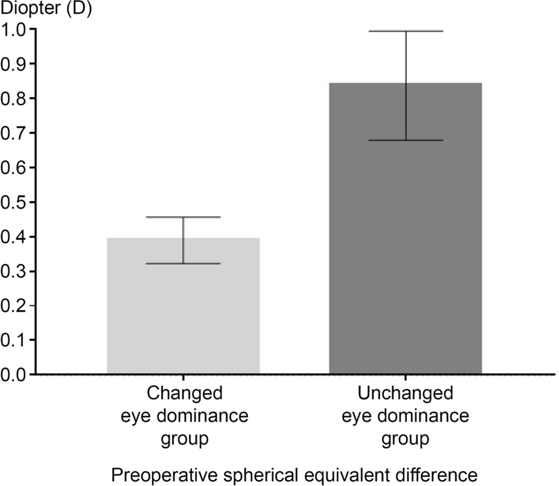 | Figure 1.Comparison of Preoperative spherical equivalent difference of both eyes (the magnitude of anisometropia) between changed eye dominance group and unchanged eye dominance group. There are significant differences between the two groups (p = 0.01, t-test). |
Table 1.
Baseline characteristics of patients
| Changed eye dominance group | Unchanged eye dominance group | p-value | |
|---|---|---|---|
| Patients | 22 (31.4) | 48 (68.6) | |
| Age (years) | 70.5 ± 11.3 | 71.6 ± 10.7 | 0.70* |
| Gender | |||
| Male | 2 (9.1) | 13 (35.1) | 0.12† |
| Female | 20 (90.9) | 35 (72.9) | |
| Dominant eye | |||
| Right | 11 (50.0) | 26 (54.2) | |
| Left | 11 (50.0) | 22 (45.8) | |
Table 2.
Comparison of spherical equivalent between changed group and unchanged group
| Changed eye dominance group | Unchanged eye dominance group | p-value* | |
|---|---|---|---|
| Number of eyes | 22 (31.4) | 48 (68.6) | |
| Time interval between cataract surgeries | 109.50 ± 125.77 | 122.56 ± 205.70 | 0.78 |
| Preoperative spherical equivalent of the dominant eye | 0.13 ± 1.29 | −0.74 ± 2.45 | 0.20 |
| Preoperative spherical equivalent of nondominant eye Preoperative spherical equivalent difference | 0.09 ± 1.35 0.39 ± 0.31 | −0.68 ± 2.87 0.84 ± 1.07 | 0.30 0.01† |
| Postoperative spherical equivalent of the dominant eye | −1.26 ± 0.51 | −0.16 ± 0.57 | <0.01 |
| Postoperative spherical equivalent of nondominant eye | −0.11 ± 0.38 | −1.23 ± 0.67 | <0.01 |
| Postoperative spherical equivalent difference | 1.15 ± 0.40 | 1.17 ± 0.59 | 0.87 |
Table 3.
Comparison of visual acuity between changed group and unchanged group
| Changed eye dominance group | Unchanged eye dominance group | p-value* | |
|---|---|---|---|
| Preoperative UCDVA (logMAR) | |||
| Dominant eye | 0.30 ± 0.24 | 0.35 ± 0.25 | 0.30 |
| Non-dominant eye | 0.31 ± 0.28 | 0.36 ± 0.27 | 0.40 |
| Preoperative UCNVA (logMAR) | |||
| Dominant eye | 0.48 ± 0.22 | 0.66 ± 0.39 | 0.08 |
| Non-dominant eye | 0.47 ± 0.26 | 0.64 ± 0.39 | 0.16 |
| Preoperative BCVA (logMAR) | |||
| Dominant eye | 0.25 ± 0.18 | 0.31 ± 0.24 | 0.50 |
| Non-dominant eye | 0.17 ± 0.13 | 0.23 ± 0.24 | 0.72 |
| Postoperative UCDVA (logMAR) | |||
| Dominant eye | 0.20 ± 0.16 | 0.10 ± 0.15 | <0.01 |
| Non-dominant eye | 0.07 ± 0.11 | 0.19 ± 0.16 | <0.01 |
| Postoperative UCNVA (logMAR) | |||
| Dominant eye | 0.20 ± 0.11 | 0.52 ± 0.34 | <0.01 |
| Non-dominant eye | 0.46 ± 0.27 | 0.29 ± 0.25 | <0.01 |
| Postoperative BCVA (logMAR) | |||
| Dominant eye | 0.03 ± 0.05 | 0.04 ± 0.05 | 0.95 |
| Non-dominant eye | 0.02 ± 0.03 | 0.04 ± 0.06 | 0.12 |
Table 4.
Comparison of stereopsis between changed group and unchanged group
| Changed eye dominance group | Unchanged eye dominance group | p-value* | |
|---|---|---|---|
| Preoperative stereopsis | 234.50 ± 216.13 | 229.21 ± 230.28 | 0.46 |
| Postoperative stereopsis | 248.00 ± 265.42 | 250.43 ± 256.64 | 0.80 |
 XML Download