

 PDF
PDF ePub
ePub Citation
 Print
Print


Abstract
Purpose
To investigate risk factors, clinical features, pathogenic organisms, and outcomes in patients with infectious scleritis.
Results
The mean patient age was 69.2 ± 8.4 years and the mean duration of hospitalization was 11.3 ± 5.8 days. Furthermore, the mean duration of symptoms before presentation was 16.8 ± 13.9 days; patients were followed for a mean duration of 23.3 ±25.4 months. All patients had prior pterygium surgery. Eighteen patients (90%) were culture-positive and Pseudomonas aeruginosa (P. aeruginosa) was identified in 12 eyes. In the acute stages, adjuvant surgical intervention was performed for 18 patients (90%) for these patients, the mean duration of hospitalization before surgery was 4.1 ± 4.4 days.
Methods
This study was a retrospective review of 20 patients with infectious scleritis who were admitted from January 2011 to December 2018 in a single tertiary hospital, with at least 3 months of follow-up. We analyzed age, risk factors, clinical manifestations, pathogenic organisms, treatment, and outcomes of infectious scleritis.
Conclusions
The most common risk factor and pathogenic organism for infectious scleritis were prior pterygium surgery and P. aeruginosa, respectively. Identification of specific causative organisms and corresponding antibiotic treatment with adjuvant surgical intervention may improve visual prognosis in patients with infectious scleritis.
Go to : 
References
1. Jabs DA, Mudun A, Dunn JP, Marsh MJ. Episcleritis and scleritis: clinical features and treatment results. Am J Ophthalmol. 2000; 130:469–76.

2. Hodson KL, Galor A, Karp CL, et al. Epidemiology and visual abdominals in patients with infectious scleritis. Cornea. 2013; 32:466–72.
3. Hsiao CH, Chen JJ, Huang SC, et al. Intrascleral dissemination of infectious scleritis following pterygium excision. Br J Ophthalmol. 1998; 82:29–34.
4. Huang FC, Huang SP, Tseng SH. Management of infectious abdominal after pterygium excision. Cornea. 2000; 19:34–9.
5. Kumar Sahu S, Das S, Sharma S, Sahu K. Clinico-microbiological profile and treatment outcome of infectious scleritis: experience from a tertiary eye care center of India. Int J Inflam. 2012; 2012:753560.
6. Jain V, Garg P, Sharma S. Microbial scleritis-experience from a abdominal country. Eye (Lond). 2009; 23:255–61.
7. Ho YF, Yeh LK, Tan HY, et al. Infectious scleritis in Taiwan-a 10-year review in a tertiary-care hospital. Cornea. 2014; 33:838–43.
8. Ahmad S, Lopez M, Attala M, et al. Interventions and outcomes in patients with infectious Pseudomonas scleritis: a 10-year perspective. Ocul Immunol Inflamm. 2017; 17:1–8.
9. Tittler EH, Nguyen P, Rue KS, et al. Early surgical debridement in the management of infectious scleritis after pterygium excision. J Ophthalmic Inflamm Infect. 2012; 2:81–7.
10. Kim YK, Kim TY. 4 cases of Pseudomonas scleritis after pterygium excision. J Korean Ophthalmol Soc. 1999; 40:2304–12.
11. Jo DH, Oh JY, Kim MK, et al. Aspergillus fumigatus scleritis abdominal with monoclonal gammopathy of undetermined significance. Korean J Ophthalmol. 2010; 24:175–8.
12. Ahn SJ, Oh JY, Kim MK, et al. Clinical features, predisposing abdominals, and treatment outcomes of scleritis in the Korean population. Korean J Ophthalmol. 2010; 24:331–5.
13. Lyne AJ, Lloyd-Jones D. Necrotizing scleritis after ocular surgery. Trans Ophthalmol Soc UK. 1979; 99:146–9.
14. Rich RM, Smiddy WE, Davis JL. Infectious scleritis after retinal surgery. Am J Ophthalmol. 2008; 145:695–9.
15. Chao DL, Albini TA, McKeown CA, Cavuoto KM. Infectious Pseudomonas scleritis after strabismus surgery. J AAPOS. 2013; 17:423–5.
16. Lin CP, Shih MH, Tsai MC. Clinical experiences of infectious scleral ulceration: a complication of pterygium operation. Br J Ophthalmol. 1997; 81:980–3.
17. Gopinathan U, Garg P, Fernandes M, et al. The epidemiological features and laboratory results of fungal keratitis: a 10-year review at a referral eye care center in South India. Cornea. 2002; 21:555–9.
18. Srinivasan M, Mascarenhas J, Rajaraman R, et al. Corticosteroids for bacterial keratitis: the steroids for corneal ulcers trial (SCUT). Arch Ophthalmol. 2012; 130:143–50.
19. Ramenaden ER, Raiji VR. Clinical characteristics and visual outcomes in infectious scleritis: a review. Clin Ophthalmol. 2013; 7:2133–22.
20. Yoo WS, Kim CR, Kim BJ, et al. Successful treatment of infectious scleritis by Pseudomonas aeruginosa with autologous abdominal graft of conchal cartilage. Yonsei Med J. 2015; 56:1738–41.
21. Moon JH, Kim JC. Efficacy of autologous tragal perichondrium graft after proper antifungal treatment in fungal necrotizing scleritis. J Korean Ophthalmol Soc. 2013; 54:1929–34.
Go to :
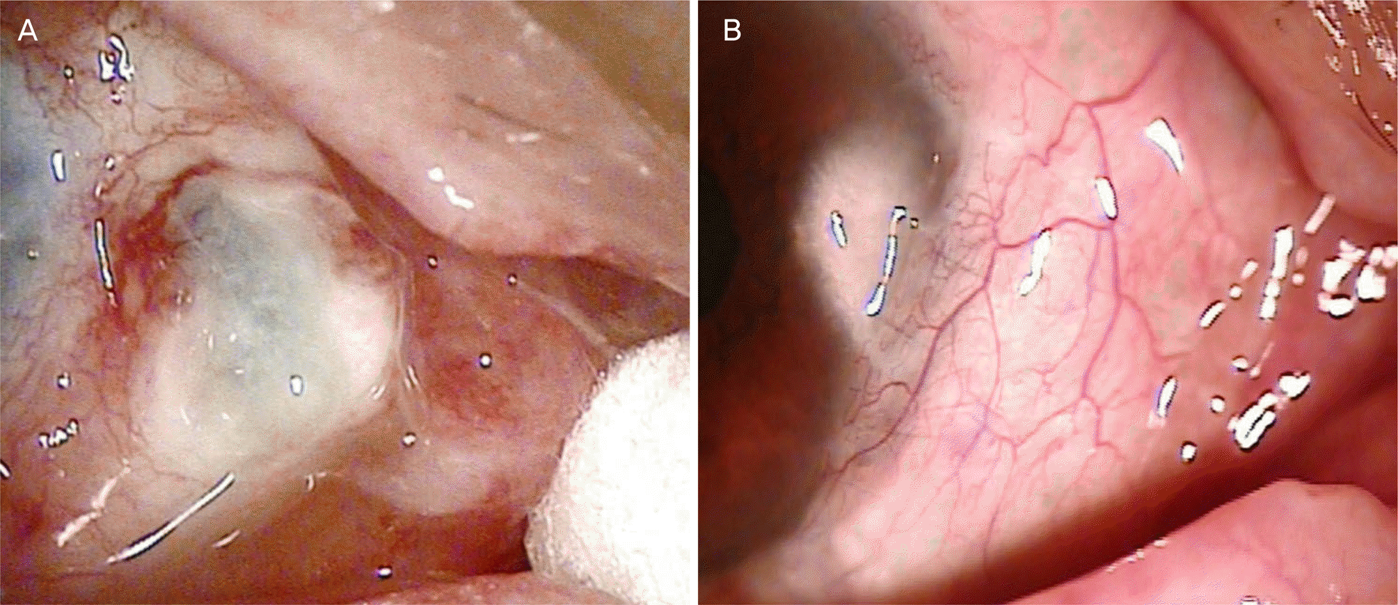 | Figure 1.Anterior segment photograph of patient with infectious scleritis before (A) and after (B) treatment. (A) Infectious scleritis caused by Pseudomonas aeruginosa developed at the previous pterygium excision area that presented with scleral necrosis. (B) Scleritis was resolved after treatment with intensive antibiotics and perichondrium graft with conjunctival flap. |
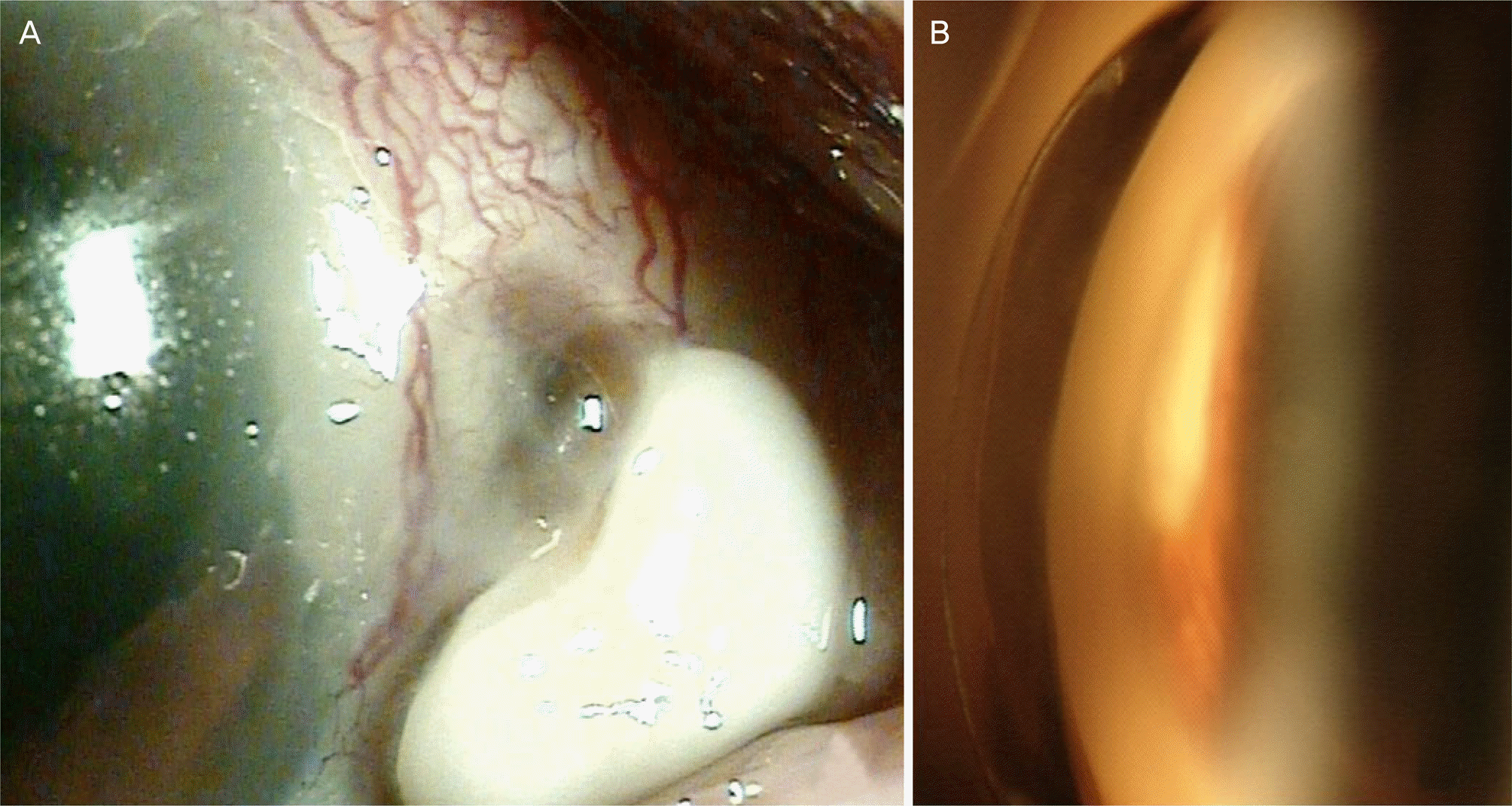 | Figure 2.Slit lamp photograph depicting Aspergillus flavus scleritis in a patient 10 years after pterygium surgery. (A) At the presentation showing scleral melting and abscess formation. (B) Fungus ball-like lesion were detected in the anterior chamber angle. |
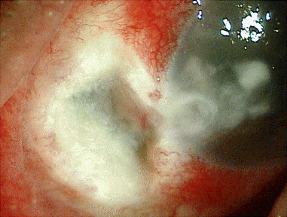 | Figure 3.Anterior segment photo of a patient with infections scleritis. Seventy-year-old woman with fungal (Fusarium species) scleritis after pterygium excision. There was a severe scleral necrosis and adjacent corneal infiltration with satellite lesions. |
Table 1.
Demographics of patients with infectious scleritis
Table 2.
Clinical characteristics of patients with infectious scleritis
Table 3.
Culture results in patients with infectious scleritis
Table 4.
Medical and surgical treatment in patients with infectious scleritis
Table 5.
Complications during treatment
Table 6.
Univariate analysis evaluating the risk factors for poor visual outcome (BCVA < 0.3) in patients with infectious scleritis
| Factor | OR (95% CI) | p-value* |
|---|---|---|
| Age | 1.014 (1.000–1.029) | 0.05 |
| Gender (female) | 0.080 (0.000–1.472) | 0.09 |
| Latency period | 1.063 (0.967–1.168) | 0.21 |
| Duration of symptoms before admission | 1.033 (0.984–1.085) | 0.19 |
| Route of admission (OPD) | 1.250 (0.971–1.610) | 0.30 |
| Duration of hospitalization | 1.070 (0.987–1.160) | 0.07 |
| Previous steroid treatment | 0.750 (0.055–2.886) | 0.71 |
| Area of necrotic scleritis involvement | 1.355 (0.960–1.911) | 0.13 |
| Presented BCVA (< 0.3) | 1.667 (1.103–2.519) | 0.10 |
| Anterior segment inflammation | 0.500 (0.166–1.506) | 0.13 |
| Calcified plaque | 0.857 (0.542–1.356) | 0.45 |
| Corneal involvement | 1.500 (0.170–13.255) | 0.60 |
| Hypopyon | 1.000 (0.132–7.570 | 0.72 |
| ESR | 1.100 (0.990–1.230) | 0.18 |
| Culture positivity of bacteria | 2.580 (0.070–9.340) | 0.61 |
| Adjuvant steroid treatment | 0.440 (0.040–5.110) | 0.51 |
| Perichondrium graft | 0.320 (0.030–3.780) | 0.36 |
| Retinal/choroidal detachment | 0.750 (0.107–5.238) | 0.63 |
| Scleral thinning | 0.429 (0.032–5.779) | 0.52 |
| Endophthalmitis | 0.857 (0.692–1.062) | 0.48 |
 XML Download