

 PDF
PDF ePub
ePub Citation
 Print
Print


Abstract
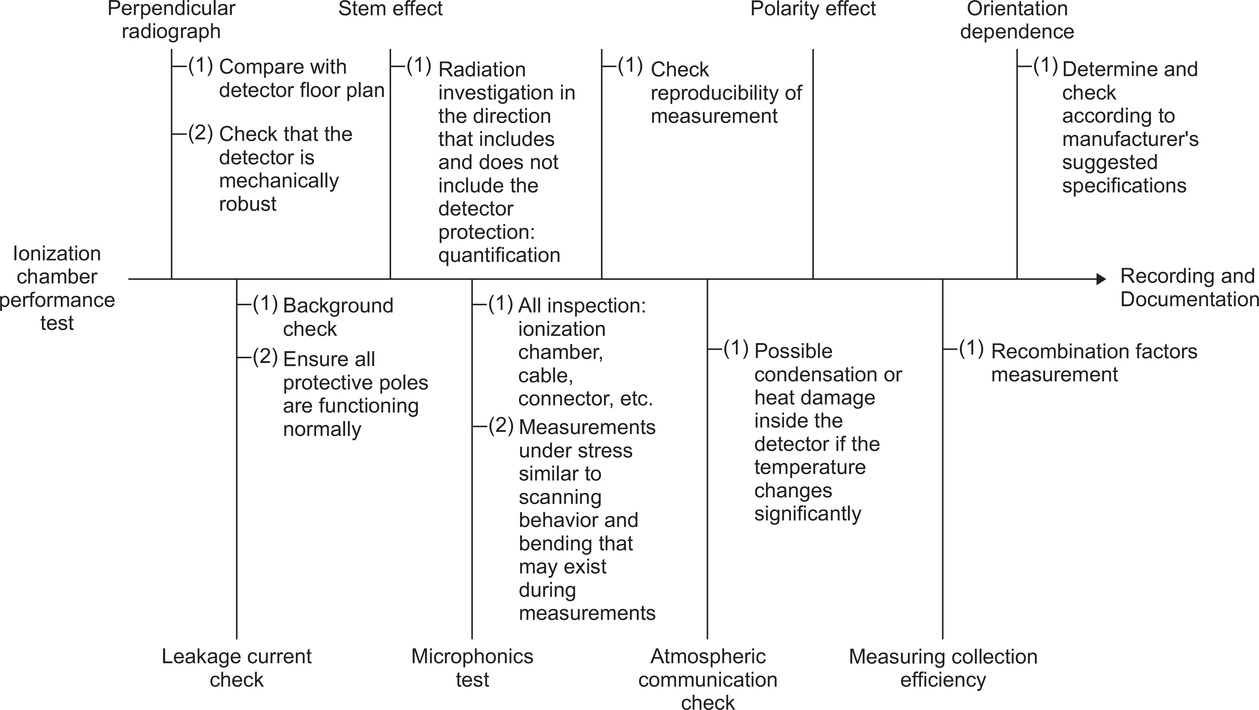
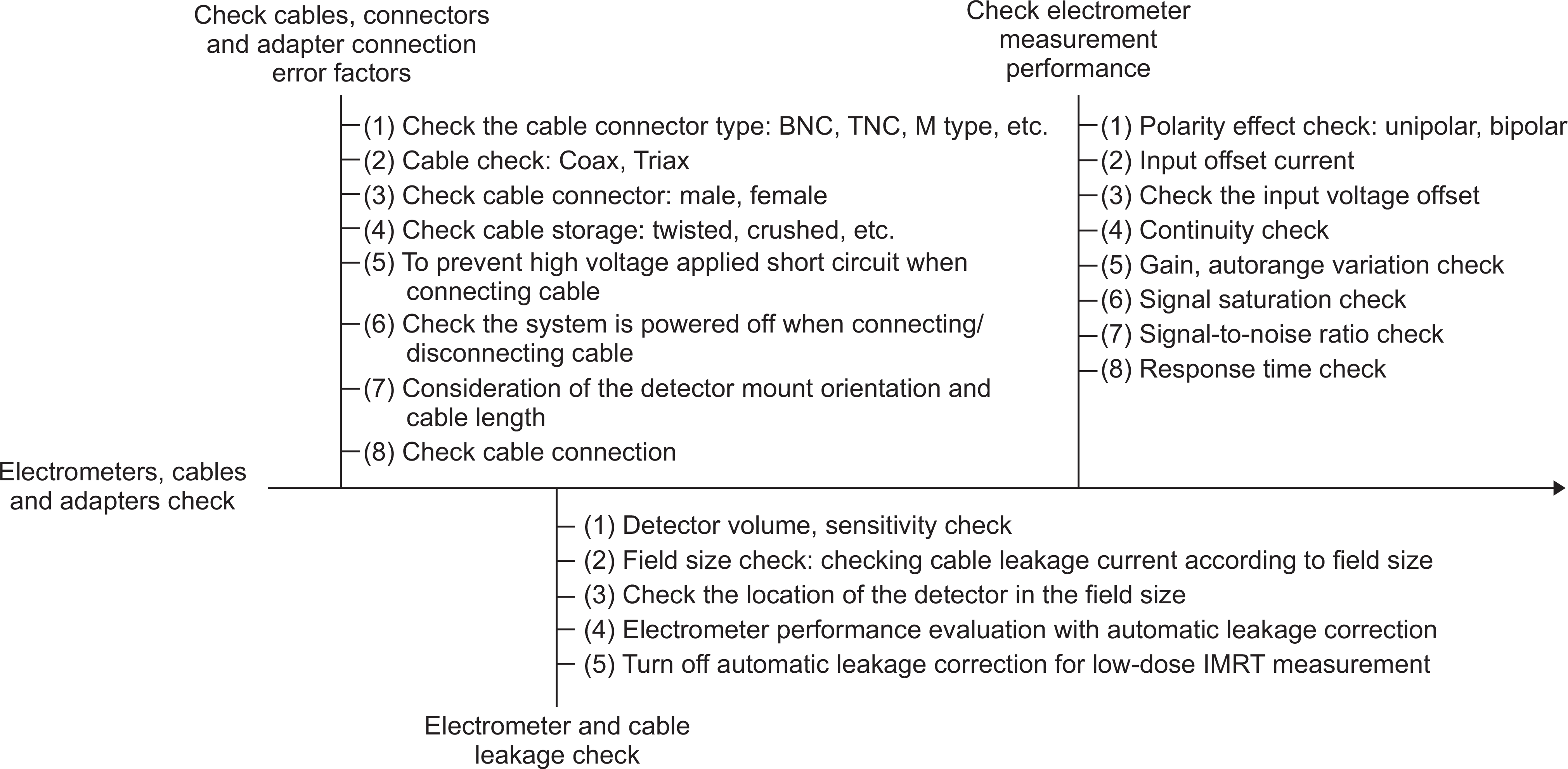
The complex dose distribution and dose transfer characteristics of intensity-modulated radiotherapy increase the importance of precise beam data measurement and review in the acceptance inspection and preparation stages. In this study, we propose a process map for the introduction and installation of high-precision radiotherapy devices and present items and guidelines for risk management at the acceptance test procedure (ATP) and commissioning stages. Based on the ATP of the Varian and Elekta linear accelerators, the ATP items were checked step by step and compared with the quality assurance (QA) test items of the AAPM TG-142 described for the medical accelerator QA. Based on the commissioning procedure, dose quality control protocol, and mechanical quality control protocol presented at international conferences, step-by-step check items and commissioning guidelines were derived. The risk management items at each stage were (1) 21 ionization chamber performance test items and 9 electrometer, cable, and connector inspection items related to the dosimetry system; (2) 34 mechanical and dose-checking items during ATP, 22 multileaf collimator (MLC) items, and 36 imaging system items; and (3) 28 items in the measurement preparation stage and 32 items in the measurement stage after commissioning. Because the items presented in these guidelines are limited in terms of special treatment, items and practitioners can be modified to reflect the clinical needs of the institution. During the system installation, it is recommended that at least two clinically qualified medical physicists (CQMP) perform a double check in compliance with the two-person rule. We expect that this result will be useful as a radiation safety management tool that can prevent radiation accidents at each stage during the introduction of radiotherapy and the system installation process.
Go to : 
REFERENCES
1.Gary A., Ezzell ., Calvin James M.., Low Daniel., Rosen Isaac., Sharpe Michael B.., Xia Ping, et al. Guidance document on delivery, treatment planning, and clinical implementation of IMRT: Report of the IMRT subcommittee of the AAPM radiation therapy committee. Med. Phys. 2003. 30(8):2089–2115.

2.IAEA Human Health Series No.25: Roles and Responsibilities, and Education and Training Requirements for Clinically Qualified Medical Physicists. International Atomic Energy Agency. 2013.
3.IAEA TRS-398 report: Absorbed Dose Determination in External Beam Radiotherapy: An International Code of Practice for Dosimetry based on Standards of Absorbed Dose to Water. International Atomic Energy Agency. 2006.
4.Almond P. R.., Biggs P. J.., Coursey B. M.., Hanson W. F.., Saiful Huq M.., Nath Ravinder, et al. AAPM's TG-51 protocol for clinical reference dosimetry of high-energy photon and electron beams. Med. Phys. 1999. 26(9):1847–1870.
5.Gerald J., Kutche ., Coia Lawrene., Gillin Michael., Hansom Wiliam F.., Leibel Steven., Morton Robert J., et al. Comprehensive QA for radiation oncology y: Report of AAPM Radiation Therapy Committee Task Group 40. Med. Phys. 1994. 21(4):581–618.
6.Eric E., Klein ., Hanley Joseph., Bayouth John., Yin Fang-Fang., Simon Wiliam., Dresser Sen, et al. Task Group 142 report: Quality assurance of medical accelerators. Med. Phys. 2009. 36(9):4197–4212.
7.Indra J., Das ., Cheng Chee-Wai., Watts Ronald J.., Ahnesjö Anders., Gibbons John., Allen Li X., et al. Accelerator beam data commissioning equipment and procedures: Report of the TG-106 of the Therapy Physics Committee of the AAPM. Med. Phys. 2008. 35(9):4186–4215.
8.Gary A., Ezzell ., Burmeister Jay W.., Dogan Nesrin., LoSasso Thomas J.., Mechalakos James G.., Mihailidis Dimitiris, et al. IMRT commissioning: Multiple institution planning and dosimetry comparisons, a report from AAPM Task Group 119. Med. Phys. 2009. 36(11):5359–5373.
9.ESTRO booklet no.9: Guidelines for the verification of IMRT. 2008.
10.Daniel A. Low, Jean M. Moran, James F. Dempsey, Lei Dong, Mark Oldham, Dosimetry tools and techniques for IMRT. Med. Phys. 2001. 38(3):1313–1338.
11.Oh Yoonjin., Shin Dong Oh., Kim Juhye., Kwon Nahye., Lee Soon Sung., Choi Sang Hyoun, et al. Proposal on Guideline for Quality Assurance of Radiation Treatment Planning System. Progress in Medical Physics. 2017. 28(4):197–206.
12.L. J. Humphries and J. A. Purdy, In Advances in Radiation Oncology Physics Dosimetry, Treatment Planning, and Brachytherapy. 1992. AAPM Monograph Vol. 19, edited by J. A. Purdy AAPM.
13.Per H., Halvorsen ., Das Indra J.., Fraser Martin., Jay Freedman D.., Robert E. Rice III., lbbott Geoffrey S., et al. AAPM Task Group 103 report on peer review in clinical radiation oncology physics. Journal of Applied Clinical Medical Physics. 2005. 6(4):50–64.
Go to :
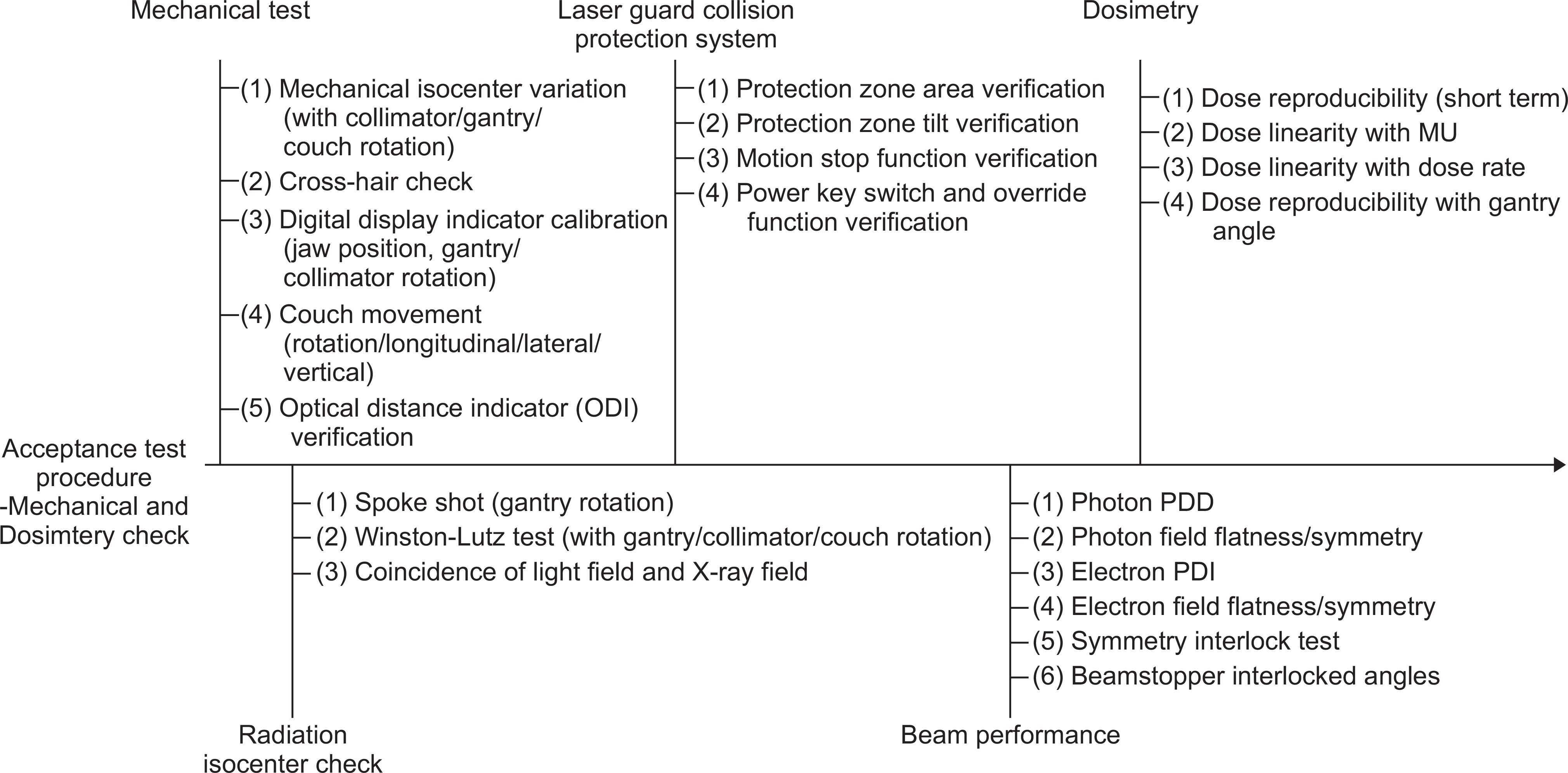 | Fig. 3The risk management items for acceptance test of external radiation therapy equipment: Mechanical and dosimetry test. |
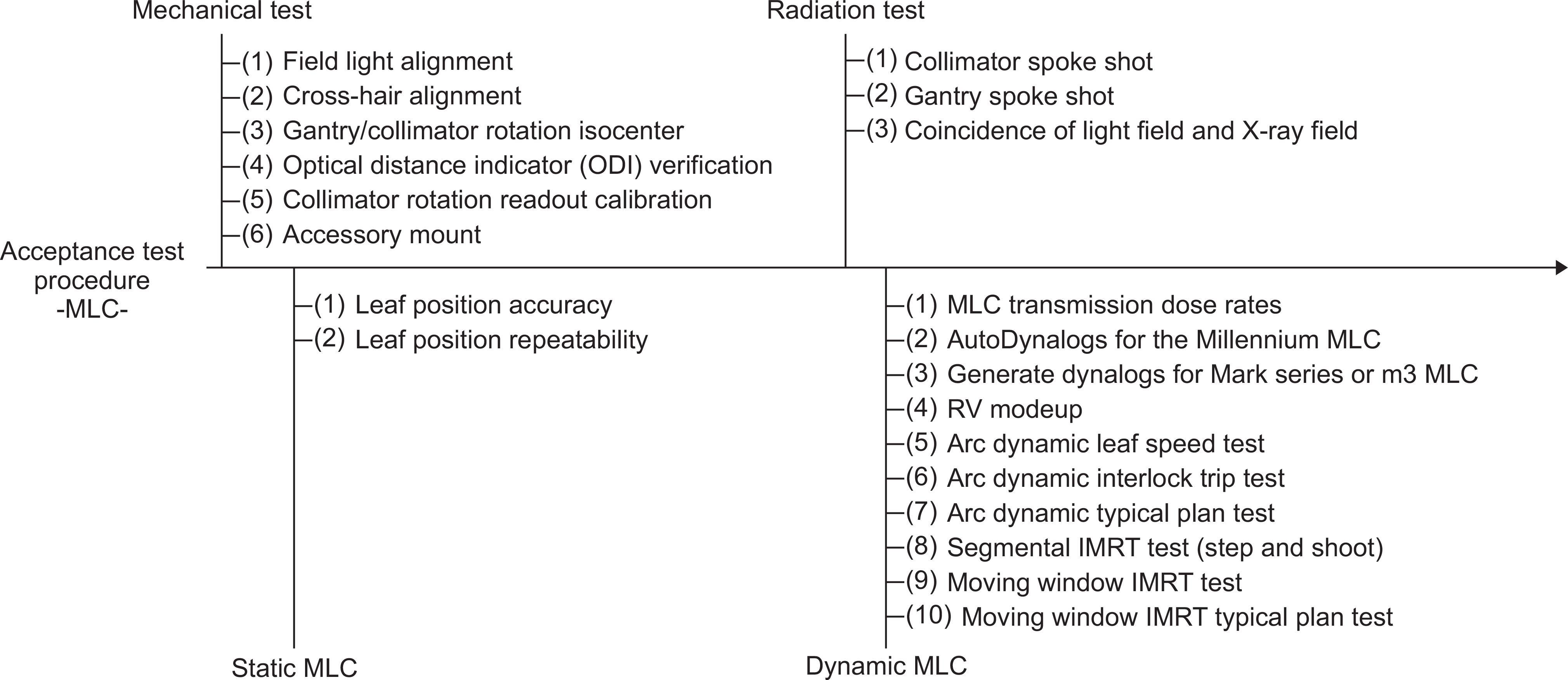 | Fig. 4The risk management items for acceptance test of external radiation therapy equipment: MLC. |
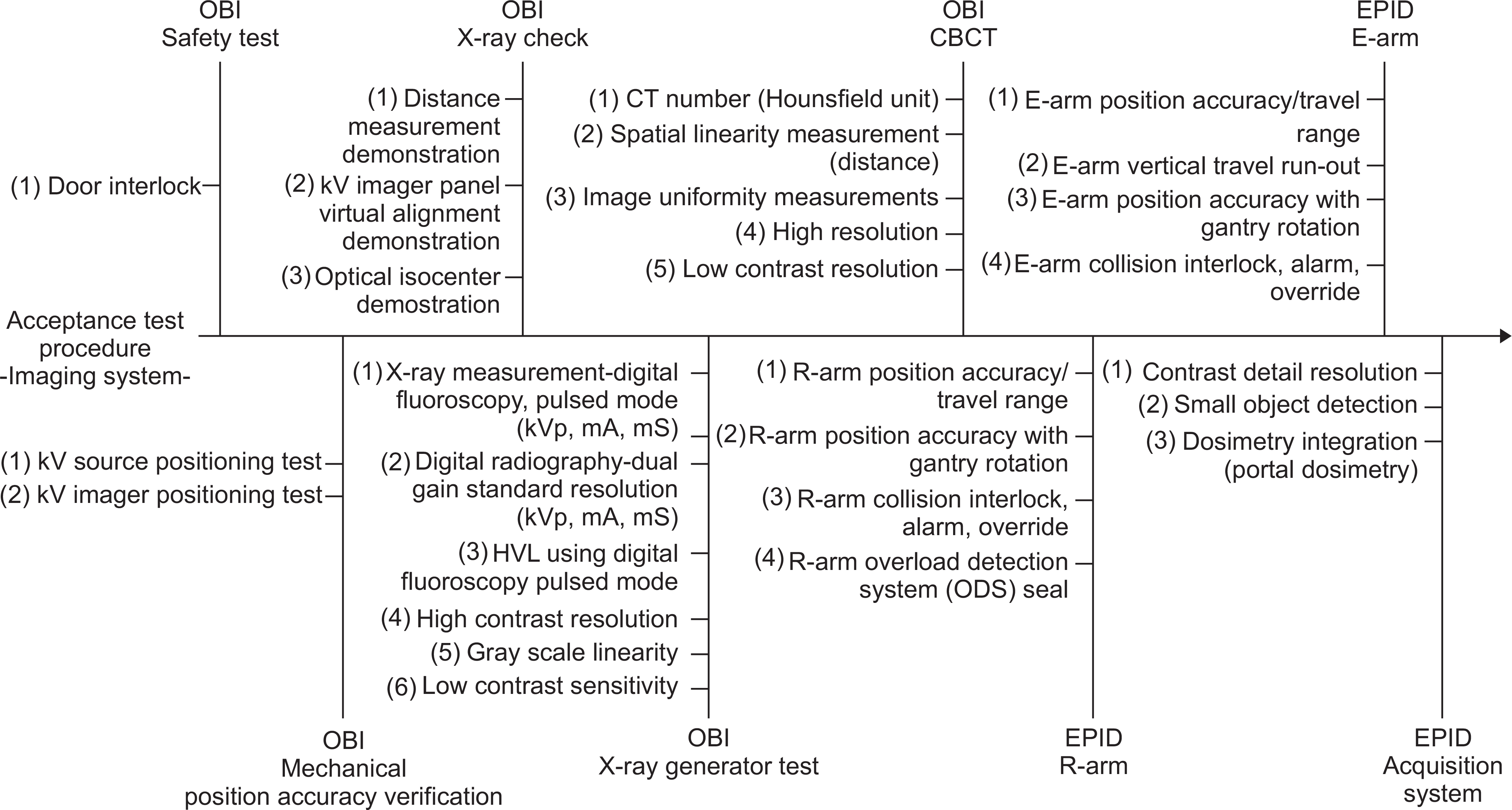 | Fig. 5The risk management items for acceptance test of external radiation therapy equipment: Imaging system. |
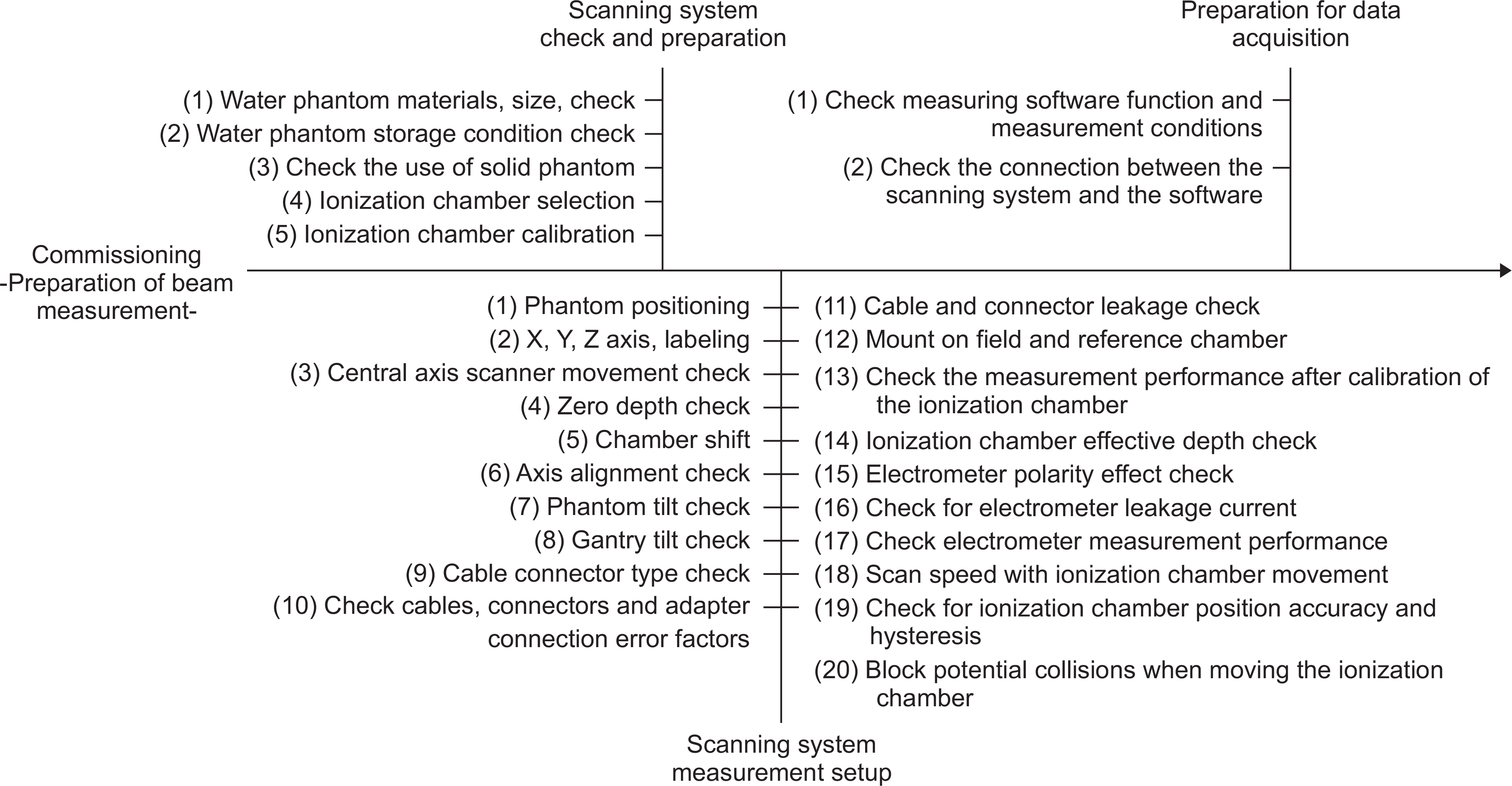 | Fig. 6The risk management items for commissioning of external radiation therapy equipment: preparation of beam measurement. |
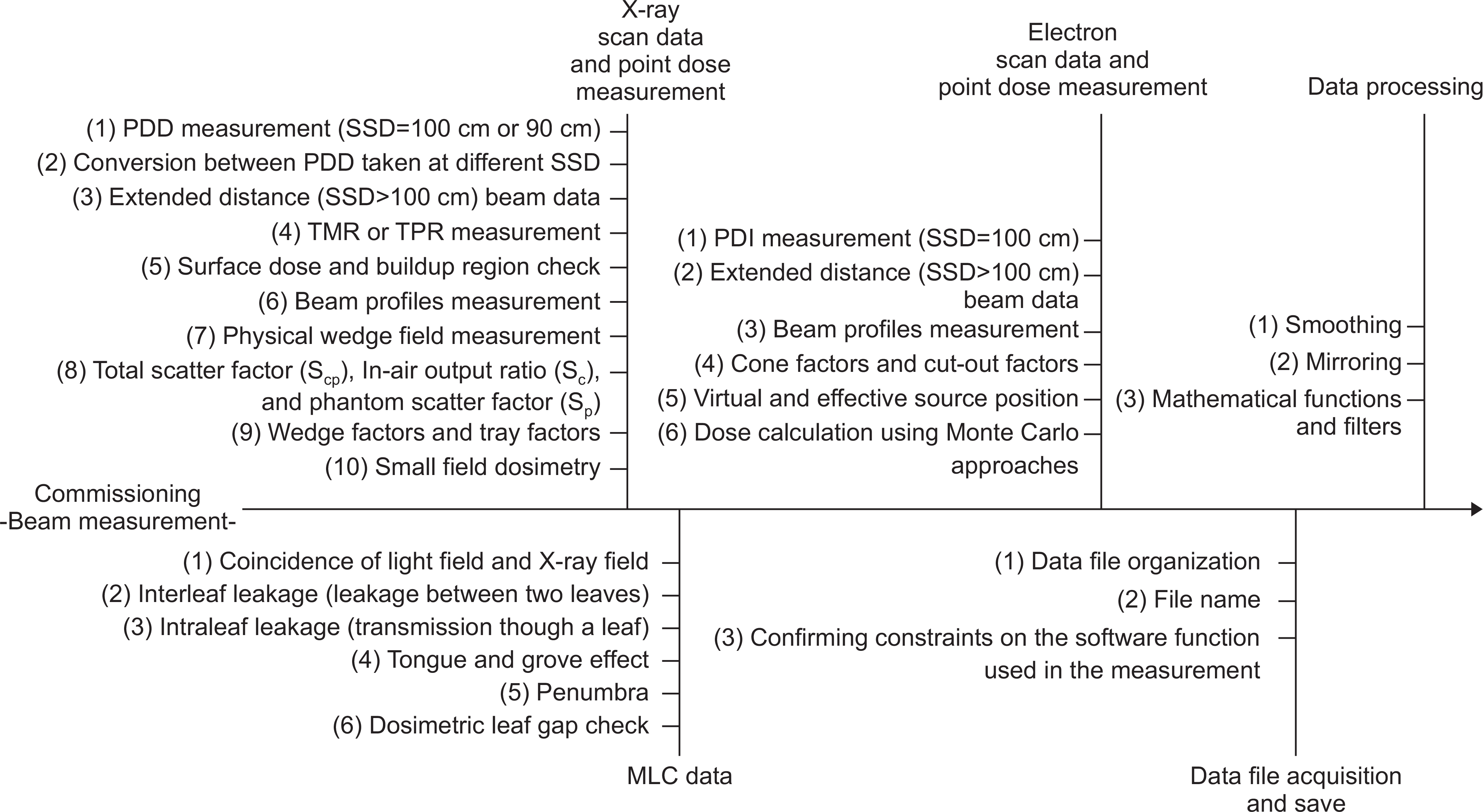 | Fig. 7The risk management items for commissioning of external radiation therapy equipment: beam measurement. |
Table 1.
The derived risk management items were compared for the correlation with TG-142 quality assurance items.
 XML Download