1. Shomer DL, Lopes da Silva FH. Niedermyer's electroencephalography: basic principles, clinical applications, and related fields. 7th ed. New York: Oxford University Press;2018.
2. Schaul N. The fundamental neural mechanisms of electroencephalography. Electroencephalogr Clin Neurophysiol. 1998; 106:101–107.

3. Niedermyer E, Lopes da silva FH. Electroencephalography: basic principles, clinical applications, and related fields. 5th ed. Philadelphia: London: Lippincott Williams & Wilkins;2005.
4. William OT. Handbook of EEG interpretation. 2nd ed. New York: New York;2014.
5. Chernecky CC, Berger BJ. Laboratory tests and diagnostic procedures. 6th ed. St. Louis: Elsevier;2013.
6. Scott NK. Infection prevention: 2013 review and update for neurodiagnostic technologists. Neurodiagn J. 2013; 53:271–288.
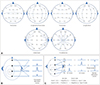
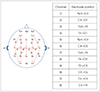
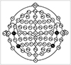
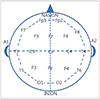
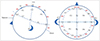
7. Jasper HH. The 10–20 electrode system of the international federation. Electroencephalogr Clin Neurophysiol. 1958; 10:371–375.
8. Sinha SR, Sullivan L, Sabau D, San-Juan D, Dombrowski KE, Halford JJ, et al. American clinical neurophysiology society guideline 1: minimum technical requirements for performing clinical electroencephalography. J Clin Neurophysiol. 2016; 33:303–307.
9. Anonymous. Recommendations for the practice of clinical neurophysiology: guidelines of the international federation of clinical neurophysiology. Electroencephalogr Clin Neurophysiol Suppl. 1999; 52:1–304.
10. O'Leary JL, Goldring S. Science and epilepsy-Neuroscience gains in epilepsy research. New York: Raven Press;1976.
11. Caton R. The electric currents of the brain. Br Med J. 1875; 2:278.
12. Coenen A, Fine E, Zayachkivska O. Adolf Beck: a forgotten pioneer in electroencephalography. J Hist Neurosci. 2014; 23:276–286.
13. Pravdich-Neminsky W. Ein versuch der registrierung der elektrischen gehirnerscheinungen. Zentralbl Physiol. 1912; 27:951–960.
14. Cybulski N, Jeleńska-Macieszyna S. Action currents of the cerebral cortex. Bull Acad Sci Cracov. 1914; 776–781.
15. Grzybowski A, Pietrzak K. Napoleon Cybulski (1854–1919). J Neurol. 2013; 260:2942–2943.
16. Haas LF. Hans Berger (1873–1941), Richard Caton (1842–1926), and electroencephalography. J Neurol Neurosurg Psychiatry. 2003; 74:9.
17. Adrian ED, Matthews BH. The Berger rhythm: potential changes from the occipital lobes in man. Brain. 1934; 57:355–385.
18. Grass AM. The electroencephalographic heritage until 1960. Am J EEG Technol. 1984; 24:133–173.
19. Berger H. Über das elektrenkephalogramm des menschen. Eur Arch Psychiatry Clin Neurosci. 1929; 87:527–570.
20. Britton JW, Frey LC, Hopp JL, Korb P, Koubeissi MZ, Lievens WE, et al. Electroencephalography (EEG): an introductory text and atlas of normal and abnormal findings in adults, children, and infants. Chicago: American Epilepsy Society;2016. p. 1–95.
22. National Academy of Engineering. Franklin FO. Biography & autobiography memorial tributes Franklin F. Offner Washington, DC: The National Academies Press;2002. 10:p. 188–192.
23. Klem GH, Lüders HO, Jasper HH, Elger C. The ten-twenty electrode system of the international federation. The international federation of clinical neurophysiology. Electroencephalogr Clin Neurophysiol Suppl. 1999; 52:3–6.
24. Bozinovski Bozinovski, Bozinovska L, Setakov M. Mobile robot control using alpha wave from the human brain. Zagreb: Proc. Symp. JUREMA;1988. p. 247–249.
25. Sutter R, Stevens RD, Kaplan PW. Continuous electroencephalographic monitoring in critically ill patients: indications, limitations, and strategies. Crit Care Med. 2013; 41:1124–1132.
26. Nield D. Scientists have connected the brains of 3 people, enabling them to share thoughts. ScienceAlert. 2018; 10. 05.
27. Kondraske GV. Neurophysiological measurements. Biomedical Engineering and Instrumentation. 1986; 138–179.
28. Fisch BJ, Spehlmann R. EG primer: basic principles of digital and analog EEG. 3rd ed. Amsterdam: Elsevier Science;1999.
29. Joseph DB. Medical devices and system: biomedical engineering handbook. 3rd ed. Boca Raton: Informa;2006.
30. Teplan M. Fundamentals of EEG measurement. Measurement Science Review. 2002; 2:1–11.
31. Fonseca C, Silva Cunha JP, Martins RE, Ferreira VM, Marques de Sá JP, Barbosa MA, et al. A novel dry active electrode for EEG recording. IEEE Trans Biomed Eng. 2007; 54:162–165.
33. Speckmann E, Elger CE. Introduction to the neurophysiological basis of the EEG and DC potentials. In : Niedermeyer E, editor. Electroencephalography. Basic principles, clinical applications and related fields. Baltimore: Williams & Wilkins;1999. p. 15–27.
34. Ebersole JS. Noninvasive localization of epileptogenic foci by EEG source modeling. Epilepsia. 2000; 41:Suppl 3. S24–S33.
35. Buzsaki G, Traub RD, Pedley TA. The cellular basis of EEG activity. In : Ebersole JS, Pedley TA, editors. Current practice of clinical electroencephalography. 3rd ed. Philadelphia: Lippincott Williams & Wilkins;2003. p. 1–11.
36. Ebersole JS. Defining epileptogenic foci: past, present, future. J Clin Neurophysiol. 1997; 14:470–483.
37. Hong SB, Jung KY. Basic electrophysiology of the electroencephalography. J Korean Neurol Assoc. 2003; 21:225–238.
38. Wong PK. Source modelling of the rolandic focus. Brain Topogr. 1991; 4:105–112.
39. Bear MF, Connors BW, Paradiso MA. Neuroscience exploring the brain. Baltimore: Lipponcott Williams & Wilkins;2001. p. 606–636.
40. Acharya JN, Hani AJ, Thirumala PD, Tsuchida TN. American clinical neurophysiology society guideline 3: a proposal for standard montages to be used in clinical EEG. J Clin Neurophysiol. 2016; 33:312–316.
41. Nunez PL, Pilgreen KL. The spline-Laplacian in clinical neurophysiology: a method to improve EEG spatial resolution. J Clin Neurophysiol. 1991; 8:397–413.
42. Hrachovy RA, Mizrahi EM. Atlas of neonatal electroencephalography. New York: Demos Medical Publishing;2016. p. 7–13.
43. Clancy RR, Bergqvist AGC, Dlugos DJ. Neonatal electroencephalography. In : Ebersole JS, Pedley TA, editors. Current practice of clinical electroencephalography. Philadelphia: Lippincott Williams & Wilkins;2003. p. 160–234.
44. Shellhaas RA, Chang T, Tsuchida T, Scher MS, Riviello JJ, Abend NS, et al. The American Clinical Neurophysiology Society's guideline on continuous electroencephalography monitoring in neonates. J Clin Neurophysiol. 2011; 28:611–617.
45. Kuratani J, Pearl PL, Sullivan L, Riel-Romero RM, Cheek J, Stecker M, et al. American Clinical Neurophysiology Society guideline 5: minimum technical standards for pediatric electroencephalography. J Clin Neurophysiol. 2016; 33:320–323.
46. Tsuchida TN, Acharya JN, Halford JJ, Kuratani JD, Sinha SR, Stecker MM, et al. American Clinical Neurophysiology Society: EEG guidelines introduction. J Clin Neurophysiol. 2016; 33:301–302.
47. Acharya JN, Hani A, Cheek J, Thirumala P, Tsuchida TN. American Clinical Neurophysiology Society guideline 2: guidelines for standard electrode position nomenclature. J Clin Neurophysiol. 2016; 33:308–311.
48. Koo DL, Kim WJ, Lee SA, Kim JM, Kim J, Park S. Fundamental requirements for performing electroencephalography. Ann Clin Neurophysiol. 2017; 19:113–117.
49. Barry W, Jones GM. Influence of eye lid movement upon electro-oculographic recording of vertical eye movements. Aerosp Med. 1965; 36:855–858.
50. Iwasaki M, Kellinghaus C, Alexopoulos AV, Burgess RC, Kumar AN, Han YH, et al. Effects of eyelid closure, blinks, and eye movements on the electroencephalogram. Clin Neurophysiol. 2005; 116:878–885.
51. Keren AS, Yuval-Greenberg S, Deouell LY. Saccadic spike potentials in gamma-band EEG: characterization, detection and suppression. Neuroimage. 2010; 49:2248–2263.
52. Epstein CM, Andriola MR. Introduction to EEG and evoked potentials. New York: Lippincott;1983.
53. Symeonidou ER, Nordin AD, Hairston WD, Ferris DP. Effects of cable sway, electrode surface area, and electrode mass on electroencephalography signal quality during motion. Sensors (Basel). 2018; 18:E1073.
54. Oliveira AS, Schlink BR, Hairston WD, König P, Ferris DP. Induction and separation of motion artifacts in EEG data using a mobile phantom head device. J Neural Eng. 2016; 13:036014.
55. Jung TP, Makeig S, Humphries C, Lee TW, McKeown MJ, Iragui V, et al. Removing electroencephalographic artifacts by blind source separation. Psychophysiology. 2000; 37:163–178.
56. Joyce CA, Gorodnitsky IF, Kutas M. Automatic removal of eye movement and blink artifacts from EEG data using blind component separation. Psychophysiology. 2004; 41:313–325.
57. Shackman AJ, McMenamin BW, Maxwell JS, Greischar LL, Davidson RJ. Identifying robust and sensitive frequency bands for interrogating neural oscillations. Neuroimage. 2010; 51:1319–1333.
58. Nolan H, Whelan R, Reilly RB. FASTER: fully automated statistical thresholding for EEG artifact rejection. J Neurosci Methods. 2010; 192:152–162.
59. Fitzgibbon SP, Lewis TW, Powers DM, Whitham EW, Willoughby JO, Pope KJ. Surface laplacian of central scalp electrical signals is insensitive to muscle contamination. IEEE Trans Biomed Eng. 2013; 60:4–9.
60. Kondylis ED, Wozny TA, Lipski WJ, Popescu A, DeStefino VJ, Esmaeili B, et al. Detection of high-frequency oscillations by hybrid depth electrodes in standard clinical intracranial EEG recordings. Front Neurol. 2014; 5:149.
61. Hämäläinen M, Hari R, Ilmoniemi RJ, Knuutila J, Lounasmaa OV. Magnetoencephalography—theory, instrumentation, and applications to noninvasive studies of the working human brain. Rev Mod Phys. 1993; 65:413–497.
62. Murakami S, Okada Y. Contributions of principal neocortical neurons to magnetoencephalography and electroencephalography signals. J Physiol. 2006; 575:925–936.
63. Nuwer M. Assessment of digital EEG, quantitative EEG, and EEG brain mapping: report of the American Academy of neurology and the American Clinical Neurophysiology Society. Neurology. 1997; 49:277–292.
64. Duffy FH, Hughes JR, Miranda F, Bernad P, Cook P. Status of quantitative EEG (QEEG) in clinical practice, 1994. Clin Electroencephalogr. 1994; 25:VI–XXII.
65. Chung HJ, Hur YJ. Diagnosis of neonatal seizures. Korean J Pediatr. 2009; 52:964–970.
66. Karl FS. The aEEG booklet a quick overview for the practical routine. 1st ed. Tokyo: Nihon Kohden Corporation;2012.
67. Maynard D, Prior PF, Scott DF. Device for continuous monitoring of cerebral activity in resuscitated patients. Br Med J. 1969; 4:545–546.
68. Azzopardi DT. Cerebral function monitoring: addition to CFM handbook for users of the olympic CFM 6000. London: Imperial College London;2004.
69. Anderson J. Cognitive psychology and its implications. 6th ed. New York: Worth;2004. p. 17.
70. Nunez PL, Srinivasan R. Electric fields of the brain: the neurophysics of EEG. Oxford: Oxford;1981.
71. Creutzfeldt OD, Watanabe S, Lux HD. Relations between EEG phenomena and potentials of single cortical cells. I. Evoked responses after thalamic and erpicortical stimulation. Electroencephalogr Clin Neurophysiol. 1966; 20:1–18.
72. Tanzer OI. Numerical modeling in electro- and magnetoencephalography [dissertation]. Espoo: Helsinki University of Technology;2006.
73. Tatum WO, Ellen R. Grass lecture: extraordinary EEG. Neurodiagn J. 2014; 54:3–21.
74. Cahn BR, Polich J. Meditation states and traits: EEG, ERP, and neuroimaging studies. Psychol Bull. 2006; 132:180–211.
75. Gerrard P, Malcolm R. Mechanisms of modafinil: a review of current research. Neuropsychiatr Dis Treat. 2007; 3:349–364.
76. Niedermeyer E. Alpha rhythms as physiological and abnormal phenomena. Int J Psychophysiol. 1997; 26:31–49.
77. Feshchenko VA, Reinsel RA, Veselis RA. Multiplicity of the alpha rhythm in normal humans. J Clin Neurophysiol. 2001; 18:331–344.
78. Pfurtscheller G, Lopes da Silva FH. Event-related EEG/MEG synchronization and desynchronization: basic principles. Clin Neurophysiol. 1999; 110:1842–1857.
79. Oberman LM, Hubbard EM, McCleery JP, Altschuler EL, Ramachandran VS, Pineda JA. EEG evidence for mirror neuron dysfunction in autism spectrum disorders. Brain Res Cogn Brain Res. 2005; 24:190–198.
80. Anderson AL, Thomason ME. Functional plasticity before the cradle: a review of neural functional imaging in the human fetus. Neurosci Biobehav Rev. 2013; 37:2220–2232.
81. Santamaria J, Chiappa KH. The EEG of drowsiness in normal adults. J Clin Neurophysiol. 1987; 4:327–382.
82. Frohlich J, Senturk D, Saravanapandian V, Golshani P, Reiter LT, Sankar R, et al. A quantitative electrophysiological biomarker of duplication 15q11.2-q13.1 syndrome. PLoS One. 2016; 11:e0167179.
83. Kisley MA, Cornwell ZM. Gamma and beta neural activity evoked during a sensory gating paradigm: effects of auditory, somatosensory and cross-modal stimulation. Clin Neurophysiol. 2006; 117:2549–2563.
84. Kanayama N, Sato A, Ohira H. Crossmodal effect with rubber hand illusion and gamma-band activity. Psychophysiology. 2007; 44:392–402.
85. Gastaut H. Electrocorticographic study of the reactivity of rolandic rhythm. Rev Neurol (Paris). 1952; 87:176–182.
86. Krishnan V, Chang BS, Schomer DL. Normal EEG in wakefulness and sleep: adult and elderly. In : Schomer DL, Lopes da Silva FH, editors. Niedermyer's electroencephalography;basic principles, clinical applications, and related fields. New York: Oxford;2018. p. 202–228.
87. Montez T, Poil SS, Jones BF, Manshanden I, Verbunt JP, Van Dijk BW, et al. Altered temporal correlations in parietal alpha and prefrontal theta oscillations in early-stage Alzheimer disease. Proc Natl Acad Sci U S A. 2009; 106:1614–1619.
88. Scher MS. Electroencephalography of the newborn. In : Niedermeyer E, Da Silva FH, editors. Electroencephalography: basic principles, clinical applications, and related fields. Philadelphia: Lippincott Williams & Wilkins;2005. p. 937–990.
89. Kellaway P. Ordely approach to visual analysis: elements of the normal EEG and their characteristics in children and adults. In : Ebersole JS, Pedley TA, editors. Current practice of clinical electroencephalography. Philadelphia: Lippincott Williams & Wilkins;2003. p. 100–159.
90. Clancy RR, Bergqvist AGC, Dlugos DJ, Nordli D. Normal Pediatric EEG: Neonates and Children. In : Ebersole JS, Husain AM, Nordli AR, editors. Current practice of clinical electroencephalography. Philadelphia: Wolters Kluwer;2014. p. 125–212.
91. Obeid R, Tsuchida TN. Treatment effects on neonatal EEG. J Clin Neurophysiol. 2016; 33:376–381.
92. Francis IC, Loughhead JA. Bell's phenomenon: a study of 508 patients. Aust J Ophthalmol. 1984; 12:15–21.
93. Markand ON. Alpha rhythms. J Clin Neurophysiol. 1990; 7:163–189.
94. Clarke AR, Barry RJ, Dupuy FE, McCarthy R, Selikowitz M, Johnstone SJ. Excess beta activity in the EEG of children with attention-deficit/hyperactivity disorder: a disorder of arousal? Int J Psychophysiol. 2013; 89:314–319.
95. Brigo F. Lambda waves. Am J Electroneurodiagnostic Technol. 2011; 51:105–113.
96. Daly DD, Pedley TA. Current practice of clinical electroencephalography. 2nd ed. Philadelphia: Lippincott Williams & Wilkins;1997.
97. Shanbao T, Nitish V. Thankor. Quantitative EEG analysis methods and applications. Norwood: Artech House;2009.
98. Tatum WO, Olga S, Ochoa JG, Munger Clary H, Cheek J, Drislane F, et al. American clinical neurophysiology society guideline 7: guidelines for EEG reporting. J Clin Neurophysiol. 2016; 33:328–332.
99. Kaplan PW, Benbadis SR. How to write an EEG report: dos and don'ts. Neurology. 2013; 80:S43–S46.
100. Halford JJ, Sabau D, Drislane FW, Tsuchida TN, Sinha SR. American clinical neurophysiology society guideline 4: recording clinical EEG on digital media. J Clin Neurophysiol. 2016; 33:317–319.


 PDF
PDF ePub
ePub Citation
 Print
Print


 XML Download