

 PDF
PDF Citation
 Print
Print


INTRODUCTION
Gout is the most common type of inflammatory arthritis among men while its incidence is gradually increasing in many countries, including Korea.12 The global burden of gout has been increasing in parallel with its prevalence.3 Without appropriate treatment, patients may experience relapsing arthritis and the disease progresses to chronic tophaceous gout, with joint destruction and renal insufficiency. However, the presenting symptoms of several diseases such as pseudogout, septic arthritis, and rheumatoid arthritis with foot involvement can mimic those of gout, which delays the diagnosis and timely intervention with adequate treatment for gout. Thus, it is important to accurately differentiate between gout and other diseases.
A definite diagnosis of gouty arthritis can be established based on the presence of monosodium urate (MSU) crystals in joint fluid extracted from the inflamed area, which is evaluated under a polarizing microscope. However, the aspiration and visualization of MSU crystals required specially trained personnel. In addition, the process of extracting joint fluid is painful to the patient. Furthermore, the first metatarsophalangeal joint, which is the most commonly involved site, is a small joint with a very small amount of synovial fluid, which makes it difficult to obtain sufficient joint fluid for analysis. Finally, the test may appear negative if the uric acid crystals have already dissolved the delayed visit to the clinic. To overcome these limitations of synovial fluid analysis, substantial effort has been done for the development of diagnostic criteria based mainly on clinical symptoms of gouty arthritis.
Since the publication of the Rome criteria in 1963,4 several sets of gout classification criteria have been developed, including the New York,5 1977 American Rheumatology Association (ARA),6 Mexico,7 and Netherlands8 criteria, although none of these scoring systems have become widely adopted in clinical practice. Meanwhile, in 2015, the American College of Rheumatology (ACR) and European League Against Rheumatism (EULAR) published a new set of classification criteria for gout,9 based on laboratory and imaging tests in addition to clinical symptoms and, if available, polarizing microscopy evaluation, to enable early diagnosis and treatment. The 2015 ACR/EULAR classification criteria were internally validated on a sample of 983 patients with arthritis (MSU positive, 509; MSU negative, 474) with available polarizing microscopy data; of these, 623 patients were White/European/Caucasian and 180 were East Asian (Taiwanese).10 To date, the clinical efficacy of the 2015 ACR/EULAR classification criteria has not been verified in Koreans.
In the present study, we aimed to determine whether the 2015 ACR/EULAR gout classification criteria can effectively distinguish gout from non-gout arthritis among Korean patients with acute arthritis in the absence of synovial fluid analysis. Additionally, we aimed to compare the performance of six currently available sets of gout classification criteria in the same population.
METHODS
Patients and study design
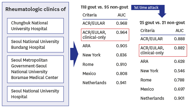
Patients with acute arthritis who underwent diagnostic and/or therapeutic arthrocentesis at any of the four participating rheumatology clinics were consecutively enrolled between February and December 2017. Those who were previously diagnosed and have been treated for arthritis were also allowed to participate if they present with acute exacerbation and need diagnostic arthrocentesis for differential diagnosis or therapeutic arthrocentesis. Polarizing microscopy examination of synovial fluid extracted from the affected joint or bursa was conducted by experienced rheumatologists and/or pathologists. Additional SF examinations, including white blood cell counts, Gram staining and cultures were performed for differential diagnosis.
The patients were stratified into the gout group (diagnosis established based on the confirmed presence of MSU crystals in the synovial fluid) and the non-gout group (diagnosis based on the clinical impression of certain non-gout disease and confirmed non-presence of MSU crystals in the synovial fluid). Patients having gout combined with non-gout arthritis were excluded from the study because their mixed clinical characteristics would have biased the clinical classification. Subgroup analyses were conducted in patients with the first episode of acute arthritis and in those with non-tophaceous disease.
Data collection
Data were collected using the predetermined questionnaire containing 12 items regarding the pattern of joint involvement (first metatarsophalangeal joint, ankle/midfoot, or other joints/polyarticular involvement), characteristics of the symptomatic episode (whether they have erythema overlying the affected joint, cannot tolerate touch or pressure to the affected joint or have great difficulty with walking or inability to use the affected joint), time course of the arthritis episodes (once or recurrent typical episodes), and clinical evidence of tophus (Supplementary Data 1, written in Korean), and the clinical record form (CRF) containing 12 items to review age, gender, clinical diagnosis, colchicine response, presence of tophus and synovial fluid culture/crystal exam, serum uric acid, radiograph, ultrasonography, and DECT data (Supplementary Data 2). The questionnaire and the CRF were compiled based on the six existing sets of gout classification criteria. The review of the medical records was done by the investigators themselves or by the fellow in the clinic (if there are any), who was also involved in the enrollment process. To avoid missing data for the analysis, the data abstraction tool (Supplementary Data 3, with instructions written in Korean) were also used.
Overviews of the ACR/EULAR gout classification criteria and other sets of gout classification criteria published to date are available in Supplementary Data 4 and 5, respectively. Since the presence of MSU crystals alone is sufficient to diagnose gout according to the ACR/EULAR, ARA, New York, and Mexico classification criteria, we considered that the results of the positive polarizing microscopy investigation were not available for the primary performance analysis. Therefore, the performance of “ACR/EULAR classification” was tested using clinical domain, serum uric acid in laboratory domain, and imaging domain (radiographic, ultrasound, or DECT imaging), while MSU results in laboratory domain scored as 0 (not done). The performance of the “ACR/EULAR—clinical only—classification” used clinical domain and serum uric acid, but not included MSU crystal results or imaging domain. In the Rome criteria, patients were considered to have gout only if at least two of three clinical criteria were satisfied (excluding the presence of MSU crystals).
Statistical analyses
Sample size was calculated using the formula n = Z2 × P(1−P)∕d2, with a 95% confidence level (Z = 1.96) and a margin of error of 5% (d = 0.05). Based on the ACR/EULAR classification criteria, the sensitivity of the clinical-only criteria was 0.85 (P = 0.85)10; therefore, a sample size of at least 49 patients was required for this study.
Categorical variables are described in terms of frequency (percentage), while data regarding continuous variables are presented as mean ± standard deviation. Comparisons between the gout and non-gout groups were performed using χ2 or Fisher's exact test for categorical variables and Student's t-test for continuous variables. The performance of each set of classification criteria was assessed in terms of sensitivity, specificity, positive predictive value, negative predictive value, and area under the receiver operating characteristic curve. Statistical analyses were performed using statistical software (MedCalc, version 18.6; MedCalc Software bvba, Ostend, Belgium). Graphs were visualized using GraphPad Prism, version 7.00, for Windows (GraphPad Software, La Jolla, CA, USA).
Ethics statement
The present study protocol was reviewed and approved by the Institutional Review Board (IRB) of the relevant hospitals (CBNUH IRB approval No., 2016-09-001; SNUH IRB approval No., 1611-055-807; SNUBH IRB approval No., B-1705/395-402; SMG-SNU Boramae Medical Center IRB approval No., 20170414/16-2017-62/051). Informed consent was submitted by all subjects when they were enrolled.
RESULTS
Baseline and clinical characteristics
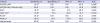
Among the 215 patients who underwent arthrocentesis, one patient having clinical rheumatoid arthritis combined with gout and another patient having spondyloarthropathy combined with gout were excluded. Finally, the study enrolled 118 patients with the clinical impression of gout and confirmed presence of MSU crystals in the synovial fluid (gout group), as well as 95 patients with a clinical impression of gout or non-gout disease but with absence of MSU crystals from the synovial fluid (non-gout group). The baseline demographics and clinical characteristics of the enrolled patients are summarized in Table 1. Conventional radiography data were available for all patients. However, ultrasonography was performed only in 35.2% of patients and DECT was not available in participating hospitals. Only 11.8% of patients had double-contour sign in the US study.
Table 1
Baseline demographics and clinical characteristics of acute arthritis patients stratified according to gout incidence
Data shown as incidence (yes/no or percentage) or mean ± standard deviation or number (%). A threshold score of ≥ 8 classifies an individual as having gout. P values are calculated by Student's t-test or χ2 test, as appropriate.
CPPD = calcium pyrophosphate dehydrate, MSU = monosodium urate, DECT = dual-energy computed tomography.
Overall performance of gout classification criteria: the 2015 ACR/EULAR criteria outperformed other gout classification criteria
The performance of the six sets of gout classification criteria was analyzed in the entire study sample (Fig. 1A and Table 2). In the primary analysis of performance, synovial fluid microscopy was considered not performed, to prevent circular reasoning. Among the six sets evaluated, the 2015 ACR/EULAR criteria showed the best performance, regardless of imaging results (Table 2). When synovial fluid microscopy results were added, the sensitivities of the ACR/EULAR, ARA, New York, and Mexico criteria improved to 100%, that of the Rome criteria improved to 97.5%, and that of the Netherlands criteria improved to 99.2%; additionally, the specificity of the ACR/EULAR criteria improved to 100% (Fig. 1A).
Fig. 1
Performance of different sets of gout classification criteria in Korean patients with acute arthritis. Receiver operating characteristic plots are shown. Box shows the results obtained when taking into consideration the positive findings on polarizing microscopy (sensitivity: 100% for the ACR/EULAR, ARA, New York, Rome, and Mexico criteria; 96.0% for the New York criteria). (A) Whole sample of patients (n = 213). The 2015 ACR/EULAR criteria showed high sensitivity and the best specificity among currently available sets of gout classification criteria. (B) Subgroup of patients with first-time arthritis (n = 46). The sensitivity of the 2015 ACR/EULAR scoring system was substantially lower in this subgroup than that noted in the overall sample, while specificity was preserved; a similar decrease in sensitivity was noted for the Netherlands criteria, whereas a more substantial decrease was noted for the ARA, New York, Rome, and Mexico criteria. (C) Subgroup of patients with non-tophaceous disease (n = 178). The diagnostic performance was preserved for all sets of existing criteria in this subgroup, except for the New York criteria.
ARA = American Rheumatism Association, ACR/EULAR = American College of Rheumatology/European League Against Rheumatism.
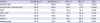
Table 2
Performance of existing sets of gout classification criteria
PPV = positive predictive value, NPV = negative predictive value, AUC = area under the receiver operating characteristic curve, ACR/EULAR = American College of Rheumatology/European League Against Rheumatism, ARA = American Rheumatism Association,
aWithout synovial fluid microscopy; dWithout synovial fluid microscopy or imaging; When taking into account the presence of monosodium urate crystals, the sensitivities of these classification criteria were b100%, e97.5%, or f99.2%, while the specificity was f100%.
Performance of gout classification criteria in subgroups: all gout classification criteria have lower sensitivity in patients with first-time arthritis
The performance of the six sets of gout classification criteria was analyzed in the subgroup of patients with first-time arthritis (Fig. 1B and Table 3) and in the subgroup of patients with non-tophaceous disease (Fig. 1C and Table 4). Once again, synovial fluid microscopy analysis was considered not performed for the primary performance analysis.
Table 3
Performance of gout classification criteria in 46 Korean patients with first-time arthritis
This analysis included 25 gout and 21 non-gout patients.
PPV = positive predictive value, NPV = negative predictive value, AUC = area under the receiver operating characteristic curve, ACR/EULAR = American College of Rheumatology/European League Against Rheumatism, ARA = American Rheumatism Association.
aWithout synovial fluid microscopy; cWithout synovial fluid microscopy or imaging; When taking into account the presence of monosodium urate crystals, the sensitivities of these classification criteria were b100%, d100%, or e96.0%.
Table 4
Performance of gout classification criteria in 178 Korean patients with non-tophaceous disease
This analysis included 83 patients with non-tophaceous gout and 95 non-gout patients.
PPV = positive predictive value, NPV = negative predictive value, AUC = area under the receiver operating characteristic curve, ACR/EULAR = American College of Rheumatology/European League Against Rheumatism, ARA = American Rheumatism Association.
aWithout synovial fluid microscopy; dWithout synovial fluid microscopy or imaging; When taking into account the presence of monosodium urate crystals, the sensitivities of these classification criteria were b100%, e96.4%, or f98.7%, while the specificity was c100%.
In patients with first-time arthritis (n = 46; gout prevalence, 54.3%), the sensitivity of the 2015 ACR/EULAR scoring system was substantially lower than that noted in the overall sample (52% vs. 80.5% for the full criteria without MSU results; 48% vs. 75.4% for the clinical-only criteria), while specificity was preserved; a similar decrease in sensitivity was noted for the Netherlands criteria, whereas a more substantial decrease was noted for the ARA, New York, Rome, and Mexico criteria (Table 3). When the synovial fluid microscopy results were added, the sensitivities of ACR/EULAR, ARA, New York, Rome, and Mexico criteria improved to 100%, while that of the Netherlands criteria improved to 96.0% (Fig. 1B). In patients with non-tophaceous disease (n = 178; gout prevalence, 46.6%), the diagnostic performance was preserved for all sets of existing criteria, except for the New York criteria (Fig. 1C and Table 4).
DISCUSSION
To our knowledge, the present investigation represents the first external validation of the 2015 ACR/EULAR gout classification criteria in Korean patients with acute arthritis. Furthermore, while validation studies for the ACR/EULAR criteria in other populations have been published,1112 our study was the first to conduct extensive analysis involving a direct comparison of the performance of the currently available gout classification criteria including the 2015 ACR/EULAR classification.
In this multicenter study enrolling over 200 Korean patients with acute arthritis, the performance of the 2015 ACR/EULAR classification criteria for gout was superior to that of the ARA, New York, Rome, Mexico, and Netherlands criteria, showing high specificity and fair sensitivity (98.5% and 80.5%, respectively) even when the synovial fluid microscopy results were not taken into consideration. These findings suggest that the 2015 ACR/EULAR classification criteria are useful for diagnosing gout even in the absence of information regarding the presence of MSU in the synovial fluid. However, we found decreased sensitivity for all six sets of gout classification criteria in the sub-group with first-time arthritis because the presence of recurrent arthritis is an important criterion in clinical diagnosis of gout, indicating that synovial fluid microscopy is recommended in such patients.
Although this may be due to the characteristics of the population participating in this study, all six existing criteria for gout showed a tendency to have lower sensitivity and higher specificity in this study, compared to previous validation studies in different population.1112 Since most of the criteria had a sufficient specificity in our study, the differences in the performance were mainly based on sensitivity. Considering the time sequence in which the diagnostic criteria were developed, it is interesting to see that the sensitivity of the diagnostic criteria gradually improved over time, from the 1963 Rome and 1966 New York criteria, with the improvement of 1977 ARA and 2010 Mexico criteria, and further improvement of the 2010 Netherlands and 2015 ACR/EULAR classification criteria. As the performance improves, the number of items of the diagnostic criteria has increased and the method of calculating scores has become increasingly complicated. However, with the exception of men gender and cardiovascular disease status in the Netherlands criteria, most items are duplicated in each clinical criterion.
Because ultrasonography or computed tomography (CT) was performed limitedly in this study, this study cannot reveal their role in the classification criteria, and this is the major limitation of this study. Second, this study was conducted at four rheumatology clinics affiliated with university hospitals (two tertiary and two secondary centers). Therefore, our study population likely included patients with more severe patterns of gout than usually seen in primary care, which represents another limitation of this study. Furthermore, the disease distribution in the non-gout group included in our study may differ from that normally encountered in the primary care setting (e.g., more than half of the non-gout patients had rheumatoid arthritis). On the contrary, all patients were clinically diagnosed (gout vs. non-gout arthritis) by experienced rheumatologists, which represents a major strength of the present study. Even when the arthrocentesis procedure is completed without complications13 and the microscopy examination is conducted by a rheumatologist and/or pathologist, there may be sampling errors, which can lead to the misclassification of MSU-negative gout patients as non-gout patients (i.e., false-negative result) if the gout diagnosis is established solely based on the presence of MSU crystals in the synovial fluid evaluated using polarizing microscopy.14 The likelihood of such an error is expected to be higher among patients with undifferentiated arthritis. The conclusions of the present study are unlikely to be biased by false-negative results associated with the inclusion of MSU-negative gout patients into the non-gout group because there were no cases of undifferentiated arthritis and because the diagnosis of non-gout arthritis in every patient allocated to the non-gout group was established by a rheumatologist.
In conclusion, our present findings suggest that the 2015 ACR/EULAR classification criteria serve as a useful diagnostic tool for gout among Korean patients with acute arthritis in the setting of specialized rheumatology clinics. This represents the first external validation of the 2015 ACR/EULAR classification criteria in this population, which was achieved using a prospective multicenter cohort covering various types of gout with mono-, oligo-, and polyarticular involvement, as well as various non-gout rheumatic diseases. Future research involving primary care clinics is warranted.
 XML Download