Abstract
Purpose
Croup is known to have epidemics in seasonal and biennial trends, and to be strongly associated with epidemics of para-influenza virus. However, seasonal and annual epidemics of croup have not been clearly reported in Korea. This study aimed to examine the seasonal/annual patterns and etiologies of childhood croup in Korea during a consecutive 6-year period.
Methods
Pediatric croup data were collected from 23 centers in Korea from 1 January 2010 to 31 December 2015. Electronic medical records, including multiplex reverse transcription polymerase chain reaction (RT-PCR) results, demographics and clinical information were cross-sectionally reviewed and analyzed.
Go to : 
References
1. Cherry JD. Clinical practice. Croup. N Engl J Med. 2008; 358:384–91.
3. Lee DR, Lee CH, Won YK, Suh DI, Roh EJ, Lee MH, et al. Clinical characteristics of children and adolescents with croup and epiglottitis who visited 146 Emergency Departments in Korea. Korean J Pediatr. 2015; 58:380–5.

4. McEniery J, Gillis J, Kilham H, Benjamin B. Review of intubation in severe laryngotracheobronchitis. Pediatrics. 1991; 87:847–53.

5. Denny FW, Murphy TF, Clyde WA Jr, Collier AM, Henderson FW. Croup: an 11-year study in a pediatric practice. Pediatrics. 1983; 71:871–6.

6. Segal AO, Crighton EJ, Moineddin R, Mamdani M, Upshur RE. Croup hospitalizations in Ontario: a 14-year time-series analysis. Pediatrics. 2005; 116:51–5.

7. Marx A, Török TJ, Holman RC, Clarke MJ, Anderson LJ. Pediatric hospitalizations for croup (laryngotracheobronchitis): biennial increases associated with human parainfluenza virus 1 epidemics. J Infect Dis. 1997; 176:1423–7.

8. Rosychuk RJ, Klassen TP, Voaklander DC, Senthilselvan A, Rowe BH. Seasonality patterns in croup presentations to emergency departments in Alberta, Canada: a time series analysis. Pediatr Emerg Care. 2011; 27:256–60.
9. Weinberg GA, Hall CB, Iwane MK, Poehling KA, Edwards KM, Griffin MR, et al. Parainfluenza virus infection of young children: estimates of the population-based burden of hospitalization. J Pediatr. 2009; 154:694–9.

10. Rihkanen H, Rönkkö E, Nieminen T, Komsi KL, Räty R, Saxen H, et al. Respiratory viruses in laryngeal croup of young children. J Pediatr. 2008; 152:661–5.

11. Wall SR, Wat D, Spiller OB, Gelder CM, Kotecha S, Doull IJ. The viral aetiology of croup and recurrent croup. Arch Dis Child. 2009; 94:359–60.

12. Kim EJ, Nam H, Sun YH, Tchah H, Ryoo E, Cho HK, et al. Comparison of etiology and clinical presentation between children with laryngotra-cheobronchopneumonitis and croup. Allergy Asthma Respir Dis. 2017; 5:274–9.

13. Jeon IS, Cho WJ, Lee J, Kim HM. Epidemiology and clinical severity of the hospitalized children with viral croup. Pediatr Infect Vaccine. 2018; 25:8–16.

14. Kim KH, Lee JH, Sun DS, Kim YB, Choi YJ, Park JS, et al. Detection and clinical manifestations of twelve respiratory viruses in hospitalized children with acute lower respiratory tract infections: Focus on human metapneumovirus, human rhinovirus and human coronavirus. Korean J Pediatr. 2008; 51:834–41.
15. Sung JY, Lee HJ, Eun BW, Kim SH, Lee SY, Lee JY, et al. Role of human coronavirus NL63 in hospitalized children with croup. Pediatr Infect Dis J. 2010; 29:822–6.

16. van der Hoek L, Sure K, Ihorst G, Stang A, Pyrc K, Jebbink MF, et al. Croup is associated with the novel coronavirus NL63. PLoS Med. 2005; 2:e240.

17. van der Hoek L, Pyrc K, Jebbink MF, Vermeulen-Oost W, Berkhout RJ, Wolthers KC, et al. Identification of a new human coronavirus. Nat Med. 2004; 10:368–73.

18. Chiu SS, Chan KH, Chu KW, Kwan SW, Guan Y, Poon LL, et al. Human coronavirus NL63 infection and other coronavirus infections in children hospitalized with acute respiratory disease in Hong Kong, China. Clin Infect Dis. 2005; 40:1721–9.

19. Han TH, Chung JY, Kim SW, Hwang ES. Human Coronavirus-NL63 infections in Korean children, 2004–2006. J Clin Virol. 2007; 38:27–31.

20. Wu PS, Chang LY, Berkhout B, van der Hoek L, Lu CY, Kao CL, et al. Clinical manifestations of human coronavirus NL63 infection in children in Taiwan. Eur J Pediatr. 2008; 167:75–80.

21. Zeng ZQ, Chen DH, Tan WP, Qiu SY, Xu D, Liang HX, et al. Epidemiology and clinical characteristics of human coronaviruses OC43, 229E, NL63, and HKU1: a study of hospitalized children with acute respiratory tract infection in Guangzhou, China. Eur J Clin Microbiol Infect Dis. 2018; 37:363–9.

22. Regamey N, Kaiser L, Roiha HL, Deffernez C, Kuehni CE, Latzin P, et al. Viral etiology of acute respiratory infections with cough in infancy: a community-based birth cohort study. Pediatr Infect Dis J. 2008; 27:100–5.
23. Camara AA, Silva JM, Ferriani VP, Tobias KR, Macedo IS, Padovani MA, et al. Risk factors for wheezing in a subtropical environment: role of respiratory viruses and allergen sensitization. J Allergy Clin Immunol. 2004; 113:551–7.
24. Manning A, Russell V, Eastick K, Leadbetter GH, Hallam N, Templeton K, et al. Epidemiological profile and clinical associations of human bocavirus and other human parvoviruses. J Infect Dis. 2006; 194:1283–90.

25. Gagliardi TB, Iwamoto MA, Paula FE, Proença-Modena JL, Saranzo AM, Criado MF, et al. Human bocavirus respiratory infections in children. Epidemiol Infect. 2009; 137:1032–6.

26. Jartti T, Lehtinen P, Vuorinen T, Koskenvuo M, Ruuskanen O. Persistence of rhinovirus and enterovirus RNA after acute respiratory illness in children. J Med Virol. 2004; 72:695–9.

27. von Linstow ML, Høgh M, Høgh B. Clinical and epidemiologic characteristics of human bocavirus in Danish infants: results from a prospective birth cohort study. Pediatr Infect Dis J. 2008; 27:897–902.
28. Williams JV, Edwards KM, Weinberg GA, Griffin MR, Hall CB, Zhu Y, et al. Population-based incidence of human metapneumovirus infection among hospitalized children. J Infect Dis. 2010; 201:1890–8.

30. Chapman RS, Henderson FW, Clyde WA Jr, Collier AM, Denny FW. The epidemiology of tracheobronchitis in pediatric practice. Am J Epidemiol. 1981; 114:786–97.
31. Waites KB, Atkinson TP. The role of Mycoplasma in upper respiratory infections. Curr Infect Dis Rep. 2009; 11:198–206.

Go to : 
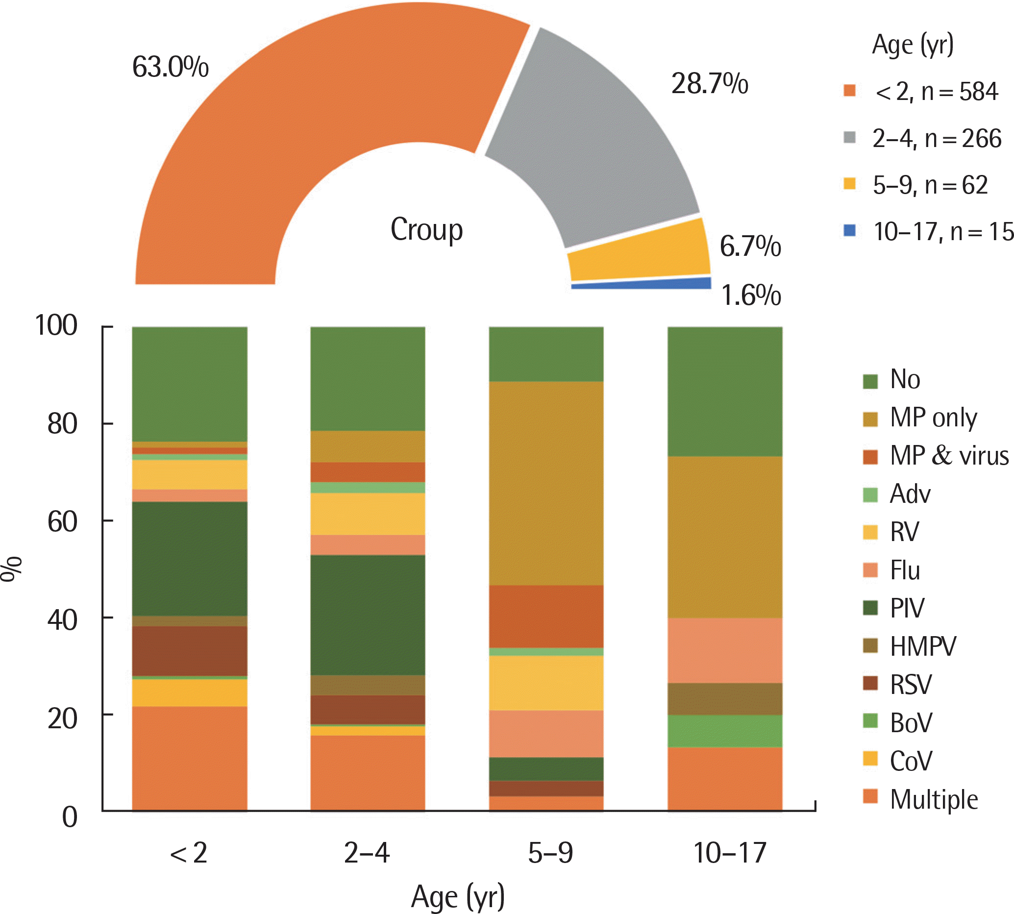
 | Fig. 1.Pathogens detected in hospitalized children with croup between 2010 and 2015. A total of 927 children with croup were hospitalized between 1 January 2010 through 31 December 2015. Among them, most prevalent age group was <2 years old (n=584, 63.0%). Most commonly detected pathogen was parainfluenza virus under the age of 4 years. MP, Mycoplasma pneumoniae; AdV, adenovirus; RV, human rhinovirus; Flu, influenza virus; PIV, parainfluenza virus; HMPV, human metapneumovirus; RSV, respiratory syncytial virus; BoV, bocavirus; CoV, coronavirus. |
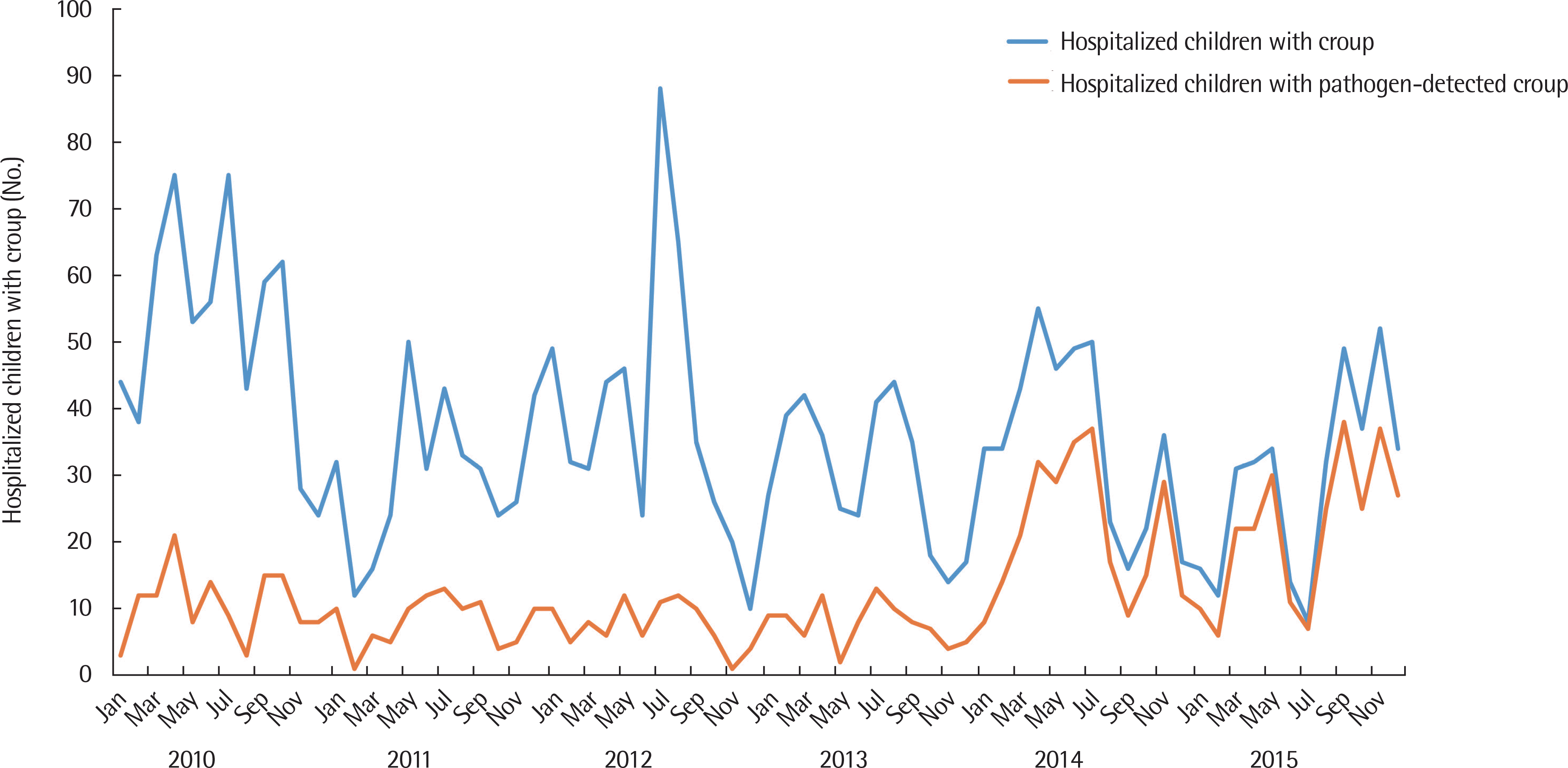
 | Fig. 2.Numbers of hospitalized children with croup and pathogen-detected croup between 1 January 2010 through 31 December 2015, according to month and year. A total of 2,598 hospitalized children with croup were identified. Among them respiratory pathogens were detected in 927 children. |
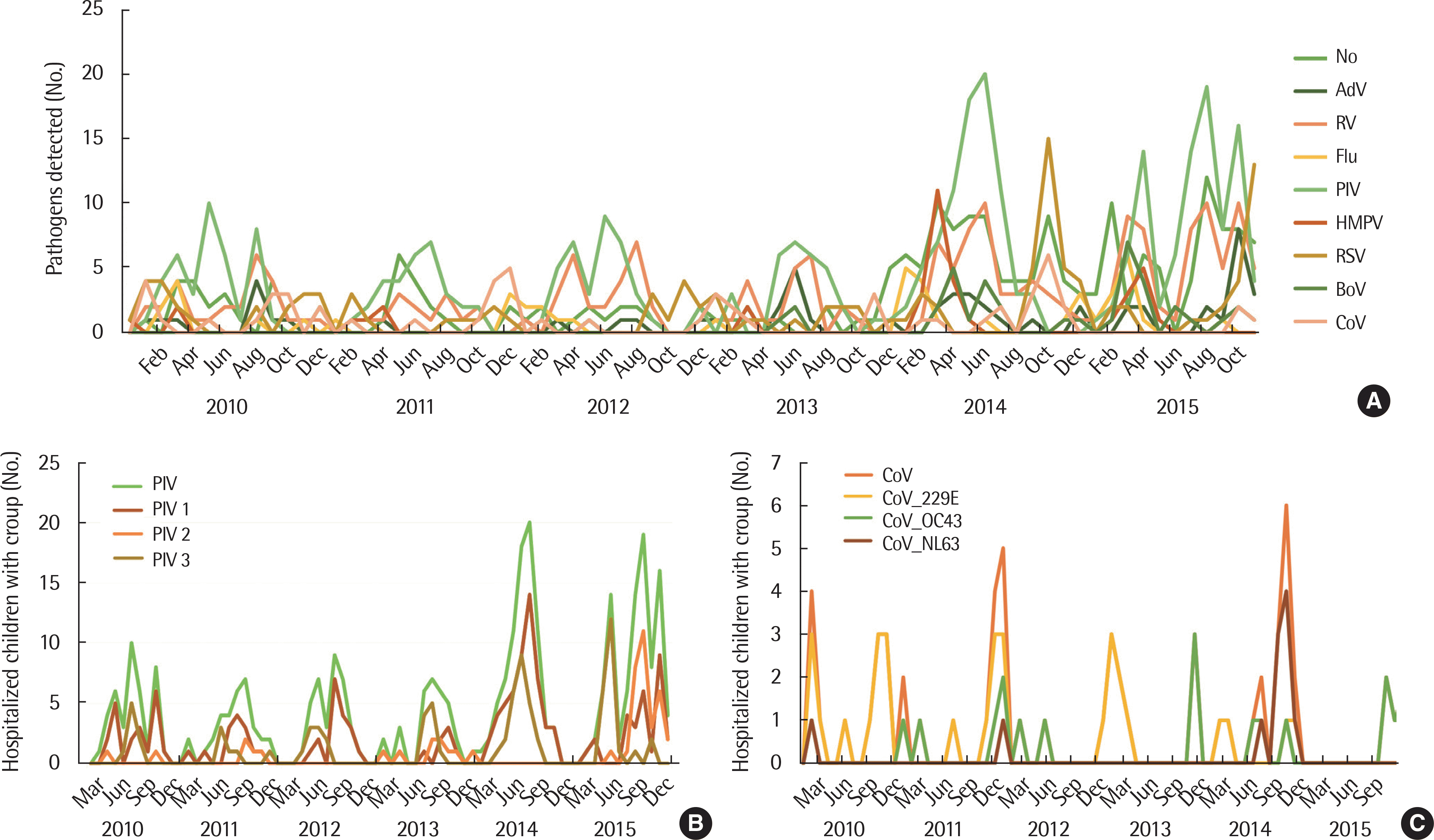
 | Fig. 3.Distribution of pathogen-detected croup between 1 January 2010 and 31 December 2015, according to month and year. (A) Pathogens detected in hospitalized children with croup. (B) Distribution of PIV, and PIV subtypes. (C) Distribution of CoV and CoV subtypes. AdV, adenovirus; HRV, human rhinovirus; Flu, influenza virus; PIV, parainfluenza virus; HMPV, human metapneumovirus; RSV, respiratory syncytial virus; BoV, bocavirus; HCoV, human coronavirus. |
Table 1.
Characteristics among 927 hospitalized childhood croup from 1 January 2010 to 31 December 2015
Table 2.
Respiratory pathogens and laboratory findings among 927 hospitalized childhood croup from 1 January 2010 to 31 December 2015
Table 3.
Comparison of clinical characteristics and laboratory findings among hospitalized children with croup due to single adenovirus, influenza virus, parainfluenza virus, respiratory syncytial virus, corona virus, and Mycoplasma pneumoniae infection
IQR, interquartile range; LOS, length of stay; AdV, adenovirus; Flu, influenza virus; PIV, parainfluenza virus; RSV, respiratory syncytial virus; CoV coronavirus; MP, Mycoplasma pneumoniae; ICU, intensive care unit; CRP, C-reactive protein; ESR, erythrocyte sedimentation rate; AST, aspartate aminotransferase; ALT, alanine aminotransferase; LDH, lactate dehydrogenase.


 PDF
PDF ePub
ePub Citation
 Print
Print


 XML Download