

 PDF
PDF ePub
ePub Citation
 Print
Print


Abstract
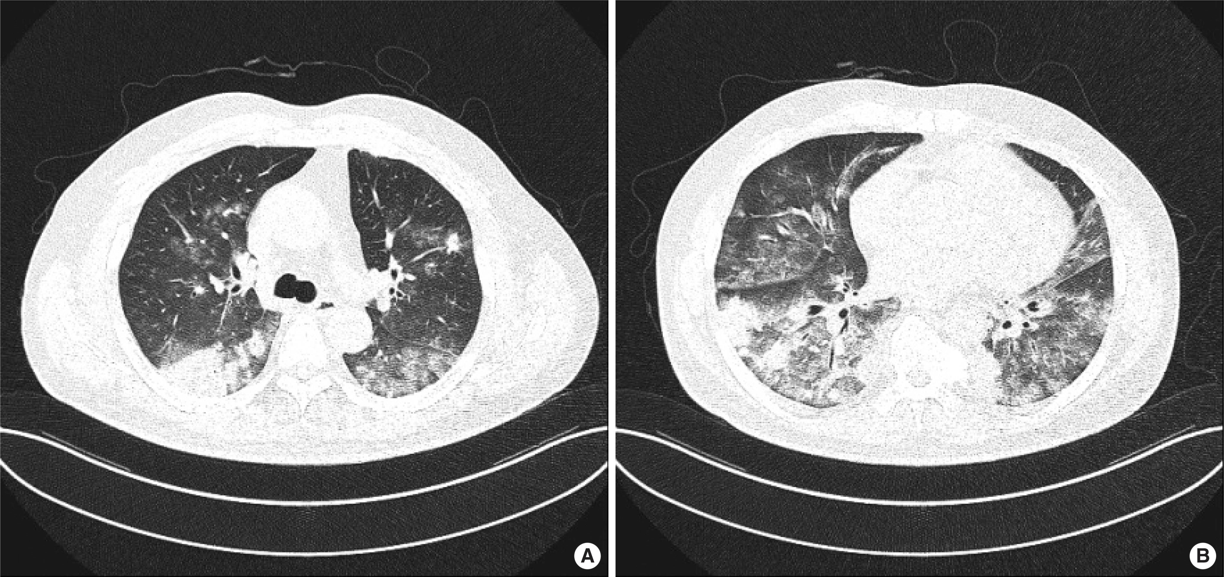
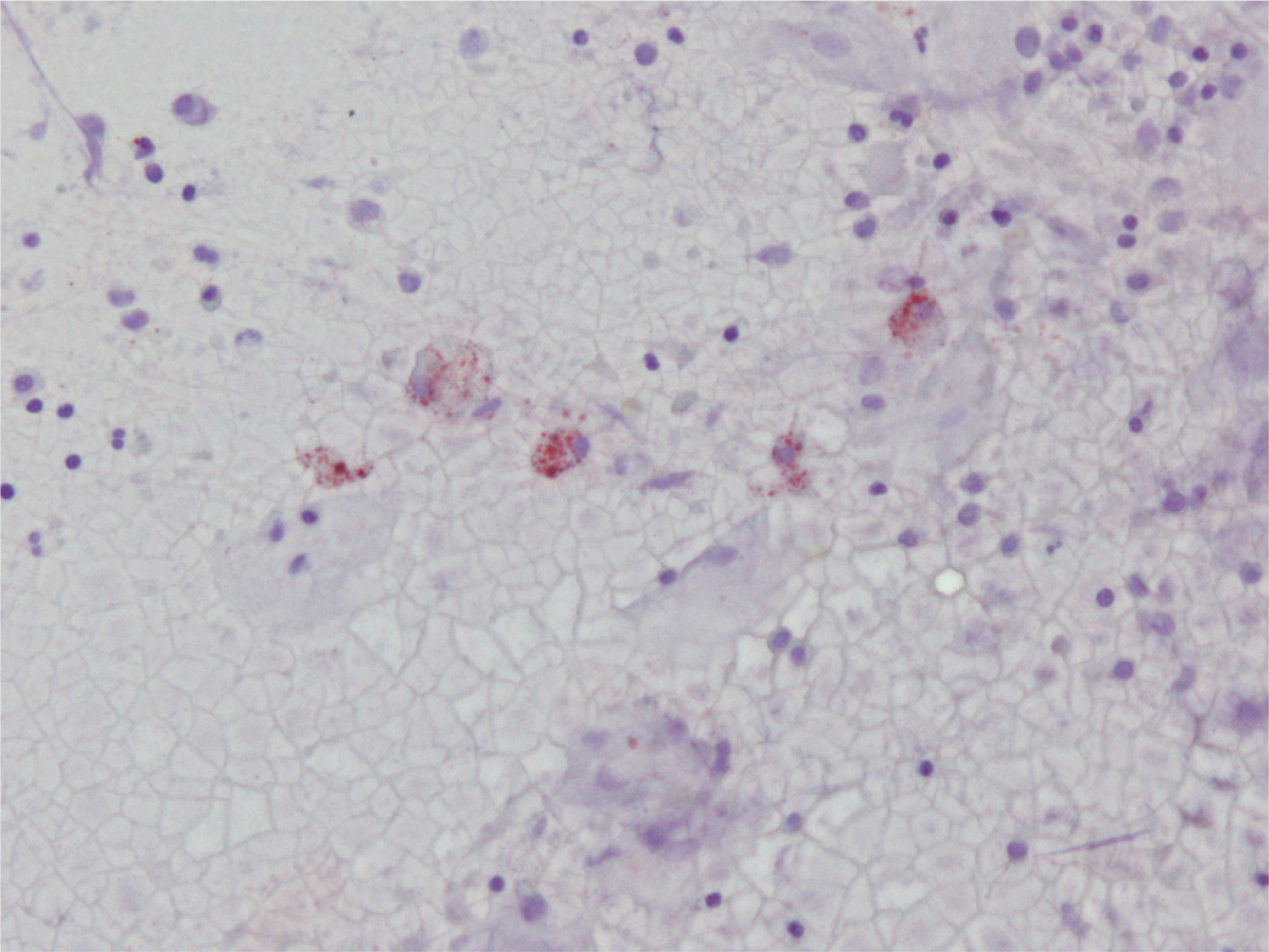
Exogenous lipoid pneumonia is an uncommon medical condition resulting from aspiration or inhalation of oily material. Generally, lipoid pneumonia has nonspecific clinical and radiological presentations, and may be misdiagnosed as bacterial pneumonia or lung cancer. We describe an unusual case of exogenous lipoid pneumonia accompanied by peripheral blood and pulmonary eosinophilia. A 63-year-old man was admitted with progressively worsening exertional dyspnea and productive cough for 5 days. A chest radiograph showed abnormalities in the lower lobe of the right lung, and a diagnosis of community-acquired pneumonia was made; intravenous antibiotics were administered. However, dyspnea and hypoxia gradually worsened and peripheral blood eosinophilia developed. A bronchoscopy was performed and bronchoalveolar lavage fluid analysis showed markedly increased numbers of eosinophils (40%). Subsequently, a comprehensive review of history revealed that he fell asleep with camellia oil in his mouth for 2 weeks to relieve foreign body sensation of the throat. Sputum and bronchoalveolar lavage fluid cytology showed the presence of lipid-laden macrophages. He was diagnosed with lipoid pneumonia and acute eosinophilic pneumonia. Chest radiograph and symptom were rapidly improved after treatment with intravenous methylprednisolone.
Go to : 
References
1. Marchiori E, Zanetti G, Mano CM, Hochhegger B. Exogenous lipoid pneumonia. Clinical and radiological manifestations. Respir Med. 2011; 105:659–66.

2. Gondouin A, Manzoni P, Ranfaing E, Brun J, Cadranel J, Sadoun D, et al. Exogenous lipid pneumonia: a retrospective multicentre study of 44 cases in France. Eur Respir J. 1996; 9:1463–9.
3. Park HP, Kwon KY, Choi WI. Lipoid pneumonia in Korea: a case report and review of the literature of Korean cases. Respir Med Extra. 2007; 3:39–43.
4. Hyun JG, Rhee CH. Clinical investigation of lipoid pneumonia in adults. Tuberc Respir Dis. 1996; 43:965–75.
5. Spickard A 3rd, Hirschmann JV. Exogenous lipoid pneumonia. Arch Intern Med. 1994; 154:686–92.
6. Felson B, Ralaisomay G. Carcinoma of the lung complicating lipoid pneumonia. AJR Am J Roentgenol. 1983; 141:901–7.
7. Betancourt SL, Martinez-Jimenez S, Rossi SE, Truong MT, Carrillo J, Eras-mus JJ. Lipoid pneumonia: spectrum of clinical and radiologic manifestations. AJR Am J Roentgenol. 2010; 194:103–9.
8. Baron SE, Haramati LB, Rivera VT. Radiological and clinical findings in acute and chronic exogenous lipoid pneumonia. J Thorac Imaging. 2003; 18:217–24.
9. Bréchot JM, Buy JN, Laaban JP, Rochemaure J. Computed tomography and magnetic resonance findings in lipoid pneumonia. Thorax. 1991; 46:738–9.
10. Gonçalves-de-Albuquerque CF, Silva AR, Burth P, de Moraes IM, Oliveira FM, Younes-Ibrahim M, et al. Oleic acid induces lung injury in mice through activation of the ERK pathway. Mediators Inflamm. 2012; 2012:956509.
11. Gonçalves-de-Albuquerque CF, Silva AR, Burth P, Castro-Faria MV, Castro-Faria-Neto HC. Acute respiratory distress syndrome: role of oleic acid-triggered lung injury and inflammation. Mediators Inflamm. 2015; 2015:260465.
12. Mirghani Z, Zein T, Annoble S, Winter J, Mostafa R. Analysis of fatty acids in ghee and olive oil and their probable causal effect in lipoid pneumonia. J Med Biochem. 2011; 30:141–7.
13. Midulla F, Strappini PM, Ascoli V, Villa MP, Indinnimeo L, Falasca C, et al. Bronchoalveolar lavage cell analysis in a child with chronic lipid pneumonia. Eur Respir J. 1998; 11:239–42.
14. Kuroyama M, Kagawa H, Kitada S, Maekura R, Mori M, Hirano H. Exogenous lipoid pneumonia caused by repeated sesame oil pulling: a report of two cases. BMC Pulm Med. 2015; 15:135.
15. De Giacomi F, Decker PA, Vassallo R, Ryu JH. Acute Eosinophilic Pneumonia: correlation of clinical characteristics with underlying cause. Chest. 2017; 152:379–85.
Go to :
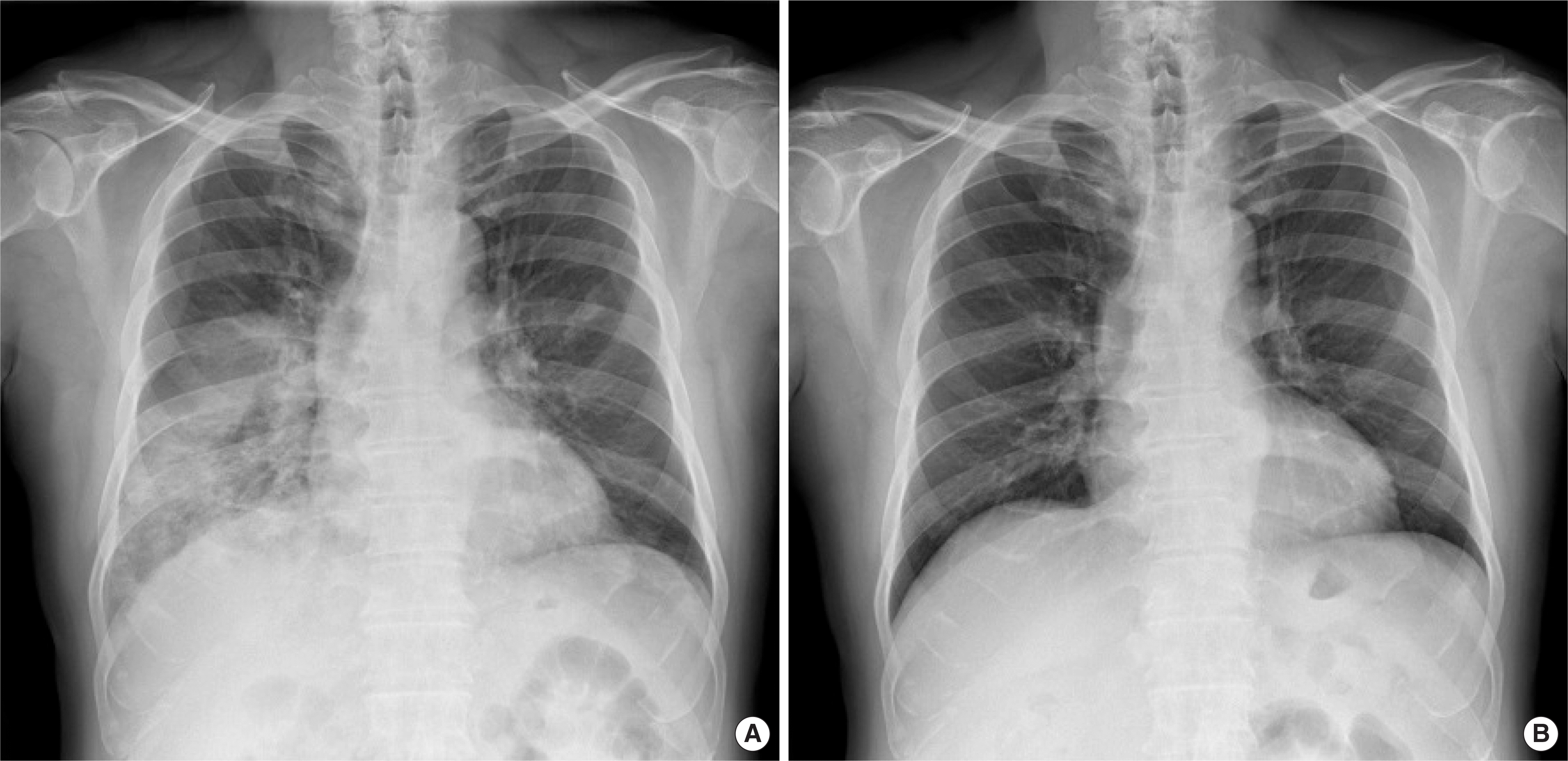 | Fig. 1.Chest X-ray of the patient showing patchy consolidation in both middle and lower lung fields (A), and almost complete resolution after 1 month (B). |
 XML Download