Journal List > Korean Circ J > v.49(6) > 1125478

Arsanjani, Agasthi, Ahn, Mookadam, and Yang: Metastatic Neuroendocrine Tumor with Cardiac Involvement Utilizing Multi-Modality Imaging
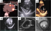
A 41-year-old female with history of benign carcinoid tumor status post nephrectomy presents with left upper quadrant abdominal pain. Computed tomography (CT) demonstrated a large retroperitoneal mass with biopsy revealing a low-grade neuroendocrine tumor. A Gallium-68 DOTATATE positron emission tomography (PET)/CT scan confirmed the retroperitoneal mass, as well as multifocal radiotracer uptake in the heart (Figure 1A). Transthoracic echocardiogram demonstrated a large echogenic mass along the inferolateral wall with minimal ultrasound contrast uptake (Figure 1B-1D). Cardiac magnetic resonance imaging (MRI) demonstrated multiple high T2 signal masses in the left and right ventricular wall with minimal gadolinium enhancement (Figure 1E and 1F). The patient subsequently underwent resection of the mass.
Myocardial metastases with neuroendocrine tumor are rare and most frequently manifest as carcinoid heart disease with right-sided valvular dysfunction.1) However, metastases can occur in other locations including intra-myocardial. Although, echocardiography is the imaging modality of choice for assessment of valvular heart disease, it may be less reliable for identification of smaller metastases. A multi-modality approach using cardiac MRI2) and Ga68-DOTATATE PET/CT imaging1) may allow for better detection of metastases.

Figures and Tables

Figure 1

Myocardial metastasis of neuroendocrine tumor. (A) A Gallium-68 DOTATATE PET/CT hybrid image demonstrating radiotracer uptake in the heart (arrows). (B) Transthoracic images demonstrating large echogenic mass (arrow) along the inferolateral wall of the LV. (C) Three-dimensional image confirming presence of large mass (arrow). (D) Contrast-enhanced echocardiographic image demonstrating minimal contrast uptake indicating minimal vascularity (arrow). (E) Turbo spin T2 sequence demonstrating multiple high T2 signal along the LV and RV wall correlating well with Gallium-68 DOTATATE PET images (arrows). (F) There is minimal late gadolinium enhancement on the cardiac MRI (arrow).

CT = computed tomography; LV = left ventricle; MRI = magnetic resonance imaging; PET = positron emission tomography; RV = right ventricular.
kcj-49-557-g001

Notes

Conflict of Interest The authors have no financial conflicts of interest.

Author Contributions

  • Conceptualization: Arsanjani R, Agasthi P, Ahn DH, Mookadam F, Yang M.

  • Formal analysis: Arsanjani R, Yang M.

  • Supervision: Arsanjani R, Ahn DH, Mookadam F.

  • Writing - original draft: Arsanjani R, Agasthi P.

  • Writing - review & editing: Arsanjani R, Agasthi P, Ahn DH, Mookadam F, Yang M.

References

1. Davar J, Connolly HM, Caplin ME, et al. Diagnosing and managing carcinoid heart disease in patients with neuroendocrine tumors: an expert statement. J Am Coll Cardiol. 2017; 69:1288–1304.
2. Bhattacharyya S, Toumpanakis C, Burke M, Taylor AM, Caplin ME, Davar J. Features of carcinoid heart disease identified by 2- and 3-dimensional echocardiography and cardiac MRI. Circ Cardiovasc Imaging. 2010; 3:103–111.
crossref
TOOLS
ORCID iDs

Reza Arsanjani
https://orcid.org/0000-0001-7081-4286

Pradyumna Agasthi
https://orcid.org/0000-0003-3067-6979

Daniel H. Ahn
https://orcid.org/0000-0002-0161-4018

Farouk Mookadam
https://orcid.org/0000-0002-9056-8956

Ming Yang
https://orcid.org/0000-0003-3001-7105

Similar articles