

 PDF
PDF ePub
ePub Citation
 Print
Print


The incidence of lower extremity defects has been increasing due to the rising number of traumatic injuries. The management of soft-tissue defect in the lower extremities is often challenging because of the few available local flap options in the area12. Thus, many reconstruction methods and protocols about lower extremity reconstruction have been introduced3. However, the choice of the right reconstruction method remains controversial4.
The cross-leg flap was the first considerable option for reconstructing leg defects after it was first described by Hamilton in 18745. However, the free flap with microsurgical technique has been the gold standard for lower extremity salvage since two decades ago6. The cross-leg flap is still being used in several centers and has proved useful7. Here, we present our experience in treating traumatic lower extremity injuries using the cross-leg flap.
CASE REPORT
1. Case 1
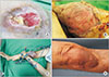
A 19-year-old male without any underlying disease presented with comminuted patellar fracture and right knee soft-tissue defect after a motorcycle accident (Fig. 1). A latissimus dorsi free flap was elevated and used to cover the defect. No appropriate vessels for anastomosis were identified during operation on the ipsilateral leg because of extensive trauma and scar tissue, so the elevated flap pedicle was anastomosed to the posterior tibial artery and vein of the contralateral leg. A split-thickness skin graft was used to cover the muscle flap. External fixation was applied for immobilization. A secondary operation for detachment was performed 22 days after pedicle training following the first operation.
Patellar osteomyelitis caused by open fracture and joint stiffness in the knee developed. Three years after surgery, no scar contracture developed and the knee range of motion was 0°–150°.
2. Case 2
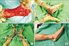
A 70-year-old male with diabetes presented with open fracture of the tibial-fibular shaft and degloving injury of the right lower leg after a motorcycle accident (Fig. 2). Internal fixation and primary repair were performed. Skin and soft-tissue necrosis occurred and were treated by debridement and negative-pressure wound therapy. Lower extremity reconstruction was performed 3 months after the trauma, when the area was infection-free.
The metal used to fix the fractured tibia and the tibia bone were exposed on the defect site. There were no suitable pedicles around the defect due to previous trauma and scar tissue. A posterior tibial artery flap from the contralateral leg was elevated. Casts, instead of external fixators, were used to fix the knees and ankles. A secondary operation for detachment was performed 23 days after pedicle training following the first operation.
The patient started physical therapy early after cast removal. Deep vein thrombosis did not occur, although osteomyelitis induced by the open fracture developed.
3. Case 3
A 7-year-old male without any underlying disease encountered a pedestrian accident and presented with soft-tissue defect and degloving injury on the ankle and heel without any fracture (Fig. 3). A posterior tibial artery flap was elevated from the contralateral leg and used to cover the defect where the calcaneus bone was exposed. A split-thickness skin graft was applied over the remaining granulated tissues. External fixation was performed for immobilization. He underwent a secondary operation for detachment 21 days after pedicle training following the cross-leg flap.
During hospitalization, the patient did not develop deep vein thrombosis, osteomyelitis, or flap necrosis. Although scar contracture could be a concern due to the patient's young age, he did not present with motion limitation or contracture 7 years after the reconstruction.
DISCUSSION
Lower extremity defects hardly heal by secondary intention, so in the past, flaps were developed from other parts of the body. Since then, various reconstruction methods have been introduced, aiming to achieve form and function at the recipient site, avoid donor site deformity, and provide safety throughout the process. Recently, microvascular surgery has been the gold standard for lower extremity salvage. However, in patients with severe blood flow insufficiency following damage of major arteries, the use of local pedicle flaps and free flaps has limitations8. In such cases, the cross-leg flap reemerges as a likely option. In this study, we witnessed success with using the cross-leg flap and thus consider it to be still a useful procedure.
The main disadvantages of the cross-leg flap are joint stiffness, deep vein thrombosis, and osteomyelitis due to external fixation or immobilization. However, early rehabilitation treatment and early detachment can minimalize such complications79. In our cases, no severe complications were found. Currently, flap detachment can be performed within 14–21 days1. In our cases, detachment was performed at 22 days on average. Although external fixators can be used to immobilize the legs, its use may not be required. In case 2, the legs were fixed with casts (Fig. 2E) as the positioning was not complicated. Two cases had osteomyelitis, but it occurred due to open fracture, not external fixation.
Severe lower extremity trauma can involve multiple vessel injuries and surrounding tissue damage. Known indications of cross-leg flap include patients with two-vessel injury in the lower extremity7. Additionally, the presence of damaged tissue and scar tissue around the defect makes it difficult for surgeons to visualize recipient vessels and make appropriate local flap advancement. Using the cross-leg flap, surgeons can avoid performing more incisions on the already traumatized and compromised limb and can improve reach of the flap9. The procedure may increase chances of success as it utilizes healthy recipient vessels outside the zone of injury9. In case 1, a pedicle of the latissimus dorsi free flap was anastomosed to the contralateral posterior tibial artery and vein, which were not injured. Using a contralateral leg vessel not only provides adequate blood supply to the flap but also shortens the time required to find a recipient vessel.
Cross-leg flaps can be best suited for young trauma patients who can tolerate complex operations and a long hospital course and can start a fast-paced physical therapy. In case 2, however, the patient was old and had diabetes. He underwent the cross-leg flap because of lack of suitable recipient vessels on the ipsilateral leg. Nonetheless, he did not have complications such as joint stiffness and deep vein thrombosis. The cross-leg flap has been also suggested as an alternate option for lower extremity salvage when no other options are available9.
Cross-leg flaps have no specific indications so far; however, they have been used in young patients under the age of 50 years and in patients with two-vessel injuries in the lower limb. In our report, all the cases showed intact vessel findings on conventional angiography or computed tomography angiography before surgery. During the operation, however, perforator vessels for local flap and recipient vessels for free flap, which were checked with Doppler sonography and directly by sight, were unreliable. All three cases had severe trauma and broad scar tissue around the defect. We recommend that cross-leg flaps be considered if the surrounding tissue has undergone severe trauma or the defect is surrounded by broad scar tissue.
This report has some limitations. As microvascular surgery is the gold standard treatment of lower limb salvage, there were only a few cases in which cross-leg flap could be performed. There were too few cases to derive statistical results. In addition, we did not make comparisons between cross-leg flaps and free flaps in our department. Additional studies are needed after obtaining sufficient numbers of cases.
Cross-leg flap can be considered not only in cases of multiple vessel injuries or when no other options are available but also in cases of broad trauma or where scar tissue is present around the defect. Cross-leg flap is a useful method because it provides not only a healthy and undamaged flap but also reliable recipient vessels for free flap. With the high success rate of the cross-leg flap procedure in our hospital, we contend that this procedure can still be a useful option.

 XML Download