

 PDF
PDF Citation
 Print
Print


INTRODUCTION
Gastro-esophageal reflux disease (GERD), defined as the abnormal reflux of gastric contents into the esophagus, is a chronic disease that results in reflux symptoms and complications, such as reflux esophagitis (RE) or Barrett's esopahgus.12 In recent decades, there has been a significant rise in the prevalence of GERD worldwide including Korea.3 The prevalence of RE was 3.4% in 1997,34 but increased to 6.2% in 2008,5 while that of symptomatic GERD was 3.5% in 2001,6 but increased to 7.1% in 2007 in Korea.7 Consequently, the clinical significance and risk factors for RE and symptomatic GERD are gaining importance.
Several studies reported that the common risk factors for RE and GERD are age,5 obesity,89 hiatal hernia,10 alcohol drinking,11 and smoking.1112 In addition, gender-related factors might contribute in the pathogenesis and development of RE and symptomatic GERD.1314 The studies of risk factors for RE and symptomatic GERD according to gender are still rare. Investigation of gender-related differences in RE and symptomatic GERD is important for providing better understanding of these pathogenesis, prevention and treatment for both men and women patients.1314 The study aimed to evaluate the gender-related differences in the prevalence and risk factors for RE and symptomatic GERD.
METHODS
Study participants
This study was retrospectively conducted in a large cohort of apparently healthy Korean men and women who underwent a comprehensive annual or biennial health examination at Ewha Womans University Mokdong Hospital from January 2015 to December 2016. In Korea, the Industrial Safety and Health Act stipulates that medical checkups are provided free of charge to all employees annually or every two years. In the case of having health examination twice during this study period, the former examination was considered here. Two-third of the participants from the health promotion center were employees of several companies and their spouses. The rest of them took a health screening at their own expense.
Among a total of 14,104 individuals, 10,680 participants (75.7%), who responded to the questionnaire and underwent upper endoscopy screening, were enrolled. Of them, 10,158 participants were finally eligible after exclusion of those with prior major abdominal surgery (n = 33), gastric cancer (n = 8), and active peptic ulcer disease (n = 481). We measured the levels of high density lipoprotein-cholesterol (HDL-C), hypertriglyceride, and fasting venous glucose with the Hitachi 7600 analyzer (Hitachi Ltd., Tokyo, Japan). According to the National Cholesterol Education Program Adult Treatment Panel III criteria, low HDL-C levels were defined as less than 40 mg/dL in men and less than 50 mg/dL in women, and hypertriglyceridemia was defined as triglyceride levels greater than 150 mg/dL. Weight (to the nearest 0.1 kg), height (to the nearest 0.1 cm), waist circumference (WC) (to the nearest 0.1 cm), and blood pressure were measured by well-trained nurses. Based on the modified criteria of Asian-Pacific guidelines of the World Health Organization, the participants were categorized according to the body mass index (BMI) as follows: normal (< 23 kg/m2), overweight (23–24.9 kg/m2) and obese (≥ 25 kg/m2). The WC was measured at the midpoint between the lower border of rib cage and iliac crest. Central obesity was identified as a WC greater than 90 cm in men and that WC greater than 85 cm in women, according to the clinical practice guidelines for overweight and obesity in Korean Society for the Study of Obesity.15 Diabetes mellitus (DM) was determined based on the diagnostic criteria of the American Diabetes Association or self-reported history of DM. Hypertension (HTN) was defined as a mean systolic blood pressure greater than 140 mmHg, a mean diastolic blood pressure greater than 90 mmHg, or a self-reported history of HTN.
Endoscopic examinations and definition of RE
Upper endoscopy was performed by experienced certified gastroenterologists. The presence and severity of RE were classified by the Los Angeles (LA) classification as grade A–D (LA-A to D) and were based on the longest length of a mucosal break and the confluence of erosions.16 Hiatal hernia was defined as a circular extension of the gastric mucosa 2 cm or more above the diaphragmatic indentation from the endoscopic findings.
Questionnaires and definition of symptomatic GERD
Symptomatic GERD was defined as heartburn or acid regurgitation at least twice per week for the past 6 months or interference with daily life within the past year.117 Korean Rome III questionnaires, which reveled good reliability and satisfactory construct validity, was used to evaluate symptomatic GERD.18 The Cronbach's alpha for heartburn was 0.82 and that for acid regurgitation was 0.84.1819 Extra-esophageal symptoms such as dysphagia, chronic cough, chest pain, and hoarseness were not considered as symptomatic GERD because of limited evidence of causal relationship with reflux.1 The participants were also instructed to complete structured questionnaires for somatization symptom checklist (SSC) scores.20 These SSC scores include 17 non-gastrointestinal (GI) symptoms or illnesses. The participants were asked to indicate how troublesome each symptom was (intensity on a scale of 0 to 4, indicating no problem to extremely troublesome) and how frequent each symptom occurred (frequency on a scale of 0 to 4, indicating no problem to daily occurrence) during the past year using the five-Likert scale. These scores indicate the degree of overall psychosomatic distress.2021 The minimum score possible was 0 and the maximum was 272. In measuring health-related quality of life (HRQoL), the Korean version of the EuroQol-5 Dimension (EQ-5D) was used.22 The EQ-5D records the degree of self-reported problems on five dimensions as follows; mobility, self-care, usual activities, pain/discomfort, anxiety/depression. Previous study has demonstrated its cross-cultural adaptation and validation on upper GI disorders and HRQoL.21
Questionnaires and lifestyle habits
Each participant completed a structured questionnaire which included demographic factors, medical history, lifestyle habits (smoking and alcohol drinking), and status of menopause (pre- vs. post-menopause). Smoking was categorized as non-smoking, past smoking and current smoking, with the latter defined as having regularly smoked more than 1.5 pack-years since the past year. Alcohol drinking was classified into none and excessive alcohol drinking, with the latter defined as more than 14 standard drinks per week in men and more than 7 standard drinks per week in women during the previous 6 months. A standard drink was defined as a cup of alcohol containing 12 grams, regardless of types of alcohol.
Statistical analysis
Differences in variables between men and women were analyzed by using the Student's t-test or Mann-Whitney U test and χ2 test. Student's t-test was used to compare continuous variables between two groups, and the χ2 test to compare categorical variables. Because HDL-C, triglyceride, and WC were skewed data, median (interquartile range) and non-parametric tests (Mann-Whitney U test) were used to compare between two groups. Multiple logistic regression was performed for assessing predictor variables selected from the univariate analyses. The odds ratios (OR) and 95% confidence intervals (CI) were computed from the estimated coefficients in the regression. The statistical analyses were carried out using SPSS version 21.0 for Windows software (SPSS Inc., Chicago, IL, USA). A two tailed P value of less than 0.05 was considered statistically significant.
Ethics statement
This study was approved by the Institutional Review Board (IRB) of Ewha University Mokdong Hospital (IRB No. 2018-08-026-002). The requirement for written informed consent was waved because we were using anonymous data and questionnaires collected regularly during the health checkup process. This study followed the principles of the Declaration of Helsinki.
RESULTS
Participant characteristics
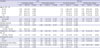
A total of 10,158 participants were enrolled in this study (Table 1). Their mean age was 47.1 ± 10.2 years and 50.9% of them were men. The overall prevalence of RE was 6.4% (651/10,158); 4.5% (457/10,158) in LA-A, 1.8% (182/10,158) in LA-B, and 0.1% (12/10,158) in LA-C. Compared to women, the incidence of obesity, DM, HTN, and hypertriglyceridemia were more common in men. Also, the lifestyle of smoking and excessive alcohol drinking was more frequent in men (Table 1). Among women participants, 1,709 of them (34.2%) had the menopause status, excluding 319 (6.4%) due to missing data. The incidence of obesity, DM, HTN, and hypertriglyceridemia in women participants with menopause were significantly higher in those with pre-menopause. Frequency of smoking and excessive alcohol drinking in pre-menopause women was also more common than those with menopause (Supplementary Table 1).
Table 1
Clinical characteristics of participants
Data are shown as number (%), mean ± standard deviation or median (IQRs).
BMI = body mass index, HDL-C = high density lipoprotein-cholesterol, IQR = interquartile range, M = men, W = women, SSC = somatization symptom checklist, LA = Los Angeles, GERD = gastro-esophageal reflux disease, NA = not available.
![]()
Prevalence of RE
The prevalence of RE in men was significantly higher than that in women (10.6% vs. 2.0%, P < 0.001) (Table 1). In women, its prevalence increased with age, but there were no significant differences with age in men. Below the age of 70, the prevalence of RE in men was significantly higher than that in women; however, above the age of 70, this difference disappeared and the prevalence became similar (Fig. 1A).
Clinical presentation of RE and symptomatic GERD according to menopause
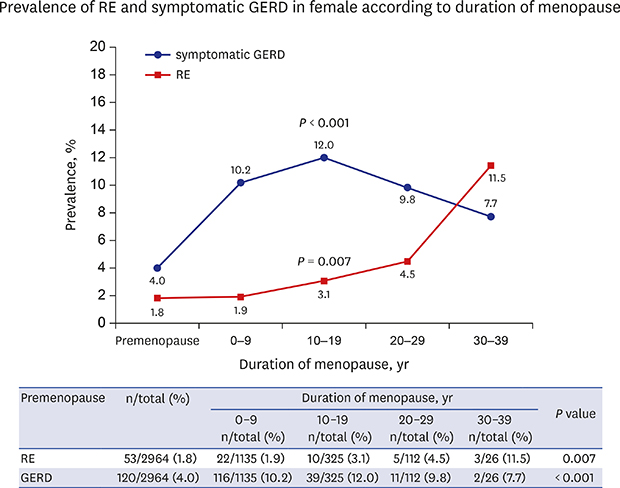
In women, the spectrum in GERD varied depending on the duration of menopause. The prevalence of RE and symptomatic GERD increased with age in women (Fig. 1A and B), particularly after menopause. During the menopausal period, the prevalence of RE was significantly increased as the duration of menopause stratified by decade became longer (P = 0.007) (Fig. 2). According to BMI adjusted logistic analysis, over 30 years of menopause was a significant risk factor in the occurrence of RE (OR, 5.29; 95% CI, 1.52–18.48; P = 0.009) (Table 2). In contrast, the prevalence of symptomatic GERD increased during the first 20 years after menopause, but tended to decrease thereafter (P < 0.001) (Fig. 2).
 | Fig. 2Prevalence of RE and symptomatic GERD with BMI according to duration of menopause. Blue line indicates symptomatic GERD, red line indicates RE, and dotted green line indicates BMI.RE = reflux esophagitis, GERD = gastro-esophageal reflux disease, BMI = body mass index.
|
Table 2
Prevalence of reflux esophagitis according to duration of menopause and adjustment for BMI

![]()
Comparison of risk factors for RE between men and women participants
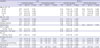
According to the univariate analysis in men, being overweight, obesity, HTN, hypertriglyceridemia, central obesity (over 90 cm of WC), past smoking, current smoking, excessive alcohol consumption, and hiatal hernia were significant risk factors for RE (Table 3); however, age was not significant. Multivariate analysis in men revealed that being overweight (OR, 1.57; 95% CI, 1.18–2.09; P = 0.002), obesity (OR, 1.81; 95% CI, 1.34–2.44; P < 0.001), hypertriglyceridemia (OR, 1.28; 95% CI, 1.06–1.55; P = 0.012), current smoking (OR, 1.67; 95% CI, 1.30–2.16; P < 0.001), and hiatal hernia (OR, 2.54; 95% CI, 1.81–3.62; P < 0.001) were all independent risk factors for RE (Table 3). By univariate analysis in women, over 70 years of age, obesity, hypertriglyceride, central obesity (over 85 cm of WC), current smoking, and hiatal hernia were significant risk factors for RE (Table 3). Multivariate analysis in women revealed that obesity and smoking were significantly independent risk factors and their ORs were about twice as high as that of men (OR, 3.17; 95% CI, 1.79–5.61; P < 0.001; OR, 3.47; 95% CI, 1.61–7.48; P = 0.001, respectively). Unlike the men participants, over 70 years of age was the moderate risk factor for RE in women (OR, 2.90; 95% CI, 1.19–7.04; P = 0.019) (Table 3).
Table 3
Comparison of risk factors for reflux esophagitis between men and women
OR = odds ratio, CI = confidence interval, BMI = body mass index, HDL-C = high density lipoprotein-cholesterol, M = men, W = women, WC = waist circumference, SSC = somatization symptom checklist.
![]()
Comparison of risk factors for symptomatic GERD between men and women
The univariate analysis revealed that current smoking, excessive alcohol drinking, and SSC scores were related to symptomatic GERD in men. By multivariate analysis, SSC scores were revealed as the scores which increased by one point, OR increased by 1.03 (95% CI, 1.02–1.04; P < 0.001). Excessive alcohol drinking (OR, 1.84; 95% CI, 1.26–2.70; P = 0.002) was also a significant risk factor for symptomatic GERD in men (Table 4). However, unlike RE, obesity, or other metabolic components were not associated with symptomatic GERD in men.
Table 4
Comparison of risk factors for symptomatic gastro-esophageal reflux disease between men and women
OR = odds ratio, CI = confidence interval, BMI = body mass index, HDL-C = high density lipoprotein-cholesterol, M = men, W = women, WC = waist circumference, SSC = somatization symptom checklist.
![]()
In univariate analysis, age, HTN, current smoking, excessive alcohol drinking, and SSC scores were significant risk factors for symptomatic GERD in women. Multivariate analysis showed that 50-59 and 60-69 years of age (OR, 1.79; 95% CI, 1.24–2.60; P = 0.002 and OR, 2.16; 95% CI, 1.29–3.63; P = 0.004, respectively) and SSC scores (OR, 1.02; 95% CI, 1.02–1.03; P < 0.001) were independent risk factors for symptomatic GERD in women (Table 4). Meanwhile, obesity and current smoking were not independent risk factors for symptomatic GERD, compared to that of RE.
Health related quality of life of GERD according to gender
The general HRQoL in woman participants was significantly lower than that in man participants. EQ5D index was 0.93 ± 0.04 in men and 0.92 ± 0.06 in woman participants, respectively (P < 0.001). EQ-5D index decreased with age for both man and woman participants (Fig. 3A). There was no significant difference of EQ-5D index according to the presence of RE in both man and woman participants (Fig. 3B). However, EQ-5D index in symptomatic GERD participants of both genders was lower than that in normal participants. EQ-5D index showed a sharp decline after 40 years of age in woman participants with symptomatic GERD, while it declined sharply after 50 years of age in man participants with symptomatic GERD (Fig. 3C).
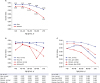 | Fig. 3Presentation of Korean version of the EQ-5D by gender and age. (A) Total participants. (B) Participants with RE. (C) Participants with Sym-GERD. Blue line indicates man and red line indicates woman participants.EQ-5D = EuroQol-5 dimension, RE = reflux esophagitis, Sym-GERD = symptomatic gastro-esophageal reflux disease, GERD = gastro-esophageal reflux disease.
|
DISCUSSION
The present study shows dynamic clinical manifestations and different risk factors for GERD according to gender. RE shows a predominance in men of about 5:1 ratio between men and women, but this difference disappeared above the age of 70. Meanwhile, symptomatic GERD exhibits a woman dominance and this difference becomes more apparent at the peri-menopausal period. GERD is the spectrum of various manifestations caused by gastro-esophageal reflux, and RE does not necessarily match the symptom as an indication of damage to the esophageal mucosa caused by backflow.1 In this study, symptomatic GERD among RE and RE among symptomatic GERD were only 5.2% and 7.8%, respectively, and the association between the two diseases was low.
Aging, especially over 70 years of age, was a woman-specific risk factor for RE. Commonly, women gain more weight after menopause because hormonal change substantially contributes to central fat deposition and obesity.23 Deleterious changes in adipokines occur with estrogen decrease at menopause, which could influence on the fatty acid metabolism and increase visceral adiposity.24 The pathophysiology of central fat deposition was also supported by previous studies in animal models. Ovariectomized mice showed adipocyte hypertrophy, adipose tissue inflammation and the development of hepatic steatosis with decreased energy expenditure.25 In fact, BMI is a well-known risk factor for RE.810 Increased BMI during menopause might affect the rise of the prevalence of RE; hence, we conducted BMI adjusted analysis in this study. As a result, longer duration of menopause still revealed stronger association with the development of RE, despite adjustment for BMI. Thus, a long duration of menopause which is related to marked reduction of estrogen level, might contribute to the pathogenesis of RE later in life.
Recent studies in experimental animal models have manifested that 17β-estradiol, one form of estrogen, may play a protective role in increasing esophageal mucosal resistance by expression of tight junction proteins, such as occludin.26 Furthermore, 17β-estradiol inhibits the transcription factor p65/relA which promotes the expression of pro-inflammatory mediators and inflammation.27 This anti-inflammatory activity of estrogen might contribute to mucosal resistance to acid. Therefore, we suggest that the development of RE in women aged over 70 years could be caused by the long duration of low estrogen level. Similarly, previous studies revealed that the roles of estrogen in animal models of GI tracts were associated with colonic epithelial permeability and mucosal immunity, affecting inflammatory bowel disease.28 Unfortunately, research on how long the low level of estrogen contribute to increase the development of RE has not been well established to date. The present study shows that the protective effects of estrogen on esophageal mucosa might be considerably decreased when the decline in estrogen level lasts for over 30 years during menopause, not at the beginning or transition of menopause.
Previous meta-analysis of 28 studies for RE showed a pooled men/women prevalence ratio of 1.57/1 (95% CI, 1.40; 1.76/1).29 Most studies included in this meta-analysis had estimated and compared the men/women ratio of RE in patients with symptomatic GERD. In the present study, however, the majority was free of symptoms and examined for regular health examination. Under the similar background of study participants who underwent regular medical check-up, several recent studies reported that the man predominance in RE has been evident, ranging from 3.1/1–4.1/1, consistent with our results.3031 They suggest that there is a gender related difference in the influence of eating habits and lifestyle factors such as high caloric intake, alcohol drinking or current smoking on the prevalence of RE.
Symptomatic GERD, which is mainly diagnosed on the basis of the presence of heartburn or acid regurgitation, presents different clinical features and risk factors from RE.67 Several previous studies presented that difference in gender distribution exists in symptomatic GERD.3233 Although there is not a significantly big difference between both genders, women have a relatively high prevalence of symptomatic GERD. However, we revealed that prevalence of symptomatic GERD in women was significantly higher compared to men in all age groups. We suggest the possibility for this discrepancy. Symptomatic GERD is diagnosed using different questionnaire methodology for each study, for instance methods of survey sampling (self-paid medical checkup, stomach cancer screening program, or random sampling), symptom definition (once to twice per week reflux symptoms or composite scores), and data acquisition (interview or self-report questionnaires).34 Therefore, the definition of GERD based on different methodology remains a limitation for most observational studies, which may affect determination of the prevalence of GERD.2134
With regard to the association between BMI and symptomatic GERD, previous studies have been controversial; some studies have noted that higher BMI is associated with symptomatic GERD,910 whereas other studies have shown negative relations between BMI and symptomatic GERD.35 The present study shows no association between obesity and symptomatic GERD.
SSC scores, which indicate the degree of psychosomatic complaints,20 is a significantly independent predictor for symptomatic GERD in both genders. Psychosomatic complaints induce each individual to lower their threshold values and increase the perception of GERD symptoms with the change of biological responses.36 Similarly, EQ-5D index tends to decrease with age in both genders. However, women aged over 50 years show sharper decline of EQ-5D index than men in symptomatic GERD. This can be interpreted as resulting primarily from the profound decrease in estrogen around menopause, based on gender difference. The median age of menopause is 51 and decline in estrogen level is maintained thereafter. The hormonal transition around menopause, especially estrogen, substantially contribute to increased menopausal symptoms including hot flushes, sleep disturbance, weight gain, mood symptoms, and somatic complaints.23 Although acid reflux is a major factor contributing to GERD symptoms, the increase in acid perception is not associated with the presence or absence of esophageal mucosal damage. These findings suggest various factors including non-acid reflux component (such as bile, pepsin or gas), increased mucosal permeability, central sensitization of spinal sensory neurons, and up-regulation of peripheral pain receptors (such as nociceptor transient receptor potential vanilloid type 1 [TRPV1]) could contribute greater to symptom perception in patients with non-erosive reflux disease.37 Regardless of pathologic acid reflux, multiple predisposing factors are associated with triggering reflux symptoms. Further studies toward cellular, molecular, and environmental factors should be required to understand the mechanisms of reflux perception in patients with non-erosive reflux disease.
On the other hand, the prevalence of symptomatic GERD tends to dramatically increase within 20 years of menopause, but that tends to decrease after 20 years of menopause. A recent report showed that menopausal symptoms could persist for an average of 10 years or longer.38 This might explain that menopause-induced symptoms are closely related to a high prevalence of symptomatic GERD within 20 years of menopause. In case of cardiovascular disease (CVD), there were similar results to this study.3940 The transition of menopause induces the vasomotor hot flushes. Particularly, severe night-time hot flushes in recently menopausal women are associated with an increase of systolic blood pressure and heart rate.39 Meanwhile, the long duration of menopause gradually induces the adverse changes in body fat distribution and vascular remodeling that can increase the prevalence of CVD later in life.40 Taken together, we propose that the duration of menopause acts on the development of RE, while the transition of menopause has an influence on symptomatic GERD. Further studies are needed to determine the role of estrogen in the pathogenesis of RE and symptomatic GERD, which might lead to effective prevention and treatment.
We have believed that excessive alcohol drinking and current smoking are closely related to gender difference. In relation to gender-role norms and stereotypes, previous studies described that alcohol drinking and smoking were generally presented as a man behavior.41 With regard to RE, current smoking is a common risk factor in both genders. However, OR of current smoking in women is about twice as high as those in men. As for symptomatic GERD, excessive alcohol drinking was the most important risk factor in men, but not in women. Overall, the present study suggests that there is a gender-related difference in the influence of alcohol drinking and smoking on the occurrence of RE and symptomatic GERD.
We identified a considerable number of silent RE patients in a large health-screening cohort. When we applied the strict definition of GERD symptoms to heartburn or acid regurgitation at least twice per week for the past 6 months or interfere with daily life within the past year, the prevalence of symptomatic GERD was 4.3% in this study. According to LA classification defined as RE grade A to D, that of RE was observed as 6.4%. Among RE patients, 94.8% had no typical GERD symptoms, which indicates silent RE. Previous study from Italy showed the silent RE in 77.9% of patients with RE (122/1,033, 11.8%).42 However, that study is based on participants who visited a hospital for dyspeptic symptom evaluation. They may not be a representative subgroup of the general population and thus, the percentage of silent RE may be relatively less common than our regular health examination study. On the other hand, several classification systems for endoscopic assessment of RE currently exist, which could have caused some confusion with diagnosis of RE.164344 Minimal change esophagitis is widely accepted as part of the spectrum of RE in Japan, using a modified LA classification system with additional grades M and N.44 In a nationwide multi-center prospective study of healthy Korea population, minimal change lesions of the esophagus, which is considered as early endoscopic findings of RE, was found in 11.9% and RE was observed in 7.91%.45 We thought that differences in the percentage of silent RE from earlier reported studies are likely to be associated with variation in endoscopic classification system of RE.
The present study has several strengths. First, this study enrolled a large sample of 10,158 ethnically homogenous participants who underwent comprehensive medical checkups. Second, the present study was conducted with a well-validated methodology. We used structured questionnaires for the diagnosis of symptomatic GERD, and assessment of SSC. RE was evaluated with upper endoscopy, the gold standard for the diagnosis of esophagitis, by experienced gastroenterologists.
We acknowledge some limitations. First, this study was conducted at only one center, which may limit the generalization. Second, two-thirds of the participants from the health promotion center were offered a medical examination from the workplace as a reward and only one-third paid for the examination themselves. Therefore, their socioeconomic status was probably better than the general population. These might lead to a potential selection bias, not identical to population-based studies. Third, there was no information about the current medication history, including the use of acid secretion inhibitory agents or nonsteroidal anti-inflammatory drugs. The use of these drugs might be a confounding factor for the analysis of RE and symptomatic GERD. Fourth, hormonal level related to menopause such as estradiol and follicle stimulating hormone were not undergone in this study. Instead of laboratory tests, questionnaire was used to identified menopause. Finally, sufficient participants were not available for analysis because only 21 women were taking the exogenous sex hormone. Therefore, we were not able to evaluate the influence of hormone replacement therapy during menopause.
In conclusion, this study is a large cross-sectional study demonstrating an association between diverse clinical manifestations of GERD and gender differences. Especially, the effect of menopause may play a critical role in the pathogenesis of chronic inflammatory condition of esophagus in women. Similar to the expression of vasomotor symptoms, the presentation of GERD symptoms is rapidly increasing during perimenopausal period. These findings are particularly relevant given the current issue in life style modification to manage GERD such as dietary controls or physical activity after menopause in women. Future research requires priori studies of the pathogenesis of sex hormones on chronic inflammation of the esophagus, in addition to the obesity that progresses with longer menopause.
 XML Download