Abstract
Objectives
We report a case of spinal subarachnoid hematoma that developed after spinal anesthesia in a female patient who had no risk factors.
Summary of Literature Review
Few case reports of spinal subarachnoid hematoma (SSH) after spinal anesthesia have been published. The incidence of SSH is much less than that of epidural hematoma.
Materials and Methods
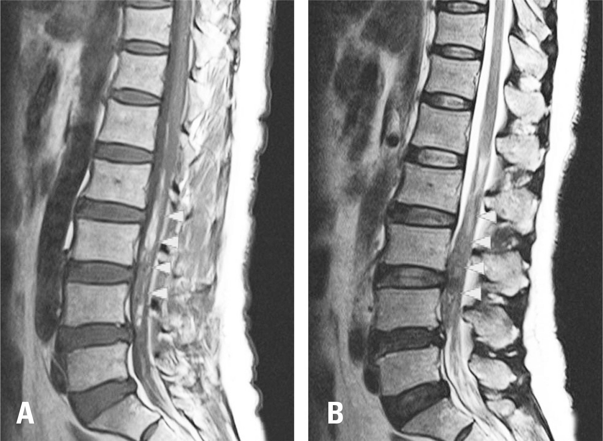
A 56-year-old female patient underwent arthroscopic surgery on her right knee under spinal anesthesia. Automated patient-controlled analgesia (PCA) was applied after surgery. On day 2, the patient complained of lower back pain, headache, nausea, and vomiting, but there were no neurological signs in the lower extremity. At day 5, she had a moderate fever (38.4°) and continuous nausea and vomiting. Magnetic resonance imaging (MRI) was conducted on day 5 and a large subarachnoid hematoma was found. We immediately performed surgical hematoma evacuation. Her low back and buttock pain improved immediately, and all symptoms disappeared in a week without any neurological sequelae.
REFERENCES
1. Horlocker TT. Complications of spinal and epidural anesthesia. Anesthesiol Clin North America. 2000 Jun; 18(2):461–85. DOI: 10.1016/S0889-8537 (05)70172-3.

2. Kreppel D, Antoniadis G, Seeling W. Spinal hematoma: a literature survey with meta-analysis of 613 patients. Neurosurg Rev. 2003 Jan; 26(1):1–49. DOI: 10.1007/s10143-002-0224-y.

3. Domenicucci M, Ramieri A, Paolini S, et al. Spinal subarachnoid hematomas: our experience and literature review. Acta Neurochir (Wien). 2005 Jul; 147(7):741–50. DOI: 10.1007/s00701-004-0458-2.

4. Gaitzsch J, Berney J. Spinal subarachnoid hematoma of spontaneous origin and complicating anticoagulation. Report of four cases and review of the literature. Surg Neurol. 1984 Jun; 21(6):534–8. DOI: 10.1016/0090-3019 (84)90265-9.

5. Lam DH. Subarachnoid haematoma after spinal anaesthesia mimicking transient radicular irritation: a case report and review. Anaesthesia. 2008 Apr; 63(4):423–7. DOI: 10.1111/j.1365-2044.2007.05368.x.

6. Jeon SB, Ham TI, Kang MS, et al. Spinal subarachnoid hematoma after spinal anesthesia. Korean J Anesthesiol. 2013 Apr; 64(4):388–9. DOI: 10.4097/kjae.2013.64.4.388.

7. Luo F, Cai XJ, Li ZY. Subacute spinal subarachnoid hematoma following combined spinal-epidural anesthesia treated conservatively: a case report. J Clin Anesth. 2012 Sep; 24(6):519–20. DOI: 10.1016/j.jclinane.2011.10.016.

8. Bruce-Brand RA, Colleran GC, Broderick JM, et al. Acute nontraumatic spinal intradural hematoma in a patient on warfarin. J Emerg Med. 2013 Nov; 45(5):695–7. DOI: 10.1016/j.jemermed.2013.04.048. Epub 2013 Aug 26.

9. Koyama S, Tomimatsu T, Kanagawa T, et al. Spinal subarachnoid hematoma following spinal anesthesia in a patient with HELLP syndrome. Int J Obstet Anesth. 2010 Jan; 19(1):87–91. DOI: 10.1016/j.ijoa.2009.05.007.

10. Sather MD, Gibson MD, Treves JS. Spinal subarachnoid hematoma resulting from lumbar myelography. AJNR Am J Neuroradiol. 2007 Feb; 28(2):220–1.
Fig 1.
Spinal subarachnoid hematoma. Magnetic resonance imaging showed subarachnoid hematoma extending from L2 to L5. Cauda equina compression with subarachnoid hematoma was most severe at the L3-4 level. (A) Hyperintense in a T1-weighted image, (B) Hypointense in a T2-weighted image.

Fig 2.
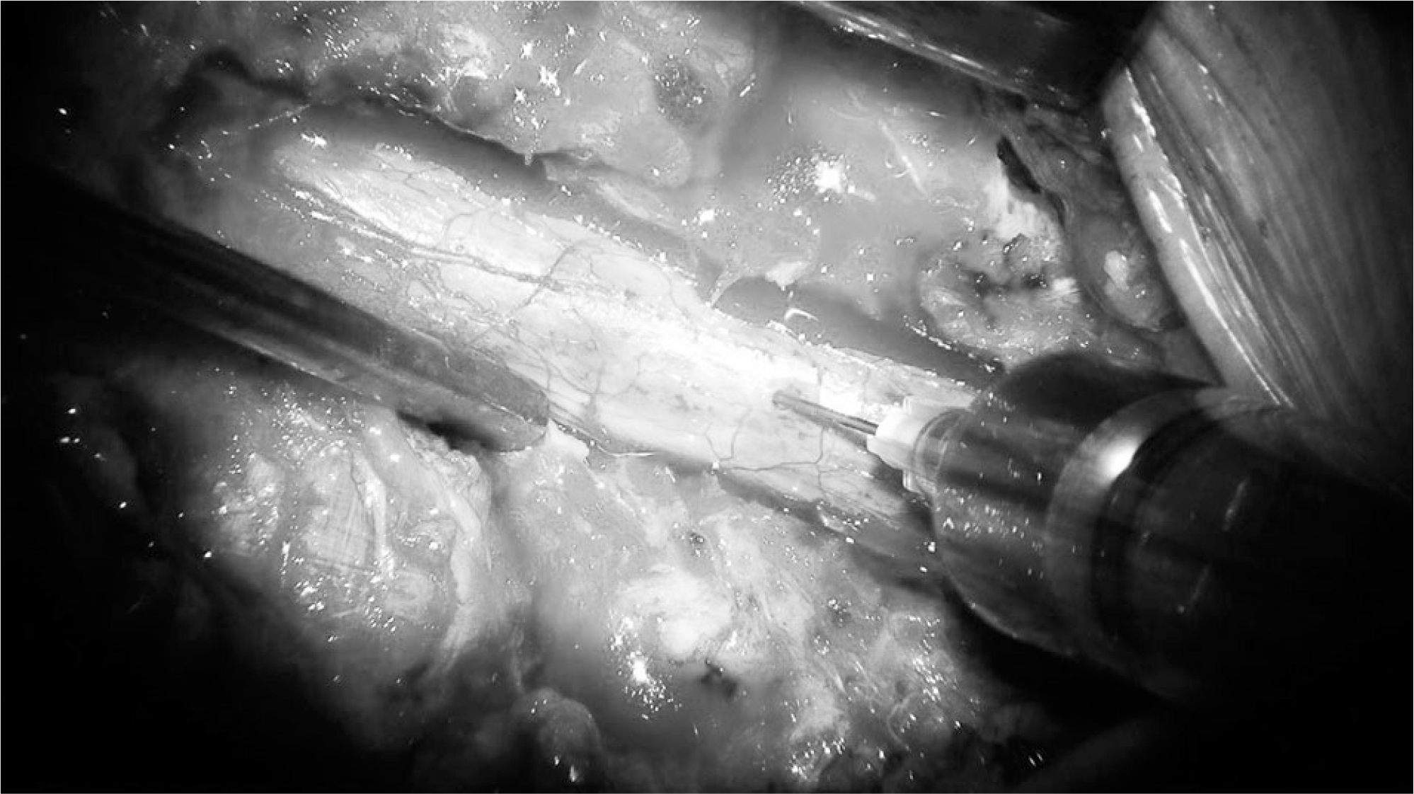
The dorsal surface of the dura mater looked reddish, but was not especially tense. Aspiration was performed, and bloody cerebrospinal fluid was drained.


 PDF
PDF Citation
 Print
Print


 XML Download