

 PDF
PDF Citation
 Print
Print


Abstract
Summary of Literature Review
Acute lumbar paraspinal compartment syndrome is very rare, but it causes muscle necrosis and acute renal failure. Therefore, it should be treated immediately.
Materials and Methods
A 31-year-old male patient and a 30-year-old male patient visited the emergency room due to severe back pain. The left paraspinal compartment pressure of the 31-year-old patient was measured as 35 mm Hg using the Whitesides technique. The paraspinal compartment pressure of the 30-year-old patient was measured as 22 mm Hg on the left side and 30 mm Hg on the right side. We diagnosed acute lumbar paraspinal compartment syndrome and performed a fasciotomy. This study received Institutional Review Board approval (ID: SC18ZESE0032).
REFERENCES
1. Matsen FA 3rd. Compartmental syndrome. An unified concept. Clin Orthop Relat Res. 1975 Nov; 113:8–14. DOI: 10.1097/00003086-197511000-00003.
2. Whitesides TE, Heckman MM. Acute compartment syndrome. Update on diagnosis and treatment. J Am Acad Orthop Surg. 1996 Jul; 4(4):209–18. DOI: 10.5435/00124635-199607000-00005.

3. Volkmann R. Ischaemic muscle paralyses and contractures. 1881. Clin Orthop Relat Res. 2007 Mar; 456:20–1. DOI: 10.1097/BLO.0b013e318032561f.
4. PECK D. Evidence for the existence of compartment syndromes of the epaxial muscles. Anat Rec. 1981; 198:199–201.
5. Carr D, Gilbertson L, Frymoyer J, et al. Lumbar paraspinal compartment syndrome. a case report with physiologic and anatomic studies. Spine (PhilaPa 1976). 1985 Nov; 10(9):816–20. DOI: 10.1097/00007632-198511000-00007.
6. Minnema BJ, Neligan PC, Quraishi NA, et al. A case of occult compartment syndrome and nonresolving rhabdomyolysis. Journal of General Internal Medicine. 2008 Mar; 23(6):871–74. DOI: 10.1007/s11606-008-0569-1.
7. Ferreira J, Galle C, Aminiam A, et al. Lumbar paraspinal rhabdomyolysis and compartment syndrome after abdominal aortic aneurysm repair. Journal of Vascular Surgery. 2003 Jan; 37(1):198–201. DOI: 10.1067/mva.2003.108.
8. Songcharoen P, Chotigavanich C, Thanapipatsiri S. Lumbar paraspinal compartment pressure in back muscle exercise. J Spinal Disord. 1994 Feb; 7(1):49–53. DOI: 10.1097/00002517-199407010-00007.
9. Bogduk N. A reappraisal of the anatomy of the human lumbar erector spinae. J Anat. 1980 Oct; 131(3):525–40.
10. Paryavi E, Jobin CM, Ludwig SC, et al. Acute exertional lumbar paraspinal compartment syndrome. Spine (Phila Pa 1976). 2010 Dec; 35(25):1529–33. DOI: 10.1097/brs.0b013e3181ec4023.
Fig. 1.
Lumbar X-ray. (A) Lumbar anteroposterior X-ray not showing ileus. (B) Lumbar lateral X-ray showing loss of lumbar lordosis.
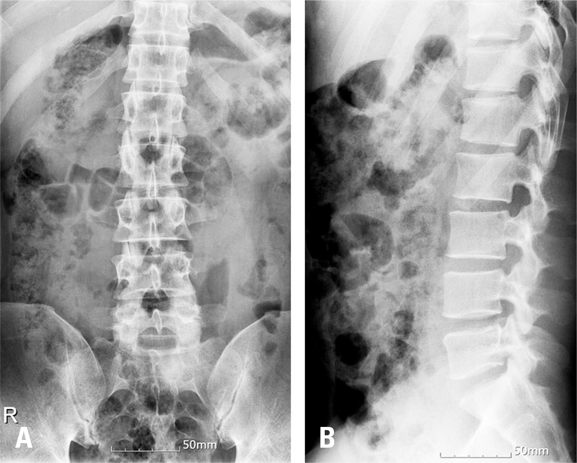
Fig. 2.
Lumbar magnetic resonance imaging. (A) T2-weighted coronal magnetic resonance imaging showing high signal intensity at the left multifidus muscle and the longissimus muscle (arrow). (B) T2-weighted axial magnetic resonance imaging showing high signal intensity at the left erector spinae muscle (arrow).
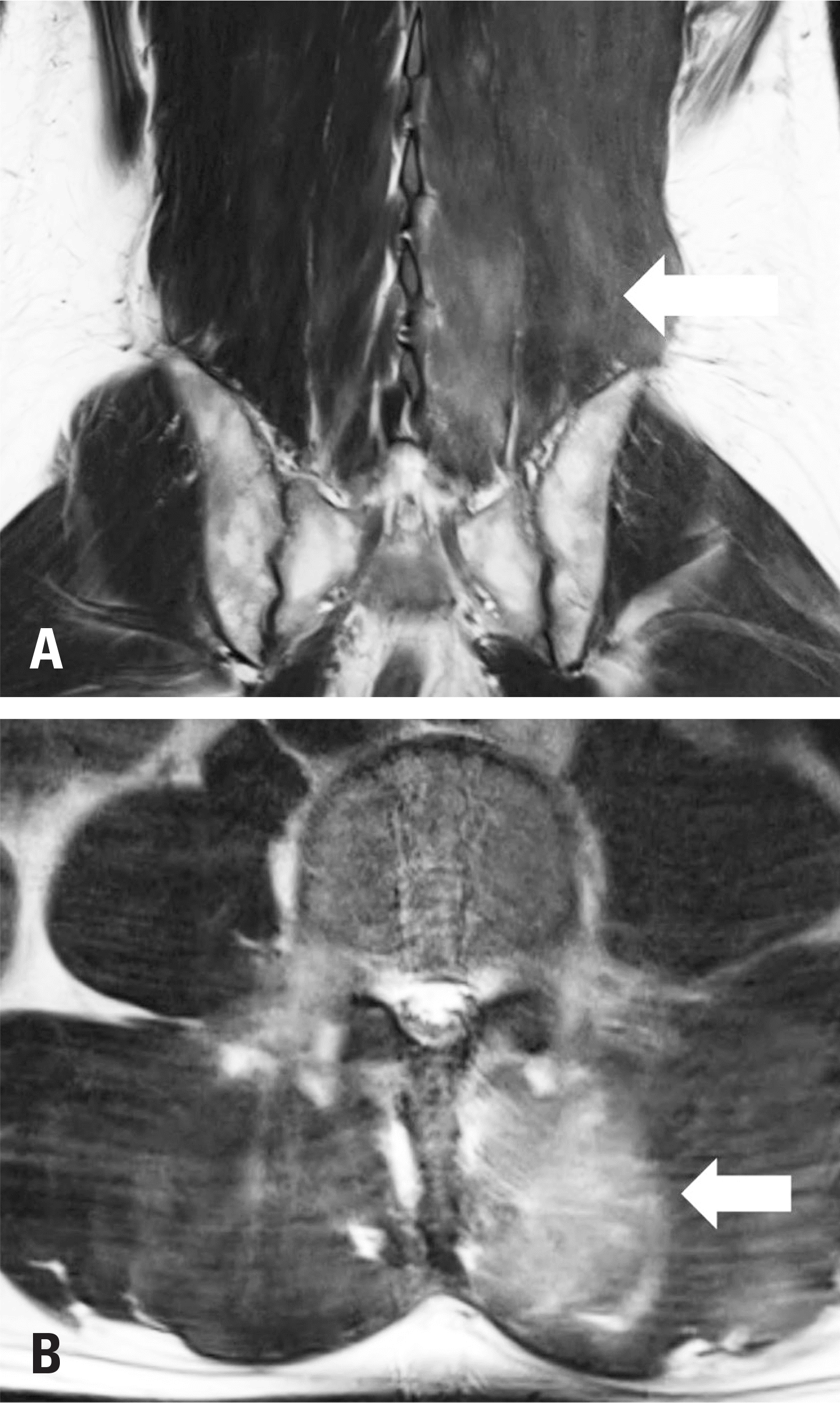
Fig. 3.
Lumbar X-ray. (A) Lumbar anteroposterior X-ray not showing ileus. (B) Lumbar lateral X-ray showing loss of lumbar lordosis.
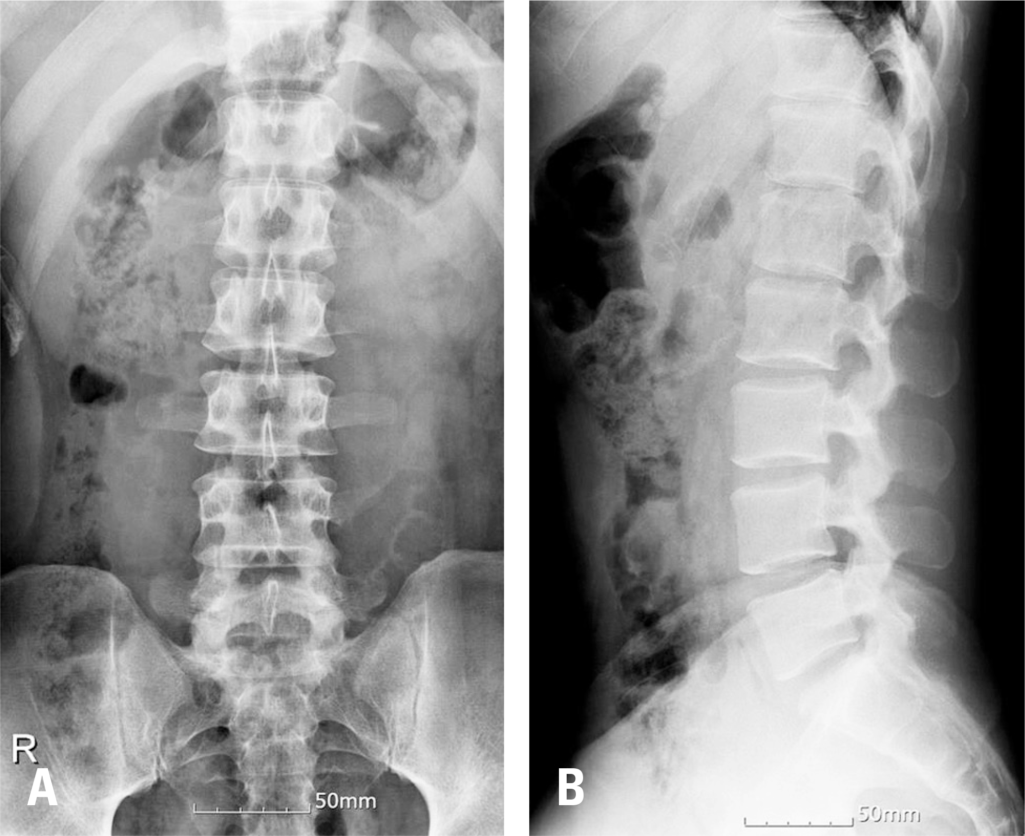
Fig. 4.
Lumbar magnetic resonance imaging. (A) T2-weighted coronal magnetic resonance imaging showing bilateral high signal intensity and edema at the multifidus, longissimus, iliocostalis lumborum, and quadratus lumborum muscles (arrows). (B) T2-weighted axial magnetic resonance imaging showing bilateral high signal intensity at the erector spinae muscles (arrows).
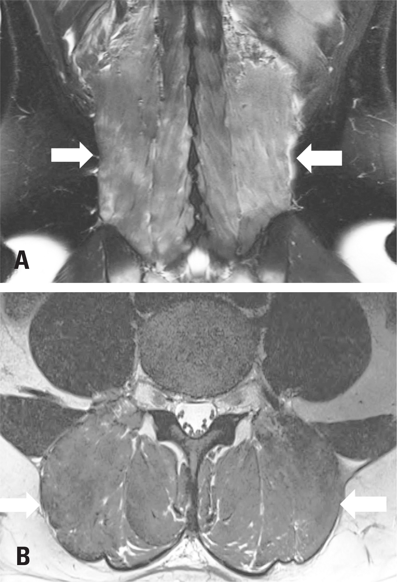
Fig. 5.
Intraoperative clinical photo of case 2. (A) The color of the erector spinae had changed in several areas. (B) The color of the erector spinae had changed to gray.
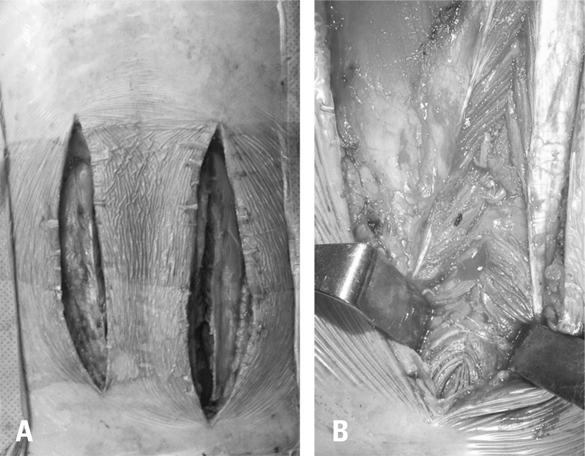
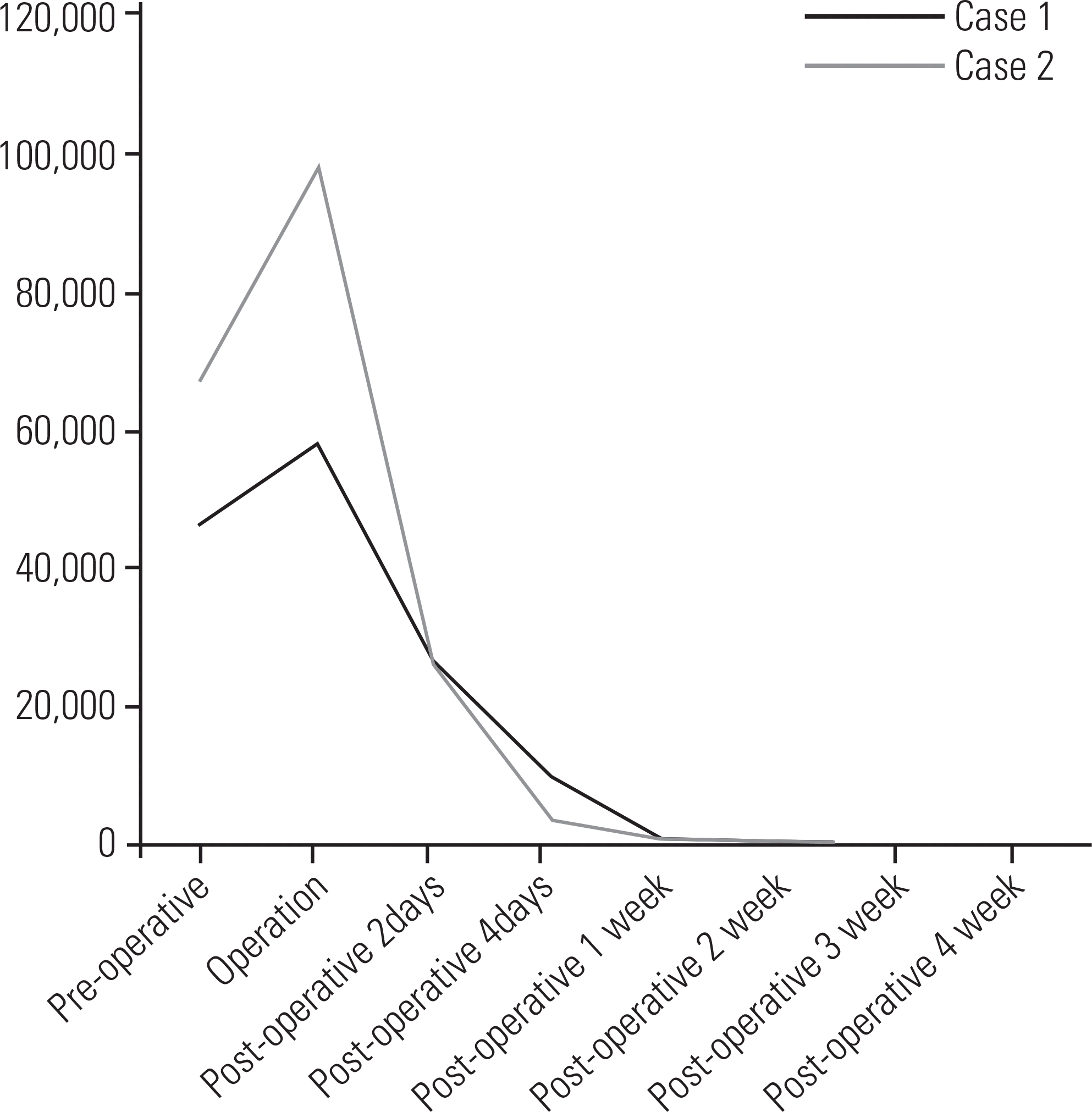
Table 1.
Changes in lab findings over time of case 1 and case 2
 XML Download