

 PDF
PDF Citation
 Print
Print


Abstract
Objectives
We report the case of a patient with C2 spondylotic radiculopathy who was treated by microscopic posterior foraminotomy.
Summary of Literature Review
C2 spondylotic radiculopathy is rare, but it can occur due to spondylosis, compression by a venous plexus or vertebral artery, or hypertrophy of the atlantoepistrophic ligament.
Materials and Methods
A 64-year-old woman was hospitalized with severe occipital pain radiating toward the left cervical area and posterior to the left ear. It started 3 years previously, and became aggravated 3 months previously. Foraminal stenosis of C1-2 was observed on magnetic resonance imaging (MRI) and degenerative changes of the facet joint of C1-2 and osteophytes originating from the left atlantoaxial joint were shown on computed tomography (CT). Dynamic rotational CT showed narrowing of the left C1-2 neural foramen when it was rotated to the left. Selective C2 root block was done, but the pain was aggravated. Thus, we decompressed the C2 nerve root by microscopic posterior laminotomy of the C1 vertebra. After surgery, the patient's occipitocervical pain mostly resolved. By the 6-month follow up, pain had not recurred, and instability was not observed on plain radiographs.
Go to : 
REFERENCES
1. Spurling RG, Scoville WB. Lateral rupture of cervical intervertebral disc. A common cause of shoulder and arm pain. Surg Gynecol Obstet. 1944; 78:350–8.
2. Ehni G, Benner B. Occipital neuralgia and the C1-2 arthrosis syndrome. J Neurosurg. 1984 Nov; 61(5):961–5. DOI: 10.3171/jns.1984.61.5.0961.

3. Star MJ, Curd JG, Thorne RP. Atlantoaxial lateral mass osteoarthritis. A frequently overlooked cause of severe occipitocervical pain. Spine (Phila Pa 1976). 1992 Jun; 17(6 Suppl):71–6.
4. Clavel M, Clavel P. Occipital neuralgia secondary to exuberant callus formation. Case report. J Neurosurg. 1996 Dec; 85(6):1170–1. DOI: 10.3171/jns.1996.85.6.1170.
5. Pakzaban P. Transarticular screw fixation of C1-2 for the treatment of arthropathy-associated occipital neuralgia. 2011 Feb; 14(2):209–14. DOI: 10.3171/2010.10. SPINE09815.
6. Pikus HJ, Phillips JM. Outcome of surgical decompression of the second cervical root for cervicogenic headache. Neurosurgery. 1996 Jul; 39(1):63–70. discussion 70-1. DOI: 10.1097/00006123-199607000-00014.
7. Lu J, Ebraheim NA. Anatomic considerations of C2 nerve root ganglion. Spine (Phila Pa 1976). 1998 Mar; 23(6):64952. DOI: 10.1097/00007632-199803150-00001.
8. Cesmebasi A, Muhleman MA, Hulsberg P, et al. Occipital neuralgia: anatomic considerations. Clin Anat. 2015 Jan; 28(1):101–8. DOI: 10.1002/ca.22468.
9. Sjaastad O, Saunte C, Hovdahl H et al “Cervicogenic” headache. An hypothesis. Cephalalgia. 1983 Dec; 3(4):24956. DOI: 10.1046/j.1468-2982.1983.0304249.x.
10. Kim CH, Kim K-T, Chung CK, et al. Minimally invasive cervical foraminotomy and diskectomy for laterally located soft disk herniation. Eur Spine J. 2015 Dec; 24(12):300512. DOI: 10.1007/s00586-015-4198-1.
Go to :
 | Fig. 2.
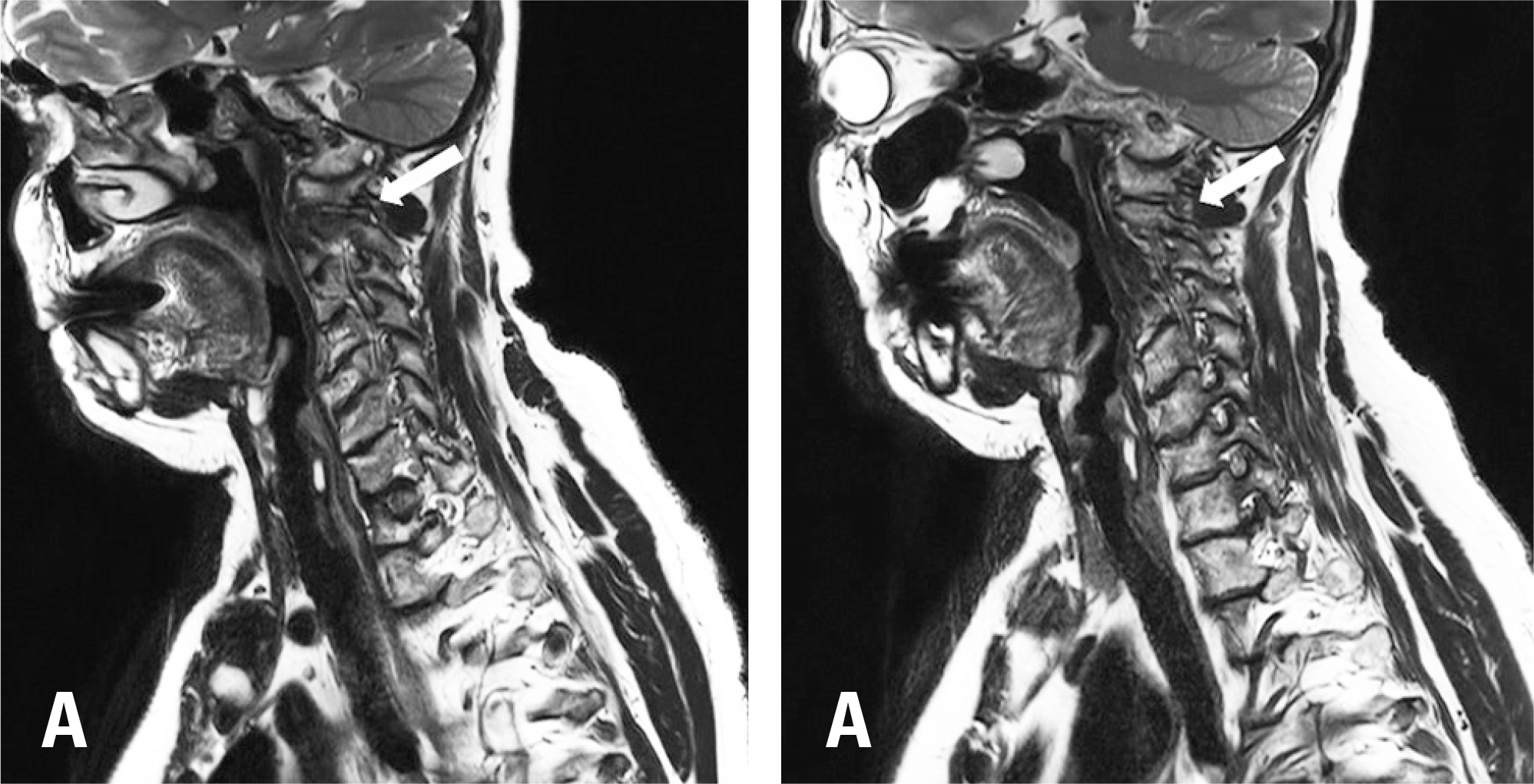
(A) The white arrow indicates the symptomatic left neural foramen with foraminal stenosis. (B) The white arrow indicates the asymptomatic right neural foramen. |
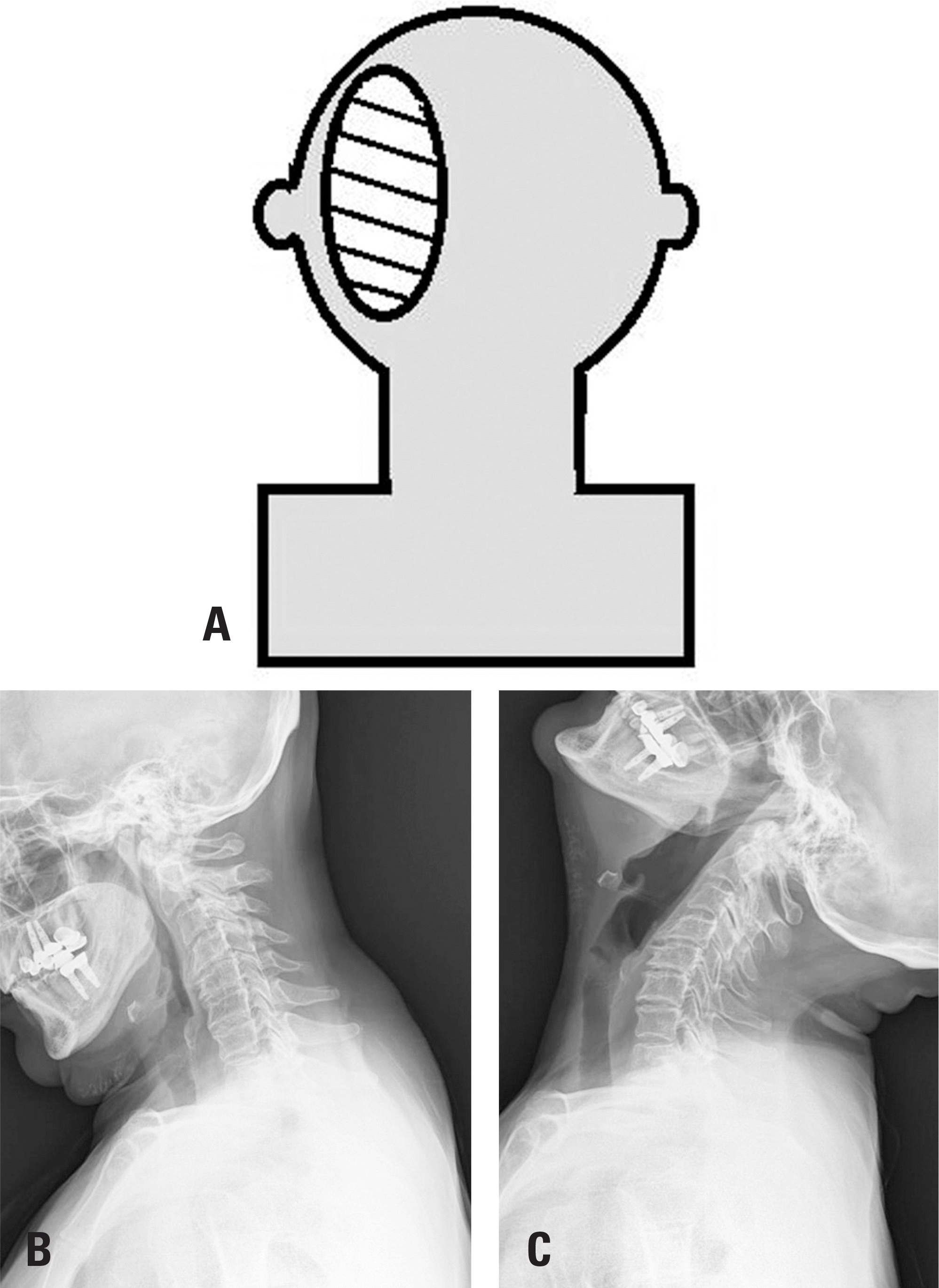
 | Fig. 1.A 64-year-old woman with the chief complaint of occipitocervical pain on the left side. (A) The patient complained of occipitocervical pain on the left side that originated from the upper cervical spine and radiated towards the left occipital area. (B, C) There was no obvious atlantoaxial instability between the flexion and extension positions. |
 | Fig. 3.
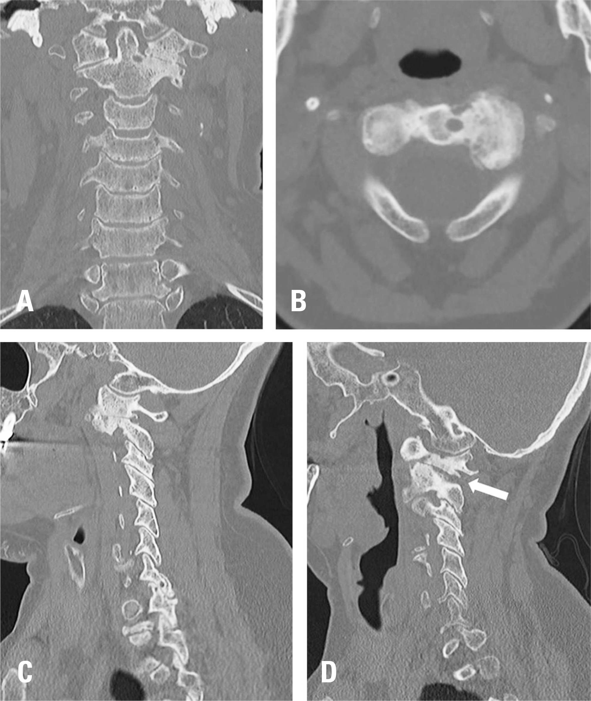
(A) a coronal image shows unilateral spondylosis. The disc space is narrowed and the vertebral body is collapsed on the left side. (B) An axial image at C1-2 shows osteoarthritis and an osteophyte. (C, D) The left C1-2 neural foramen (white arrow) is narrower in the left rotation position (d) than the neutral position (c). |
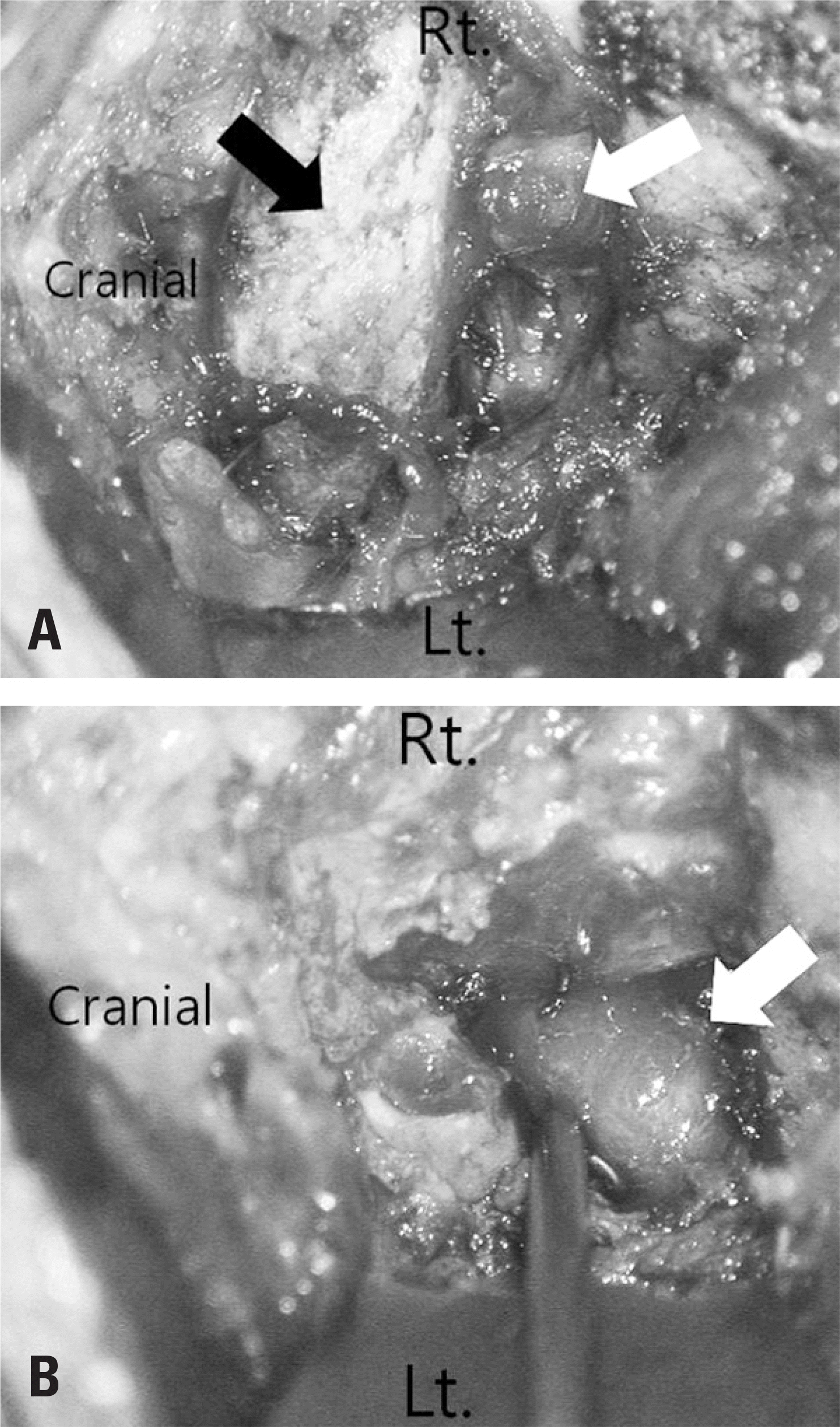 | Fig. 4.Intraoperative photos from the surgical microscope. (A) The C2 nerve root was found to be compressed by the inferior part of the C1 lamina. (black arrow: C1 posterior arch, white arrow: diffuse venous engorgement above the C2 nerve root) (B) Partial resection of the inferior edge of the C1 posterior arch was performed and nerve root impingement disappeared (white arrow: C2 nerve root). |
 XML Download