Abstract
Dynamic contrast enhanced (DCE) magnetic resonance (MR) imaging plays an important role in noninvasive detection and characterization of primary and metastatic lesions in the liver. Recently, efforts have been made to improve spatial and temporal resolution of DCE liver MRI for arterial phase imaging. Review of recent publications related to arterial phase imaging of the liver indicates that there exist primarily two approaches: breathhold and free-breathing. For breathhold imaging, acquiring multiple arterial phase images in a breathhold is the preferred approach over conventional single-phase imaging. For free-breathing imaging, a combination of three-dimensional (3D) stack-of-stars golden-angle sampling and compressed sensing parallel imaging reconstruction is one of emerging techniques. Self-gating can be used to decrease respiratory motion artifact. This article introduces recent MRI technologies relevant to hepatic arterial phase imaging, including differential subsampling with Cartesian ordering (DISCO), golden-angle radial sparse parallel (GRASP), and X-D GRASP. This article also describes techniques related to dynamic 3D image reconstruction of the liver from golden-angle stack-of-stars data.
References
1. Gomaa AI, Khan SA, Toledano MB, Waked I, Taylor-Robinson SD. Hepatocellular carcinoma: epidemiology, risk factors and pathogenesis. World J Gastroenterol. 2008; 14:4300–4308.

2. Mittal S, El-Serag HB. Epidemiology of hepatocellular carcinoma: consider the population. J Clin Gastroenterol. 2013; 47(Suppl):S2–6.
3. Ringe KI, Husarik DB, Sirlin CB, Merkle EM. Gadoxetate disodium-enhanced MRI of the liver: part 1, protocol optimization and lesion appearance in the noncirrhotic liver. AJR Am J Roentgenol. 2010; 195:13–28.

4. Cruite I, Schroeder M, Merkle EM, Sirlin CB. Gadoxetate disodium-enhanced MRI of the liver: part 2, protocol optimization and lesion appearance in the cirrhotic liver. AJR Am J Roentgenol. 2010; 195:29–41.

5. Bruix J, Sherman M. American Association for the Study of Liver Diseases. Management of hepatocellular carcinoma: an update. Hepatology. 2011; 53:1020–1022.

6. Nakamura S, Nakaura T, Kidoh M, et al. Timing of the hepatic arterial phase at Gd-EOB-DTPA-enhanced hepatic dynamic MRI: comparison of the test-injection and the fixed-time delay method. J Magn Reson Imaging. 2013; 38:548–554.

7. Pietryga JA, Burke LM, Marin D, Jaffe TA, Bashir MR. Respiratory motion artifact affecting hepatic arterial phase imaging with gadoxetate disodium: examination recovery with a multiple arterial phase acquisition. Radiology. 2014; 271:426–434.

8. Saranathan M, Rettmann DW, Hargreaves BA, Clarke SE, Vasanawala SS. DIfferential Subsampling with Cartesian Ordering (DISCO): a high spatiotemporal resolution Dixon imaging sequence for multiphasic contrast enhanced abdominal imaging. J Magn Reson Imaging. 2012; 35:1484–1492.

9. Hope TA, Saranathan M, Petkovska I, Hargreaves BA, Herfkens RJ, Vasanawala SS. Improvement of gadoxetate arterial phase capture with a high spatiotemporal resolution multiphase three-dimensional SPGR-Dixon sequence. J Magn Reson Imaging. 2013; 38:938–945.

10. Ichikawa S, Motosugi U, Oishi N, et al. Ring-like enhancement of hepatocellular carcinoma in gadoxetic acid-enhanced multiphasic hepatic arterial phase imaging with differential subsampling with cartesian ordering. Invest Radiol. 2018; 53:191–199.

11. Clarke SE, Saranathan M, Rettmann DW, Hargreaves BA, Vasanawala SS. High resolution multi-arterial phase MRI improves lesion contrast in chronic liver disease. Clin Invest Med. 2015; 38:E90–99.

12. Ikram NS, Yee J, Weinstein S, et al. Multiple arterial phase MRI of arterial hypervascular hepatic lesions: improved arterial phase capture and lesion enhancement. Abdom Radiol (NY). 2017; 42:870–876.

13. Fujinaga Y, Ohya A, Tokoro H, et al. Radial volumetric imaging breathhold examination (VIBE) with k-space weighted image contrast (KWIC) for dynamic gadoxetic acid (Gd-EOB-DTPA)-enhanced MRI of the liver: advantages over Cartesian VIBE in the arterial phase. Eur Radiol. 2014; 24:1290–1299.

14. Chandarana H, Feng L, Block TK, et al. Free-breathing contrast-enhanced multiphase MRI of the liver using a combination of compressed sensing, parallel imaging, and golden-angle radial sampling. Invest Radiol. 2013; 48:10–16.

15. Feng L, Grimm R, Block KT, et al. Golden-angle radial sparse parallel MRI: combination of compressed sensing, parallel imaging, and golden-angle radial sampling for fast and flexible dynamic volumetric MRI. Magn Reson Med. 2014; 72:707–717.

16. Feng L, Axel L, Chandarana H, Block KT, Sodickson DK, Otazo R. XD-GRASP: Golden-angle radial MRI with reconstruction of extra motion-state dimensions using compressed sensing. Magn Reson Med. 2016; 75:775–788.

17. Chandarana H, Feng L, Ream J, et al. Respiratory motion-resolved compressed sensing reconstruction of free-breathing radial acquisition for dynamic liver magnetic resonance imaging. Invest Radiol. 2015; 50:749–756.

18. Choi JY, Lee JM, Sirlin CB. CT and MR imaging diagnosis and staging of hepatocellular carcinoma: part I. Development, growth, and spread: key pathologic and imaging aspects. Radiology. 2014; 272:635–654.

19. Choi JY, Lee JM, Sirlin CB. CT and MR imaging diagnosis and staging of hepatocellular carcinoma: part II. Extracellular agents, hepatobiliary agents, and ancillary imaging features. Radiology. 2014; 273:30–50.

20. Motosugi U, Bannas P, Sano K, Reeder SB. Hepatobiliary MR contrast agents in hypovascular hepatocellular carcinoma. J Magn Reson Imaging. 2015; 41:251–265.

21. Yoon JH, Lee JM, Yu MH, Kim EJ, Han JK. Triple arterial phase MR imaging with gadoxetic acid using a combination of contrast enhanced time robust angiography, keyhole, and viewsharing techniques and two-dimensional parallel imaging in comparison with conventional single arterial phase. Korean J Radiol. 2016; 17:522–532.

22. Davenport MS, Bashir MR, Pietryga JA, Weber JT, Khalatbari S, Hussain HK. Dose-toxicity relationship of gadoxetate disodium and transient severe respiratory motion artifact. AJR Am J Roentgenol. 2014; 203:796–802.

23. Huh J, Kim SY, Yeh BM, et al. Troubleshooting arterial-phase MR images of gadoxetate disodium-enhanced liver. Korean J Radiol. 2015; 16:1207–1215.

24. Davenport MS, Viglianti BL, Al-Hawary MM, et al. Comparison of acute transient dyspnea after intravenous administration of gadoxetate disodium and gadobenate dimeglumine: effect on arterial phase image quality. Radiology. 2013; 266:452–461.

25. Davenport MS, Caoili EM, Kaza RK, Hussain HK. Matched within-patient cohort study of transient arterial phase respiratory motion-related artifact in MR imaging of the liver: gadoxetate disodium versus gadobenate dimeglumine. Radiology. 2014; 272:123–131.

26. Yoon JH, Lee JM, Yu MH, et al. Evaluation of transient motion during gadoxetic acid-enhanced multiphasic liver magnetic resonance imaging using free-breathing golden-angle radial sparse parallel magnetic resonance imaging. Invest Radiol. 2018; 53:52–61.

27. Min JH, Kim YK, Kang TW, et al. Artifacts during the arterial phase of gadoxetate disodium-enhanced MRI: multiple arterial phases using viewsharing from two different vendors versus single arterial phase imaging. Eur Radiol. 2018; 28:3335–3346.

28. Hope TA, Petkovska I, Saranathan M, Hargreaves BA, Vasanawala SS. Combined parenchymal and vascular imaging: high spatiotemporal resolution arterial evaluation of hepatocellular carcinoma. J Magn Reson Imaging. 2016; 43:859–865.

29. Lustig M, Donoho D, Pauly JM. Sparse MRI: the application of compressed sensing for rapid MR imaging. Magn Reson Med. 2007; 58:1182–1195.

30. Kaltenbach B, Bucher AM, Wichmann JL, et al. Dynamic liver magnetic resonance imaging in free-breathing: feasibility of a cartesian T1-weighted acquisition technique with compressed sensing and additional self-navigation signal for hard-gated and motion-resolved reconstruction. Invest Radiol. 2017; 52:708–714.
31. Weiss J, Notohamiprodjo M, Martirosian P, et al. Self-gated 4D-MRI of the liver: initial clinical results of continuous multiphase imaging of hepatic enhancement. J Magn Reson Imaging. 2018; 47:459–467.

32. Peters DC, Derbyshire JA, McVeigh ER. Centering the projection reconstruction trajectory: reducing gradient delay errors. Magn Reson Med. 2003; 50:1–6.

33. Block KT. Advanced methods for radial data sampling in MRI. Ph.D. thesis, Georg-August-Universitaet Goettingen,. 2008.
34. Block KT, Uecker M, Frahm J. Undersampled radial MRI with multiple coils. Iterative image reconstruction using a total variation constraint. Magn Reson Med. 2007; 57:1086–1098.

35. Song HK, Dougherty L. Dynamic MRI with projection reconstruction and KWIC processing for simultaneous high spatial and temporal resolution. Magn Reson Med. 2004; 52:815–824.

36. Winkelmann S, Schaeffter T, Koehler T, Eggers H, Doessel O. An optimal radial profile order based on the golden ratio for time-resolved MRI. IEEE Trans Med Imaging. 2007; 26:68–76.

37. Chan RW, Ramsay EA, Cheung EY, Plewes DB. The influence of radial undersampling schemes on compressed sensing reconstruction in breast MRI. Magn Reson Med. 2012; 67:363–377.

38. Hedderich DM, Weiss K, Spiro JE, et al. Clinical evaluation of free-breathing contrast-enhanced T1w MRI of the Liver using pseudo golden angle radial k-space sampling. Rofo. 2018; 190:601–609.

39. Kajita K, Goshima S, Noda Y, et al. Thin-slice free-breathing pseudo-golden-angle radial stack-of-stars with gating and tracking T1-weighted acquisition: an efficient gadoxetic acid-enhanced hepatobiliary-phase imaging alternative for patients with unstable breath holding. Magn Reson Med Sci. 2019; 18:4–11.
40. Brodsky EK, Bultman EM, Johnson KM, et al. High-spatial and high-temporal resolution dynamic contrast-enhanced perfusion imaging of the liver with time-resolved three-dimensional radial MRI. Magn Reson Med. 2014; 71:934–941.

41. Block KT, Frahm J. Spiral imaging: a critical appraisal. J Magn Reson Imaging. 2005; 21:657–668.

42. Agrawal MD, Spincemaille P, Mennitt KW, et al. Improved hepatic arterial phase MRI with 3-second temporal resolution. J Magn Reson Imaging. 2013; 37:1129–1136.

43. Xu B, Spincemaille P, Chen G, et al. Fast 3D contrast enhanced MRI of the liver using temporal resolution acceleration with constrained evolution reconstruction. Magn Reson Med. 2013; 69:370–381.

44. Breuer FA, Blaimer M, Heidemann RM, Mueller MF, Griswold MA, Jakob PM. Controlled aliasing in parallel imaging results in higher acceleration (CAIPIRINHA) for multislice imaging. Magn Reson Med. 2005; 53:684–691.

45. Breuer FA, Blaimer M, Mueller MF, et al. Controlled aliasing in volumetric parallel imaging (2D CAIPIRINHA). Magn Reson Med. 2006; 55:549–556.

46. Kim BS, Lee KR, Goh MJ. New imaging strategies using a motion-resistant liver sequence in uncooperative patients. Biomed Res Int. 2014; 2014:142658.

47. Wright KL, Harrell MW, Jesberger JA, et al. Clinical evaluation of CAIPIRINHA: comparison against a GRAPPA standard. J Magn Reson Imaging. 2014; 39:189–194.

48. Michaely HJ, Morelli JN, Budjan J, et al. CAIPIRINHA-Dixon-TWIST (CDT)-volume-interpolated breathhold examination (VIBE): a new technique for fast time-resolved dynamic 3-dimensional imaging of the abdomen with high spatial resolution. Invest Radiol. 2013; 48:590–597.
49. Yu MH, Lee JM, Yoon JH, Kiefer B, Han JK, Choi BI. Clinical application of controlled aliasing in parallel imaging results in a higher acceleration (CAIPIRINHA)volumetric interpolated breathhold (VIBE) sequence for gadoxetic acid-enhanced liver MR imaging. J Magn Reson Imaging. 2013; 38:1020–1026.

50. Park YS, Lee CH, Kim IS, et al. Usefulness of controlled aliasing in parallel imaging results in higher acceleration in gadoxetic acid-enhanced liver magnetic resonance imaging to clarify the hepatic arterial phase. Invest Radiol. 2014; 49:183–188.

51. Beck GM, De Becker J, Jones AC, von Falkenhausen M, Willinek WA, Gieseke J. Contrast-enhanced timing robust acquisition order with a preparation of the longitudinal signal component (CENTRA plus) for 3D contrast-enhanced abdominal imaging. J Magn Reson Imaging. 2008; 27:1461–1467.

52. Eggers H, Bornert P. Chemical shift encoding-based water-fat separation methods. J Magn Reson Imaging. 2014; 40:251–268.

53. Eggers H, Brendel B, Duijndam A, Herigault G. Dual-echo Dixon imaging with flexible choice of echo times. Magn Reson Med. 2011; 65:96–107.

54. Berglund J, Ahlstrom H, Johansson L, Kullberg J. Two-point dixon method with flexible echo times. Magn Reson Med. 2011; 65:994–1004.

55. Benkert T, Feng L, Sodickson DK, Chandarana H, Block KT. Free-breathing volumetric fat/water separation by combining radial sampling, compressed sensing, and parallel imaging. Magn Reson Med. 2017; 78:565–576.

56. Pandharipande PV, Krinsky GA, Rusinek H, Lee VS. Perfusion imaging of the liver: current challenges and future goals. Radiology. 2005; 234:661–673.

57. Thng CH, Koh TS, Collins DJ, Koh DM. Perfusion magnetic resonance imaging of the liver. World J Gastroenterol. 2010; 16:1598–1609.

58. Sourbron S, Sommer WH, Reiser MF, Zech CJ. Combined quantification of liver perfusion and function with dynamic gadoxetic acid-enhanced MR imaging. Radiology. 2012; 263:874–883.

59. Chen Y, Lee GR, Wright KL, et al. Free-breathing liver perfusion imaging using 3-dimensional through-time spiral generalized autocalibrating partially parallel acquisition acceleration. Invest Radiol. 2015; 50:367–375.

Fig. 1.
DCE liver MRI scan protocols. (a) Schematic of a conventional DCE liver imaging protocol. All acquisition sequences have the same single-phase 3D fast gradient echo (GRE), which is referred to as eTHRIVE for Philips, VIBE for Siemens, and LAVA for GE, acquired within a breathhold (BH), typically lasting for 15 seconds. (b) Schematic of a modified protocol where a multiphase imaging method (green box) is substituted for the conventional single-phase acquisition of arterial phase. BH: breathhold; FB: free-breathing. (c) Schematic of a modified protocol where a multiphase FB imaging (green box) is used and the duration of FB imaging is extended to cover the portal venous phase. (d) Time versus MR signal curves from normal liver region of interest (ROI) and tumor ROI. The orange box indicates the acquisition time interval of single-phase BH imaging and the green box shows that of multiphase FB imaging. In single-phase BH imaging, start time of the acquisition is critical for differentiation of tumor enhancement from normal tissue.
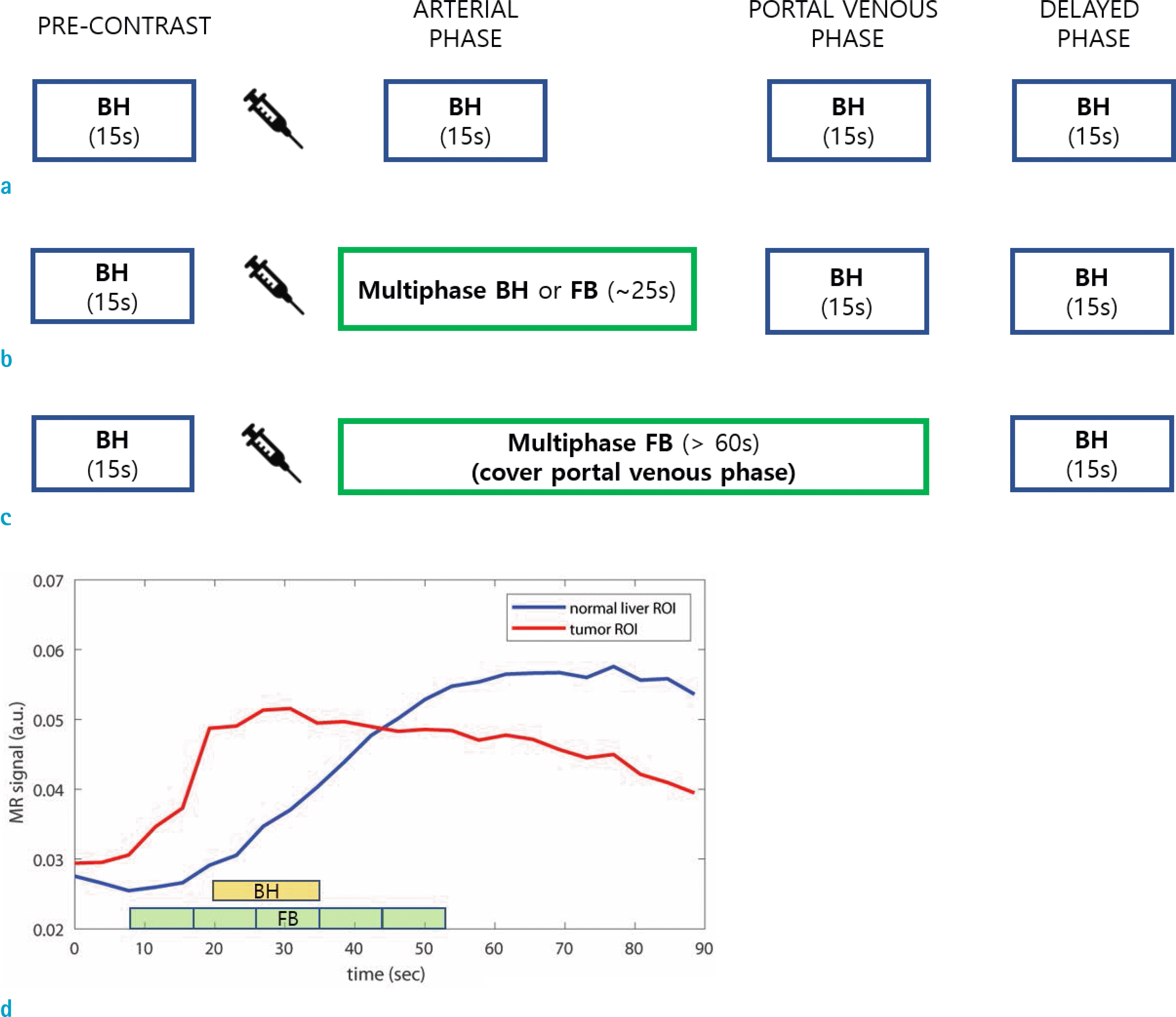
Fig. 2.
Schematic illustration of DISCO. (a) Sampling distribution. Section A is fully sampled in (ky, kz) while sections B, C, and D are undersampled in a pseudo-random manner. The union of samples from sections B, C, and D is fully sampled. (b) Temporal acquisition order of the k-space sections. Note that section A is acquired more frequently in time than sections B, C, and D.
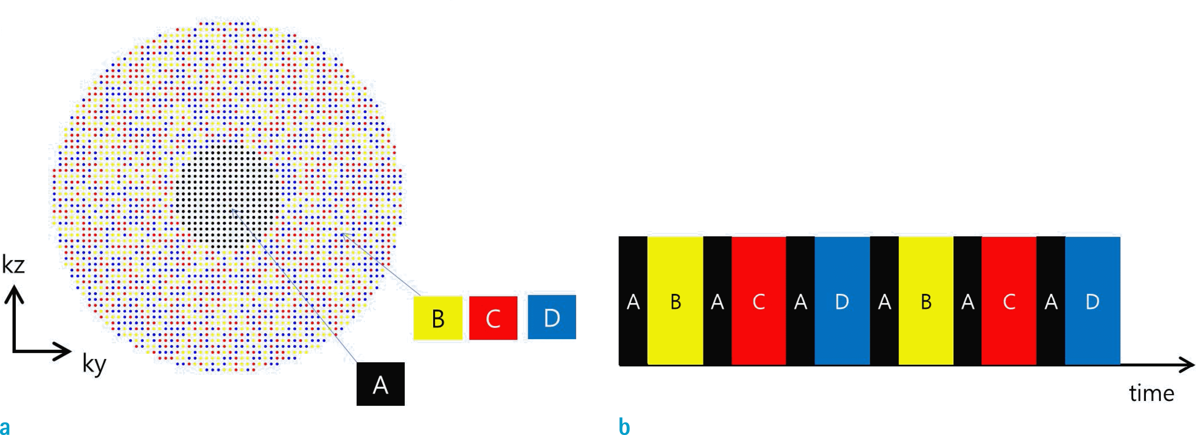
Fig. 3.
Schematic illustration of 3D GRASP. (a) 3D view of k-space sampling pattern. (b) 2D view in (kx, kz). (c) 2D view in (kx, ky). For a given angle spoke, it samples uniformly in kz axis until it fills out the kz space. The spoke angle is then incremented by golden angle α ≈ 180°/1.618 ≈ 111°, followed by filling out the kz space. This process is repeated until it reaches the prescribed number of radial spokes. (d) Continuous imaging sequence in 2D golden-angle radial sequence. The same principle can also be applied to 3D GRASP without loss of generality. The number in the green box indicates spoke order. (e, f) Sampling patterns after retrospective selections of temporal window by grouping (e) five consecutive spokes per frame and (f) three consecutive spokes per frame. For both cases (one with grouping of 5 spokes and the other with grouping of 3 spokes), patterns of angle spacing in radial spokes are similar in all frames. This is relevant to the fact that golden angle radial sampling guarantees temporal stability. Per frame basis, (e) is more densely sampled (less spatial aliasing) than (f). (e) has a lower frame rate than (f).
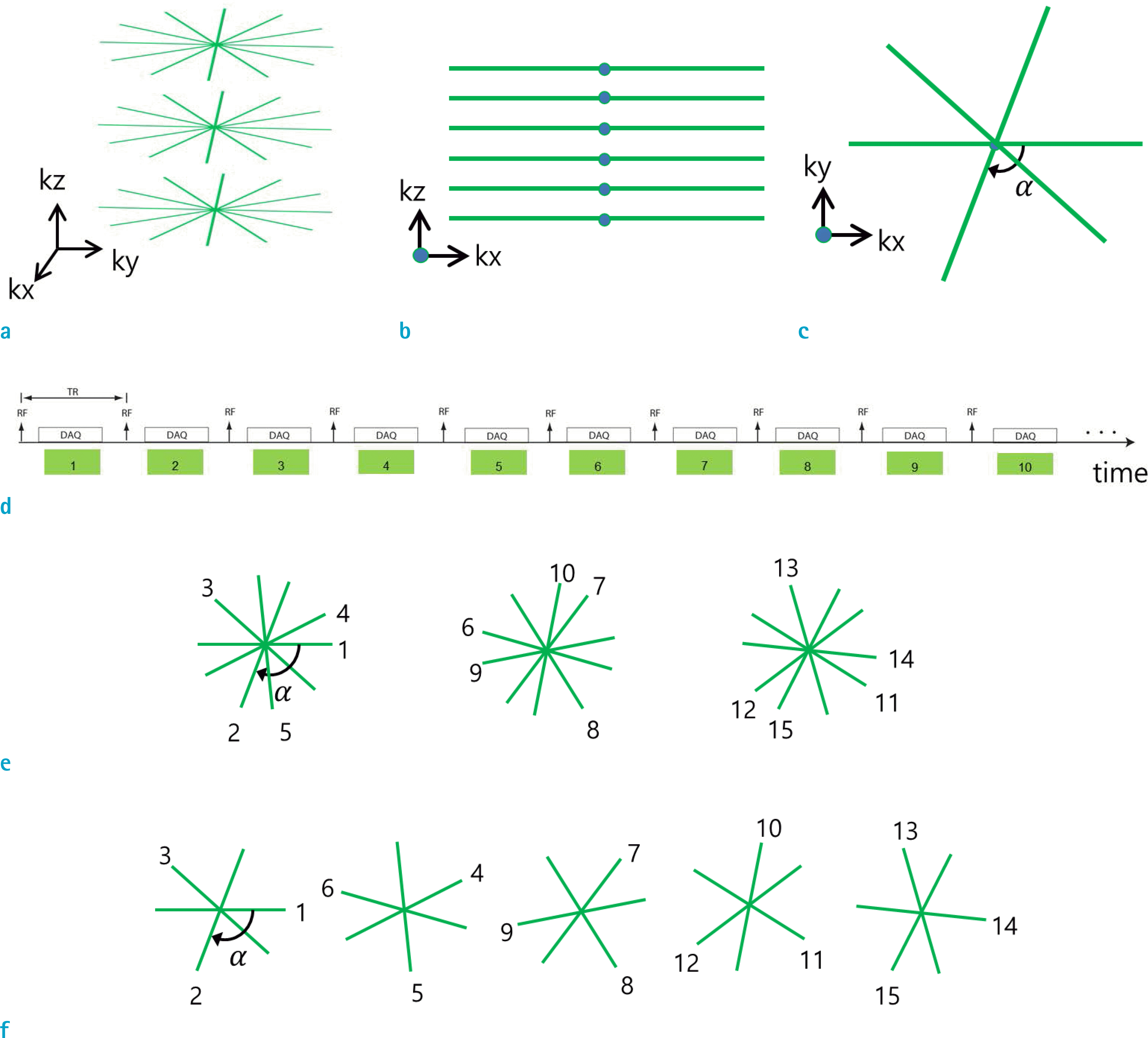
Fig. 4.
Retrospective reconstructions from GRASP data. (a) Schematic of retrospective adjustment of a starting point (red dashed line) and temporal window (green and blue) from continuously acquired raw data in GRASP. (b) Retrospective reconstructions with choices of 40, 55, and 70 spokes. Results exhibit stable image reconstruction quality in all frames for the three cases.
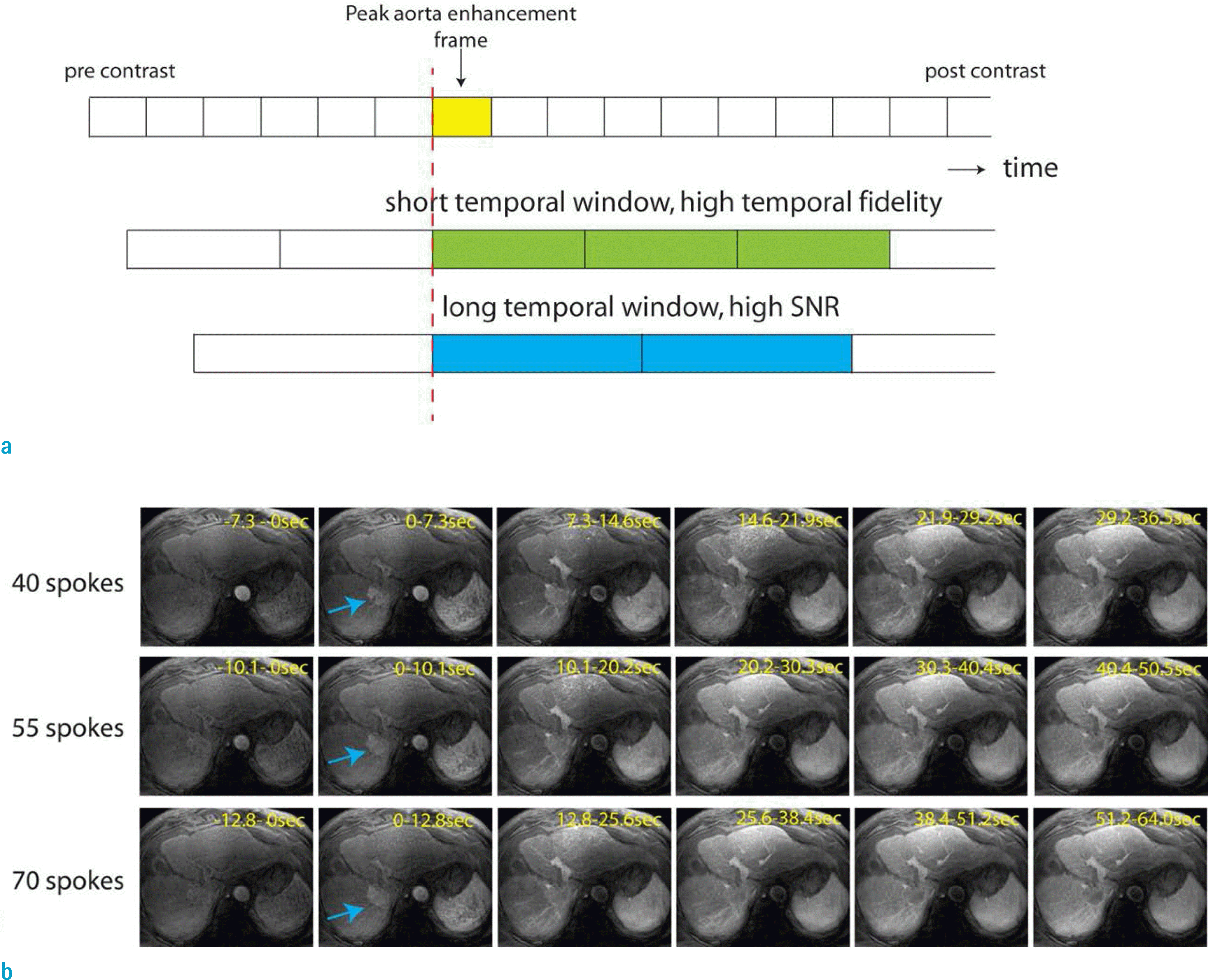
Fig. 5.
Illustration of data binning procedures in XD-GRASP. (a) Raw data samples at the center in (kx, ky) are extracted for each spoke t’ and are denoted by d1(kz, t’). (b) One-dimensional Fourier transform is performed along kz to transform data d1(kz, t’) to d2(z, t’) for each spoke t’. d2(z, t’) represents 1D projection signal projected to superior-inferior (S-I) direction. (c) Construction of a 2D image d3(z, t) by concatenating 1D projection data for all coils and all radial spokes. (d) A result of the first principal component after performing principal component analysis (PCA) of the 2D image d3(z, t). (e) A result of smooth version of (d) after taking a median filter in time. (f) Estimation of the envelope (red line) after a spline fitting to peak points in (e). (g) Flattened curve after division of (e) by (f). Final motion state assignment is indicated by different colors (state 1: red, state 2: blue, state 3: black, state 4: green). Contrast phases are differentiated by background colors in the plot.
Fig. 6.
An example of reconstructed images using XD-GRASP. (a) Final reconstructed images of XD-GRASP. These images contain an additional respiratory motion-resolved dimension. (b) Comparison of GRASP and XD-GRASP for contrast phase 5. GRASP exhibits an image of averaged motion from all respiration states. XD-GRASP contains images, each of which is from a respiration state. Note that there are differences in image appearance between respiration states 1 and 4 (yellow and green arrows). Also note motion-averaged image appearance in the GRASP image (red and white arrows).
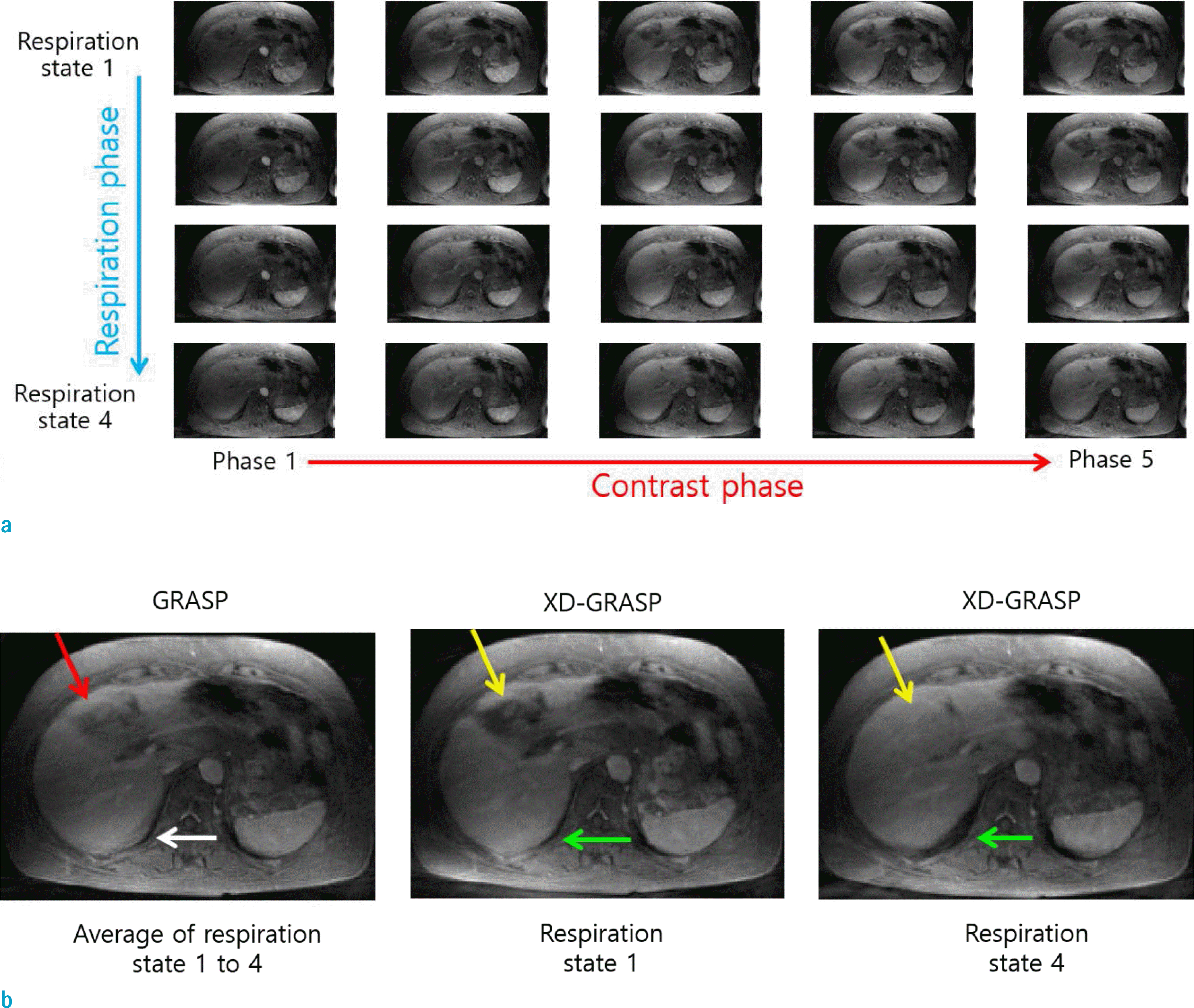
Fig. 7.
Illustration of water fat separation from continuously acquired dual-echo 3D stack-of-stars golden-angle radial raw data. (a) Schematic of a radial dual-echo pulse sequence. (b) TE1 and TE2 images can be separately reconstructed using compressed sensing and parallel imaging. These complex-valued images are input to a 2-point Dixon water fat separation algorithm. The output is a set of water and fat images. Water-fat swap artifact is indicated by red arrows. (c) Final fat suppressed dynamic image frames are shown.
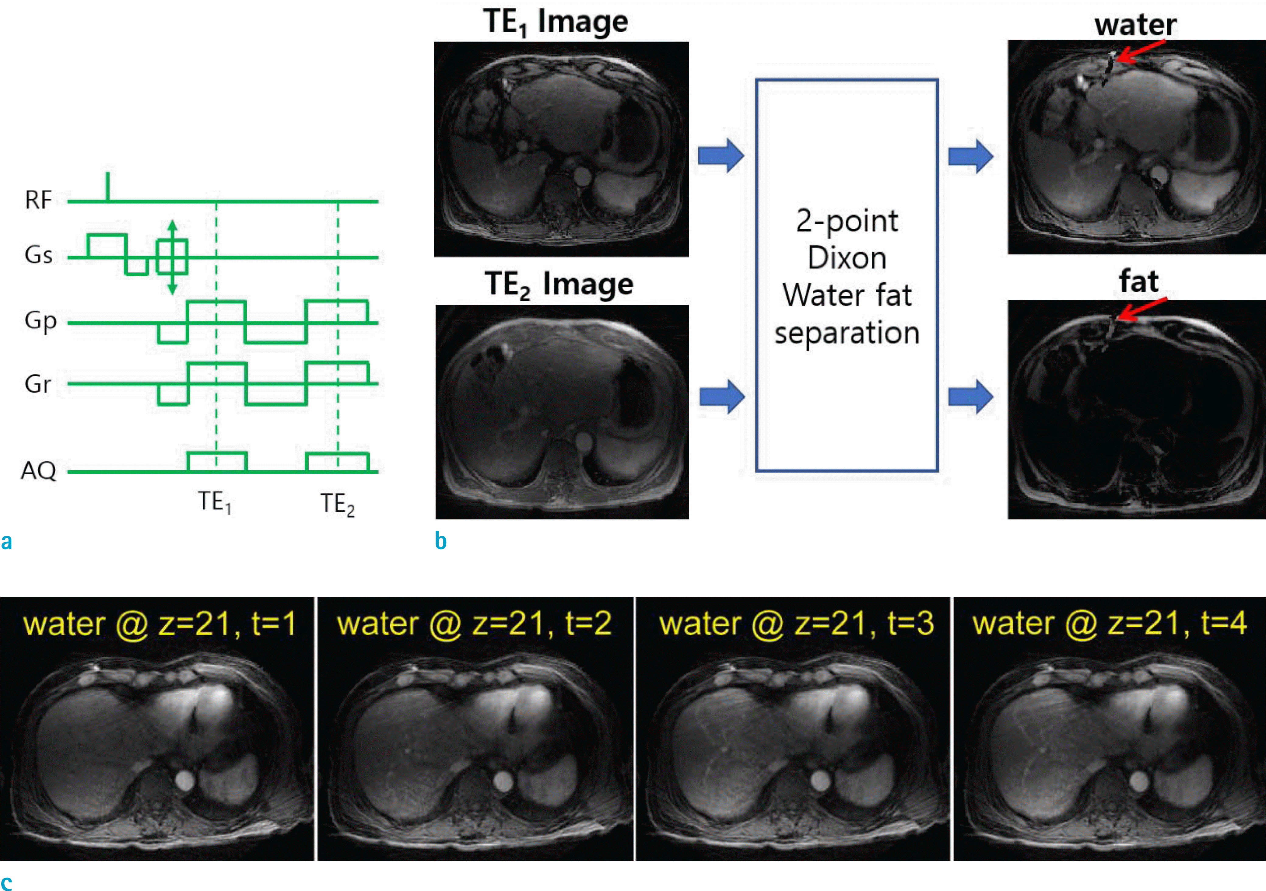
Table 1.
Summary of Selected Image Acquisitions for Arterial Phase Imaging
ACQ = acquisition; BH = breathhold; CDT-VIBE = CAIPIRINHA-dixon-twist volume-interpolated breathhold examination; CS = compressed sensing; DISCO = differential subsampling with cartesian ordering; FB = free breathing; GRASP = golden-angle radial sparse parallel imaging; HCC = hepatocellular carcinoma; KWIC = k-space weighted image contrast; N/A = not available; T = tesla; TSM = transient severe motion


 PDF
PDF ePub
ePub Citation
 Print
Print


 XML Download