



 PDF
 ePub
 Citation
 Print


Wan Soo Kim, Hyun Jin Kim
Figures and Tables
Fig. 1
Contrast enhanced computed tomography scan (A: axial section view, B: coronal section view) of epiploic appendagitis reveals fat containing ovoid nodular lesion with rim enhance adjacent to the ascending colon (arrows).

Fig. 2
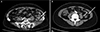
Contrast enhanced computed tomography scan cases of epiploic appendagitis. (A) A 42 years old female admit the emergency room with left lower quadrant pain for 5 days, the axial view showed the rim enhanced fat infiltration adjacent descending colon (arrow). (B) The another axial section view of the same patients. It shows fatty density oval mass surrounded by hyperattenuated rim around the sigmoid colon (arrow).
References
1. Harrigan AH. Torsion and inflammation of the appendices epiploicae. Ann Surg. 1917; 66:467–478.
2. Park JJ, Jung SA, Noh YW, et al. Clinical characteristics of primary epiploic appendagitis. Intest Res. 2009; 7:47–51.
3. Dockerty MB, Lynn TE, Waugh JM. A clinicopathologic study of the epiploic appendages. Surg Gynecol Obstet. 1956; 103:423–433.
4. Hong CS, Kwon OY, Lee JS, Choi HS, Hong HP, Ko YG. Clinical study of primary epiploic appendagitis diagnosed in an emergency department. J Korean Soc Emerg Med. 2011; 22:494–502.
5. Patel H, Abdelbaki A, Steenbergen P, Chanana C, Li S. Know the name: acute epiploic appendagitis-CT findings and review of literature. AME Case Rep. 2018; 2:8.

6. Singh AK, Gervais DA, Hahn PF, Sagar P, Mueller PR, Novelline RA. Acute epiploic appendagitis and its mimics. Radiographics. 2005; 25:1521–1534.
7. Schnedl WJ, Krause R, Tafeit E, Tillich M, Lipp RW, Wallner-Liebmann SJ. Insights into epiploic appendagitis. Nat Rev Gastroenterol Hepatol. 2010; 8:45–49.
8. Rao PM, Wittenberg J, Lawrason JN. Primary epiploic appendagitis: evolutionary changes in CT appearance. Radiology. 1997; 204:713–717.
 XML Download