

 PDF
PDF Citation
 Print
Print


INTRODUCTION
Cervical cancer is the fourth leading cause of cancer death among women in the world, and most of cases occurs in low- and middle-income countries [1]. In Brazil, it is the third cancer most frequent in the female population, with an estimate of 16,370 new cases in 2018 [2]. Early diagnosis of precursor lesions through screening may allow timely treatment, preventing the burden of invasive disease [3]. A Brazilian national guideline recommends screening with cytology every three years from 25 to 64 years old [4]. The efficiency of the program varies, which is reflected in different rates of incidence and mortality by regions [25].
Squamous cell carcinoma is the most common histological type, corresponding to about 90% of all cases, followed by adenocarcinoma. Other types such as adenosquamous carcinoma, small cell carcinoma, sarcomas, and metastatic carcinomas are less frequent [6]. Clinical staging is recommended by the International Federation of Gynecology and Obstetrics (FIGO), that describes 4 stages: from I to IV [7]. In Brazil 5-year survival is around 90% in stage I and 30% in stage IV [8].
Only 1.2% of cervical cancer cases affect women under 25 years old in Brazil [9]. Due to the low incidence of this tumor in young people, the evidence on prognosis is conflicting [1011]. Some argue that these tumors are more aggressive in younger women, but it is not known whether this is due to the fact that these women have more aggressive histological types or because of the higher involvement of high-risk pathological factors [10111213].
This study aims to evaluate the histological and stage characteristics of cervical cancer diagnosed in women under 25 years old, comparing them with older women. This is an analysis of the hospital cancer registry (RHC) database of São Paulo state, an extremely populous region, corresponding to 22% of the Brazilian population. We expect that the results of this study might contribute to improve strategies for screening and management of cases of cervical cancer in this age group.
MATERIALS AND METHODS
This was a cross-sectional study that used cervical cancer cases registered in the database of the RHC of the Oncocenter Foundation of São Paulo [14], from 2000 to 2015. Cases were women during their primary treatment, registered under code C53 of the International Classification of Diseases for Oncology (ICD-O).
Data is available for the public on line. Access was in September 2017, when 18,423 cases from 77 hospitals in the state of São Paulo were available. In the period only 471 cases were coded as C55 (UTERUS, NOS), which corresponded to 2,6% of cases coded as C53 (CERVIX UTERI). Cases are included in the database by trained hospital technicians, who systematically review medical records and place them on an online platform. All hospitals licensed for cancer care in the state fill in the database. Completeness is high. From 2000 to 2013 616,877 cases (all cancer types) were included in the database. The diagnosis was morphologically verified in 98.3% and “unknown and ill-defined primary site” was very low-ICD-O C80.9 in 1.5% and C76 in 0.4% [15].
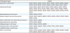
The variables were age, FIGO staging and histological type. Histological types were recorded under the morphological codes of the ICD-O-3rd edition [16] for cervical cancer. There are about 60 morphological types grouped into the following categories (Table 1): squamous carcinomas, adenocarcinomas, adenosquamous carcinomas, mesenchymal tumors, mixed epithelial and mesenchymal tumors, neuroendocrine carcinomas, malignant germ cell tumors, hematopoietic and lymphoid tumors, undifferentiated carcinomas, and others. Cases classified as stage 0 (cervical intraepithelial neoplasia grade 3 and/or adenocarcinoma in situ) were excluded.
Table 1
Cervical cancer morphological codes for histological grouping, International Classification of Diseases for Oncology-3rd edition
![]()
For histological type analysis, 2 large groups were created: women aged under or older than 25 years old. For stage analysis, we chose to categorize age into 3 groups: younger than 25 years old, between 25 and 34 years old and greater than or equal to 35 years old, so that the differences between the younger and older group would be highlighted, reducing the influence of young age (25–34) in the older group. This was not the case for histology analysis due to the low frequency of some categories.
The distribution of variables was measured in absolute numbers and frequencies. To compare the prevalence of cancer, the prevalence ratio (PR) and its 95% confidence interval (CI) were calculated by clinical stage and histological type. For the analysis of PR among the age groups according to stage, the group of women under 25 years old was the reference group. The SAS System for Windows (SAS Institute Inc., Cary, NC, USA) v9.2 was used. The study was approved by the Research Ethics Committee of Unicamp (CAAE 90764617.4.0000.5404) and followed the national guidelines. As a retrospective analysis of a database, informed consent was not needed.
RESULTS
A total of 18,423 cases were included in the database between 2000 and 2015. The mean age at diagnosis of cervical cancer was 52.62 years (standard deviation 14.78), and 204 (1.1%) cases were under 25 years old at diagnosis.
Table 2 shows the distribution of the stages at diagnosis according to the age group. Stage I (IA+IB) was the most frequent in women under 25 years old (36.2%) and also in women between 25 and 34 years old (43.4%). In women older than 34 years old, the most frequent stage was stage III (IIIA+IIIB) (31.8%). There was no statistically significant difference in the distribution of stages according to the age group. A secondary analysis of the data with the pooled stages (I, II, III, and IV) also showed no statistically significant difference between groups (Supplementary Table 1).
Table 2
Distribution of cervical cancer cases by age group and stage in 18,423 cases in São Paulo state, Brazil

![]()
In Table 3 the absolute and relative frequency of histological types are demonstrated according to the age group. Squamous carcinomas were more frequent in both age groups, corresponding to 73.5% of the cases in women under 25 years old and 78.5% of cases in women over 25 years old. There was a higher frequency in women under 25 years old in the following histological types: neuroendocrine carcinomas (PR=6.10; 95% CI=2.03–18.35), malignant germ cell tumors (PR=54.98; 95% CI=26.53–113.95), mesenchymal tumors (sarcomas) (PR=5.67; 95% CI=2.58–12.45) and hematopoietic and lymphoid tumors (PR=10.72; 95% CI=2.90–36.69).
Table 3
Distribution of cervical cancer by age group and histological type in 18,423 cases in São Paulo, Brazil

Values are presented as number (%).
CI, confidence interval, Hematopoietic and lymphoid, tumors of the hematopoietic and lymphoid tissues; Mixed epit/mesenc tumors, mixed epithelial/mesenchymal tumors; PR, prevalence ratio.
![]()
DISCUSSION
In this prevalence study, it was observed that the diagnosis of cervical cancer was an uncommon event before 25 years old, occurring in only 1.1% of the total cases. The most frequent stage in women under 25 years old and in those between 25 and 34 years old was stage I, and in women above 35 years old, stage III. There were no significant differences in the distribution of stages between age groups. The most common histological type was squamous cell carcinoma regardless of age, but women younger than 25 years old showed a higher frequency of more rare histological types, as sarcomas, neuroendocrine carcinomas, malignant germ cell tumors and hematopoietic and lymphoid tumors.
Younger women had a higher frequency of diagnosis at the earlier stage I, albeit non-significantly. Those are cases with a better prognosis. Cases diagnosed at stage IA (micro-invasive) usually are those detected through screening, which in Brazil is extensively performed in young women opportunistically. Around 15% of all screening test (cytology) performed in São Paulo state in the public system in 2014 were in women under 25 years old [17]. Because IA cases are micro-invasive tumors, and the natural history of cervical cancer is long, it is questionable whether these tumors diagnosed under age 25 would become frankly invasive if they were discovered by screening starting at age 25.
In England, where screening is organized and reaches high coverage [18], it is observed that most tumors are diagnosed in early stages in all age-groups. There, in women under 25 years old, the proportion of cases in advanced stages were higher than in older women, which may indicate a greater aggressiveness of these tumors in young women [19]. This was not observed in our study, where young women had early tumors. In our study only 25.7% of the cases in women older than 35 years old were diagnosed in the initial stage (stage I). Young women may have been diagnosed at early stages because they usually have better access to health system through primary care (pre-natal care, family planning, and common gynecologic complains). Furthermore, these cases probably were diagnosed after loop electrosurgical excision procedure for a high-grade precursor lesion.
The evidence that cervical cancer has a more aggressive behavior when comparing young and older women is due to a higher incidence of parametrial, lymph node and distant metastases observed [111213], characteristics not evaluated in this study. Age-related survival studies are needed to elucidate this issue [1011].
Squamous cell carcinoma was the most common histological type observed in all age groups. A proportional reduction in the frequency of this type of tumor has been observed with the emergence of non-squamous types [2021]. This proportional increase in adenocarcinomas and other non-squamous histological types can be explained by the lower effectiveness of the cytological screening in detecting those tumors, which in addition have a natural history not as well defined as the squamous ones. We did not observe differences in the frequency of adenocarcinomas by age groups. According to the literature, young women generally present a higher frequency of non-squamous tumors [10111322]. A higher proportion of neuroendocrine carcinomas, malignant germ cell tumors, mesenchymal tumors and hematopoietic and lymphoid tumors were observed, although the number of cases was very small. A common feature of these tumors (except neuroendocrine carcinomas) is the non-association with human papillomavirus (HPV). Neuroendocrine carcinoma is the most common rare type, and is usually associated with HPV. It is recognized as having a worse prognosis in relation to squamous carcinoma and adenocarcinoma, with a higher proportion of lymph node involvement and a higher frequency of metastases with lower survival [2324].
In relation to other rare histological types there is a shortage of data demonstrating its higher when compared to the most frequent types. An issue that makes difficult to evaluate these rare types is that, in addition to being rare, there is no uniformity in the use of the nomenclature and sometimes can be difficult to define the primary site.
Usually data quality in low- and middle-income countries is poor, so the evidence is weak. In São Paulo state, a high-middle income state in Brazil, the high data completeness in the registry of the hybrid coronary revascularization facilitated the study. The sample of more than 18,000 cases allowed us to carry out statistically significant analyzes, even when data were distributed by age groups. The main limitation is in relation to the non-population nature of the registry, preventing the estimation of incidence rates. In addition, we do not know if the cancers in the registry were detected by screening or not. Efforts should be made to improve quality of data.
A better understanding of the clinical presentation of this cancer may help to plan strategies for early detection of cancer and management in young women. Screening in women under 25 years should not be recommended as a public health policy, since it is a rare disease [9], the effectiveness is questionable [1925] and the morbidity related to overtreatment is significant [26]. However, symptomatic young women should be promptly investigated and a gynecological routine exam can be performed when women seeks for a clinical care or counseling after sexual debut.
In conclusion the proportion of cervical cancer cases diagnosed in women under 25 years old was 1.1%. In this group, early tumors (stage I) were the most frequent. Squamous carcinoma was the most frequent histological type regardless age, but rare histological types were more frequent in young women.
 XML Download