

 PDF
PDF ePub
ePub Citation
 Print
Print


INTRODUCTION
Percutaneous sacroplasty (PSP) has emerged as an effective treatment in the setting of both osteoporotic and metastatic sacral insufficiency fractures (SIFs), with patients experiencing nearly full pain relief and mobility improvement immediately and longitudinally (123). Analogous to percutaneous vertebroplasty (PVP) in the cervical and thoracolumbar spine, PSP involves two equally important procedures: precise needle placement and bone cement deposition in the fractured sacrum and/or metastatic lesion. Although PSP was first utilized in a metastatic lesion of the sacrum for pain alleviation in the year 2000 (4), PSP for osteoporotic SIFs is well-described and widely reported (567). However, published literature describing the treatment of patients with painful sacral tumors is limited (891011121314), with even scarcer descriptions for sacral metastases (9111214).
The surgical approach for PSP is determined by the location of the lesion and professional preference. Five primary approaches have been reported previously: the short-axis approach, the long-axis approach, the transiliac approach, the anterior-oblique approach, and the interpedicular approach (115161718). As for sacral metastases, the goal is to precisely place the bone needle within the sacral lesion, with each puncture approach having its own advantages and disadvantages. However, there is limited evidence on patient outcomes and complications following the interpedicular approach for sacral tumors (16), especially for metastases involving multiple sacral vertebral bodies. The purpose of this study was to report our experience with the use of PSP by an interpedicular approach for the treatment of painful sacral metastases involving multiple sacral vertebral bodies.
MATERIALS AND METHODS
Patient Population
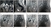
This prospective study was approved by the institutional ethics committee of our hospital, and informed consent was obtained from all patients. From March 2017 to September 2018, patients with painful sacral metastases involving multiple sacral vertebral bodies were recruited from our department for treatment with PSP by an interpedicular approach. Ten patients (six male and four female; mean age, 56.3 ± 13.8 years) with painful sacral metastases involving multiple sacral vertebral bodies were included in this study. All of the patients presented with severe pain without neurological deficits and were referred to our institution because of pain that was resistant to conventional treatments, including opioids, chemotherapy, and radiotherapy. All patients showed survival longer than 3 months following the performance of PSP. All patients referred for treatment were asked to complete a short questionnaire regarding the presence, severity, and duration of pain and disability. All patients had pathologically confirmed primary cancer lesions and computed tomography (CT), magnetic resonance imaging (MRI), or both CT and MRI findings showing the presence of osteoblastic lesions in the painful area; all patients received imaging examinations before the procedure to determine the degree of lesion invasion with or without pathological fracture, and to rule out other chronic diseases that could cause back pain. After the procedure, a noncontrast CT examination to assess cement leakage was performed in all patients.
Procedural Details
The procedures were performed under biplane fluoroscopic guidance (Innova IGS630; GE Healthcare, Fairfield, CT, USA) in all the cases. The patient was placed in a prone position on an operating table. Heart rate monitoring and pulse oximetry were carried out continuously throughout the procedure. The blood pressure was monitored externally and recorded automatically every five minutes.
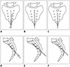
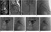
Strict aseptic technique was employed throughout the procedures with skin sterilization and standard draping techniques. The appropriate access point, puncture angle, and distance from the skin to the lesion were determined in advance based on CT or MRI images. The dural sac was imaged on MRI, and the puncture approach was selected appropriately to minimize the risk of cerebrospinal fluid (CSF) leakage and nerve damage. The skin incision site was generally chosen in the midline over the sacral hiatus at the level of the third or fourth sacral vertebral body. After local anesthesia, a 13-gauge bevel needle (Cook Inc., Bloomington, IN, USA) was punctured through the skin and subcutaneous tissue parallel to the long axis of sacrum, and advanced until the tip reached the posterior wall of the sacral spinal canal. After penetrating the posterior wall of the sacral spinal canal, the needle was carefully advanced through the canal until it approached the anterior border of the sacral canal at the level of the third and fourth sacral vertebral bodies. During penetration of the sacral canal, the patient's response was continuously monitored, and the approach was adjusted if necessary. At that point, the needle was angled cranially or caudally along an optimal path. The needle was then advanced through the sacrum until it approached the anterior wall of the sacral vertebral body, parallel to the anterior border of the sacrum. Anteroposterior and lateral fluoroscopy were used throughout the procedure, and the bevel tip allowed the needles to be “steered” during placement along the required course to avoid penetration through the anterior wall of the sacral body and the sacral foramen. Then, mixed bone cement (polymethyl methacrylate [PMMA]) (PALACOS® V; Heraeus Medical GmBH, Hanau, Germany) was injected into the target sacral body when it became doughy and could stand at the tip of the bone cement inserter. The injection process was monitored continuously under fluoroscopic control in the anteroposterior and lateral planes. During cement injection, the bone needle cannula was gradually withdrawn as needed to allow a greater area of cement deposition. Injection was ceased when substantial resistance was met or when the PMMA reached the margin of the sacral foramen or sacral vertebral body (Figs. 1, 2, 3). The amount of bone cement used for PSP was noted. All patients received CT scans immediately after the procedure to assess cement distribution and to determine whether there was any PMMA leakage.
Data Collection
This study was a prospective, single-arm, paired comparison observational study with patients serving as their own controls. Data regarding technical success, PMMA volume injected, pain relief, pain medication changes, functional outcomes, length of hospitalization, and complications were evaluated at follow-up consultations or at patient death. Technical success was defined as successful puncture of the sacral body with an interpedicular approach, and sequential injection of bone without major complications. Any potential complications after PSP, such as cement leakage, wound infection, nerve injury, or pulmonary embolism, were recorded. We defined major complications as accidental nerve root injury, cauda equina syndrome, pulmonary embolism, intestinal rupture, or perioperative mortality, and minor complications as postoperative urinary retention, wound hematoma, and infection.
Outcomes were assessed according to the Oswestry disability index (ODI) (19), visual analogue scale (VAS) score (20), and pain medication changes for mobility and pain. The VAS is a 10-point scale that patients are asked to mark based on their level of back and/or leg pain, with scores of zero indicating no pain and 10 indicating the worst pain possible. The ODI is a score on a 10-item ordinal scale on which each item has six possible responses. The score is measured as a percentage (0–100%), with an increasing score indicating increasing disability. ODI scores are given throughout this article in terms of percentages. The number of opioids prescribed was compared before and after the procedure. Efficacy was determined by a combination of a descriptive or a numerical reduction (defined as any decrease in the pre-procedural pain level) of post-procedural pain compared with pre-procedural pain. A difference in VAS ≥ 3 points, meaning at least a 30% reduction in pain, was considered a clinically significant result, as reported in previous studies (2122).
Statistical Analysis
All statistical analyses were performed using commercially available software (SPSS Version 16; SPSS Inc., Chicago, IL, USA). Data were expressed as mean ± standard deviation. The results at all of the study time points were compared using a paired t test, with a p value of less than 0.05 considered statistically significant.
RESULTS
Patient Demographics
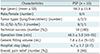
Patient demographics and procedural information are shown in Table 1. Primary tumor types were lung tumors (n = 4); liver tumors (n = 3); and thyroid tumor, malignant neurilemmoma, and epithelioid angiosarcoma (n = 1 for each). All the 10 patients underwent CT, MRI, or both before PSP. The number of treated lesions ranged from two to three; 8 of the 10 patients had two lesions ranging from S1 to S2, 1 patient had two lesions ranging from S2 to S3, and 1 patient had three lesions ranging from S1 to S3, for a total of 22 metastases. In addition, the interval between the completion of imaging examination and PSP was 4.0 ± 3.6 days (range, 1–13 days) and that between presentation with sacral symptoms and PSP was 1.70 ± 2.23 months (range, 0.5–8 months).
Procedure Safety
PSP was technically successful and well-tolerated in all patients. The mean procedure duration was 48.5 ± 3.0 minutes (range, 45–55 minutes). The mean volume of PMMA injected was 7.80 ± 2.44 mL (range, 5–12 mL), and length of hospitalization was 7.1 ± 2.4 days (range, 3–10 days) (Table 1). There were no documented occurrences of surrounding tissue damage (no nerve damage or spinal damage), and there were no other complications such as bacterial infections, anesthesia-related adverse events, CSF leakage, bleeding, or their combinations. PMMA leakage was observed in 30% (3/10) of the patients during the operation under real-time fluoroscopic guidance, which occurred in the presacral soft tissue (n = 3). All instances of leakage were asymptomatic and required no special treatment.
Clinical Evaluation
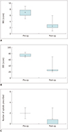
Changes in VAS, ODI, and pain medication usage following PSP are shown in Figure 2. The average preoperative VAS was 6.90 ± 1.20, which decreased to 2.70 ± 1.34 immediately after the operation (p < 0.01) (Fig. 4A). Meanwhile, ODI collected from the 10 patients decreased from 74.40 ± 5.48 pre-procedure to 29.60 ± 14.57 immediately post-procedure (p < 0.01) (Fig. 4B). The median number of opioids prescribed per patient decreased from 2 (interquartile range [IQR] 1–3) pre-procedure to 1 (IQR 0–3) post-procedure (p < 0.01) (Fig. 4C). Four of the 10 patients had no need for opioid usage; 5 of the 10 patients showed decreased opioid usage; and only 1 patient showed unchanged usage.
DISCUSSION
The bone is the most common site for metastatic disease. Approximately 66% of bone metastases are extraspinal, with the major proportion occurring in the pelvis and sacrum (2324). Meanwhile, metastases are the most common malignant lesions in the sacrum (24), accounting for 1% to 7% of all spinal tumors (2526). Anatomically, the sacrum is a weight-bearing structure that dissipates vertical axial forces, which are transmitted along the spinal axis. When the sacrum is involved by metastases, a series of symptoms may present, including neurological deficits, severe disability, and debilitating local or radiating pain that is exacerbated by sitting, which may render such an essential resting position excruciating (27).
Adequate treatment for symptomatic bone metastases is as important as the treatment of the primary tumor in order to maintain patient quality of life. In recent years, image-guided PSP has emerged as a feasible and effective treatment for painful and disabling sacral metastases, and this technique aims to alleviate pain and regain mobility in a manner similar to PVP or percutaneous kyphoplasty (PKP) of the cervical and thoracolumbar spine. Several reports and small series have demonstrated this technique's efficacy in providing pain relief and bone stabilization, although the mechanism underlying its analgesic effect remains incompletely understood (9111214282930).
PSP may be technically more challenging and arduous than PVP or PKP due to the complexity of the sacral anatomy (including its pyramidal shape and porous structure), difficulty in visualizing the anterior sacral cortical margin with fluoroscopy, poorly defined fluoroscopic landmarks for bone needle placement, and the risk of sacral nerve root or spinal canal compromise due to cement migration (1531). Therefore, a thorough understanding of the radiologic anatomy of the sacrum is critical for avoiding a major complication during this procedure. Nonetheless, there are still five primary approaches for sacrum lesions elaborated in the published literature: the short-axis approach, the long-axis approach, the transiliac approach, the anterior-oblique approach, and the interpedicular approach (11518). According to Denis' classification, there are three zones of sacral fractures: zone I, sacral ala lateral to neural foramina; zone II, foramina area; and zone III, sacral body and spinal canal (32). Generally, zone I fractures solely involve the sacral ala and can be treated by the short-axis approach, the long-axis approach, the transiliac approach, and the anterior-oblique approach; zone II fractures are restricted to the sacral foramina and can be treated only by the long-axis approach; and zone III fractures are midline and involve the sacral vertebral body, and can be treated by the transiliac approach and the interpedicular approach. However, when multiple sacral vertebral bodies are involved by tumor infiltration, the interpedicular approach seems to be the only feasible approach for PSP.
This is the first report, to the best of our knowledge, to demonstrate PSP on this cohort of patients using the interpedicular approach. In the present study, PSP was found to be feasible and highly efficacious, and pain relief and functional improvement were early and adequate. After PSP, 90% of the patients experienced immediate and significant improvement of pain and mobility, similar to other previously published literature (9111214). Therefore, PSP is a minimally invasive procedure that may be useful for patients with painful sacral metastases involving multiple sacral vertebral bodies–particularly for those in poor general condition–and offers several advantages over conventional treatments. First and most importantly, sacral body lesions can be directly accessed and treated without passing through the foramina zone, decreasing the risk of injury to sacral nerves. Second, multiple sacral body lesions can be managed in one session by withdrawing the bone needle, reducing the risk of repeat and inadequate puncture. Third, PSP with the interpedicular approach can be performed under fluoroscopy only without the need for additional CT guidance, reducing operation time, cost, and radiation exposure.
Although we did not encounter any clinical complications related to needle puncture and cement leakage in the present study, potential complications of PSP do remain, which mainly include nerve damage and cement leakage, which may migrate into the neural foramina, the spinal canal, or pulmonary circulation. Other potential complications include gastrointestinal injury, bleeding, and infection. Moreover, CSF leakage is another severe complication of PSP with an interpedicular approach, which should be avoided during needle placement. The dural sac should be preoperatively imaged on MRI and the puncture site should be selected below the subarachnoid cavity level, generally in the midline over the sacral hiatus at the level of the third or fourth sacral vertebral body. In addition, a thorough understanding of the radiologic anatomy of the sacrum is critical for avoiding a major complication during this portion of the procedure.
There are several limitations in this study. First, the sample size was not large enough to make broad generalizations. Second, there is no comparison with other therapeutic options such as surgical treatment or radiotherapy. Third, the study did not present the exact amount of radiation dose during the PSP procedure, although it did provide the operation time. However, the results of this study are promising, and a larger prospectively controlled comparison study is necessary to confirm our findings.
In conclusion, PSP with the interpedicular approach is an effective, safe, and minimally invasive procedure for treating painful sacral metastases involving multiple sacral vertebral bodies, which can provide marked reduction of pain and improvement of mobility. Further large-scale prospective studies are needed to confirm these findings.
 XML Download