

 PDF
PDF Citation
 Print
Print


Sinus of Valsalva aneurysm (SVA) is a relatively rare heart disease in humans that is often congenital [1]. Overall, the disease accounts for 0.6% of congenital heart abnormalities [2]. Interestingly, it occurs most often in the right coronary cusp and least often in the left coronary cusp [34]. SVA is more common in men [5] and in the Asian population [67]. Furthermore, it frequently involves a ventricular septal defect (VSD), aortic regurgitation, and pulmonary artery stenosis. The normal diameter of the sinus of Valsalva is 4.0 cm for men and 3.6 cm or less for women [8]; this reference value is useful for diagnosing SVA in humans. Although SVA seldom presents with clinical symptoms before rupturing, a thrombus can form quickly within the aneurysm [9], leading to fatal heart failure when it ruptures [10]. Therefore, treatment is recommended even before rupture [11].
Kleine et al. [12] first reported a ruptured SVA in a dog in 1966. Since then, there have been some cases of rupture [1314] and a report of a giant unruptured aneurysm with a thrombus [15], but knowledge of this condition is quite limited. Moreover, there is also no prescribed reference size for the sinus of Valsalva in dogs, and no criteria exist for diagnosing SVA.
The present case was diagnosed with an unruptured right SVA due to a suspected displacement and dilation of the sinus of Valsalva accompanied by a VSD on echocardiography and a dilation of the right sinus of Valsalva on cardiac catheterization. As the present case was a juvenile small breed of dog, weighing 1.0 kg, follow-up observations were chosen because open-heart surgery with a cardiopulmonary bypass would be too risky. After the dog died, the necropsy also confirmed the dilated sinus of Valsalva.
A 2-month-old male Maltese terrier weighing 1.0 kg was found to have a heart murmur by a home veterinarian and was brought to the authors' hospital for examination (day 1).
His general condition at the first examination was favorable; he was active, had a healthy appetite, and had no respiratory problems. He had a heart rate of 184 bpm, respiratory rate of 24/min, and body temperature of 38.8°C. The visible mucous membrane was pink, and the femoral artery pressure was normal. The systolic blood pressure/diastolic blood pressure (mean arterial pressure) was 138/107 (120) mmHg. Upon auscultation, a Levine 4/6 systolic heart murmur was heard that was loudest at the base of the heart. An electrocardiographic examination revealed sinus rhythm and no arrhythmias. The chest X-ray did not show any abnormalities in the lung field, and the dog had a vertebral heart size of 9.0 and a cardiothoracic ratio of 72.0%.
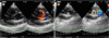
The right parasternal/left ventricular outflow tract transaxial section and short-axis view on echocardiography revealed a VSD (flow rate 3.6 m/s) near where the aortic valve attaches to the anterior wall of the aorta (AO), as well as the right sinus of Valsalva at a location that covered the defect hole (Fig. 1). Cardiac catheterization was planned with a provisional diagnosis of VSD with a displacement of the sinus of Valsalva.
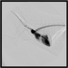
On day 15, cardiac catheterization was conducted. Anesthesia was induced with atropine (0.05 mg/kg, subcutaneous injection), ampicillin (Ampicillin sodium injection 1 g; Kyoritsu Seiyaku Corporation, Japan) (30 mg/kg, intravenous injection [iv]), butorphanol (Vetorphale; Meiji Seika Pharma, Japan) (0.2 mg/kg, iv), midazolam (Dormicum injection 10 mg; Astellas Pharma, Japan) (0.2 mg/kg, iv), and propofol (Mylan injection 1%; Mylan, Japan) (to effect, iv). Anesthesia was maintained with isoflurane (isoflurane for animals; Intervet K.K., Japan) (1.5–2.2%). Under X-ray fluoroscopic guidance, a 0.018 guide wire and a multipurpose angled catheter (Goodtec angiographic catheter; Goodman, Japan) were inserted from the left common carotid artery, and an iodine-based contrast agent (Oypalomin 300; Konica Minolta Japan, Japan) was injected into the left ventricle (LC), AO, and right ventricle (RC). Contrast enhancement from the LC revealed a dilated right sinus of Valsalva (Fig. 2). The RC was not enhanced distinctly through the defect hole, and the ventral side of the right sinus of Valsalva was enhanced only slightly.
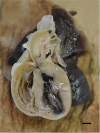
This procedure gave a diagnosis of a right sinus of SVA accompanied by a VSD. The wound was closed using the standard method, and the dog recovered from anesthesia uneventfully. Unfortunately, the dog died due to a worsening of his condition, and a necropsy was performed. After the heart and lungs were fixed in formalin, the left ventricular outflow tract was opened, showing that the sinus of Valsalva had a 7 mm diameter and a VSD defect hole on its opposite surface (Fig. 3).
The present case was confirmed to have a VSD by cardiac ultrasound on day 1. On the other hand, contrast enhancement of the LC during cardiac catheterization on day 15 revealed no enhancement of the RC through the defect hole, and the ventral side of the right sinus of Valsalva was enhanced only slightly. This finding suggested that the VSD hole had been covered by a dilation of the sinus of Valsalva, possibly weakening the shunt from the LC to the RC.
In dogs, the disease affects males almost exclusively, including the present case. Many studies have reported involvement of the left coronary cusp in larger dogs [131415]. This report is the first to describe an unruptured SVA in the right coronary cusp of a Maltese terrier dog. SVA occurs rarely, making it difficult to accumulate data, but as observed with humans, the disease appears to affect males more often than females in large and small breeds of dogs.
 XML Download