 PDF
PDF ePub
ePub Citation
 Print
Print


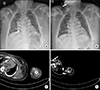
A 35-year-old man with end-stage renal disease was admitted for chronic respiratory failure. During hospitalization, arteriovenous graft malfunction occurred and a computed tomography (CT) angiography was planned. A 5 French power injectable peripherally inserted central catheter (PICC, Turbo-Ject®; Cook Medical, Bloomington, IN) was inserted under fluoroscopic guidance, and located in the distal superior vena cava and fixed to the skin with sutures (Fig. 1A). Surprisingly, two days after catheter insertion, chest X-rays showed the catheter had become displaced into the ipsilateral internal jugular vein (Fig. 1B). The insertion site was clear, and no evidence of other external force was applied. During the review of CT performed one day ago, we found that the position of the catheter changed after injection of the contrast agent (Fig. 1C, D). An upper extremity CT angiography was performed with 150 mL of iodinated contrast infused at a rate of 5 mL/s. There was no problem with contrast injection, but the position of the catheter was changed in the delayed image performed three minutes later. The catheter was removed after positional changes were noted and there were no complications. This case highlights that displacement of power injectable PICC can occur even within the authorized infusion rate and reminds us again that it is essential to compare previous chest x-rays for detection of complications associated with indwelling devices.
- About Synapse
- How to Participate
- KAMJE Databases


- About Synapse
- How to Participate
- KAMJE Databases
Journal List > Chonnam Med J > v.55(2) > 1124738
- TOOLS
 XML Download
- Similar articles