

 PDF
PDF Citation
 Print
Print


INTRODUCTION
Periapical diseases may persist even after completion of root canal treatment due to persistent or recurrent microbial infection [12]. Non-surgical retreatment is usually the first choice when endodontic treatment has failed because it provides an additional opportunity to eradicate the intra-radicular source of infection. Endodontic retreatment procedures include complete removal of the existing root filling material to the apical foramen followed by biomechanical preparation, disinfection, and subsequent re-filling of root canals [3]. However, some filling material may remain on the canal walls which can harbor micro-organisms, resulting in less favorable treatment outcomes.
Numerous new endodontic root canal sealers have been recently introduced, but there is scant information regarding these materials. First, EndoSeal MTA (Maruchi, Wonju, Korea), a finely pulverized pozzolan-based mineral trioxide aggregate (MTA), was introduced. Pozzolan cement, the main component of this sealer, acquires cementitious properties after pozzolanic reaction with calcium hydroxide and water, and allows sufficient flow of pre-mixed substrate through the injection tip with an adequate working consistency. Favorable mechanical characteristics, such as a fast setting time (approximately 4 minutes), higher washout resistance than other commercially available MTAs, and biologic effects including biocompatibility, mineralization potential, and an odontogenic effect of the pozzolan cement were reported [45]. Second, EndoSequence BC Sealer (BC sealer; Brasseler USA, Savannah, GA, USA), also known as iRoot SP (Innovative Bio-ceramix, Vancouver, Canada), has been more recently introduced as a premixed and injectable root canal sealer that uses bioceramic technology. According to the manufacturer, this highly biocompatible calcium silicate-based sealer does not shrink, can form strong chemical bonding with the dentin hydroxyapatite, and can be used predictably even with a single-cone technique [6].
In some retreatment studies, the precise volume of residual filling material was determined using micro-computed tomography (micro-CT), which allows a non-invasive three-dimensional quantitative evaluation of the efficacy of treatment [7]. By allowing observation of the canal during various stages of root canal retreatment, this method overcomes the limitations of other assessment methods.
The purpose of this study was to compare the amount of residual filling material after retreatment in canals filled with gutta-percha (GP) and various sealers (EndoSeal MTA, BC sealer, and AH Plus [Dentsply Maillefer, Ballaigues, Switzerland]), using micro-CT imaging.
MATERIALS AND METHODS
Sample preparation
This study was approved by the Institutional Review Board of Seoul St. Mary's Hospital, the Catholic University of Korea (KC 16SISI0109). Extracted human permanent teeth were radiographed and examined to check the canal morphology, and we excluded teeth with severe root curvature (> 25°) [8] or canal obliteration. Finally, we selected 21 single-rooted mandibular premolars with one canal, 21 double-rooted maxillary premolars with two canals, and 15 mandibular second molars with class I (using the classification of Fan et al. [9]) C-shaped root canals. All teeth were stored in 5.25% sodium hypochlorite (NaOCl) solution for soft tissue removal, and the root surfaces were cleaned with scalers. All samples were decoronated to standardize root length (10–12 mm in single-root teeth, 9–11 mm in double-root teeth, 8–9 mm in C-shaped root teeth) and the teeth were stored in distilled water.
Root canal preparation
An access cavity was prepared with a size 4 round bur and an Endo-Z bur (Dentsply Maillefer) on each tooth. A size 10 K-file (Dentsply Maillefer) was placed in the root canal until it could be observed at the apex, and the working length (WL) was then determined by subtracting 1 mm from this length. The root canals were instrumented with an X-Smart electronic motor (Dentsply Maillefer) and ProFile system (Dentsply Maillefer) in a crown-down sequence to a master apical file (MAF) size of 35/0.06 in single-rooted teeth and 30/0.06 in multi-rooted teeth. One milliliter of 3% NaOCl was used to irrigate each canal between each file, and the final irrigation was conducted with 1 mL of NaOCl and 1 mL of 17% ethylenediamine tetraacetic acid (EDTA) for 1 minute, followed by 5 mL of NaOCl. A plastic syringe with a 27 G needle (Monoject endodontic needle, Tyco/Kendall, Mansfield, MA, USA) was used and inserted deep down, but not reaching the binding point. After the root canal walls were dried with paper points (Dentsply Maillefer), master GP cones (Diadent, Cheongju, Korea) were selected according to the MAF size of each canal and evaluated for fitness by checking the tug back sensation.
Root canal obturation
We randomly subdivided the single- and double-rooted teeth (n = 7/each) and the C-shaped root teeth (n = 5) into three groups according to the sealers used: 1) an epoxy-resin based sealer (AH Plus), 2) a calcium silicate-based sealer containing zirconium oxide (BC sealer), and 3) an MTA-derived calcium silicate-based sealer (EndoSeal MTA).
In the AH Plus group, the master cone was coated with AH Plus sealer and placed at an appropriate length. The root canal was obturated by the continuous wave technique with System B (SybronEndo, Orange, CA, USA) and Obtura (Obtura Spartan, Fenton, MO, USA). In the BC sealer and EndoSeal MTA groups, the root canals were obturated according to the manufacturers' recommendations. The sealers were delivered through their injection tips, and the master cones were inserted and pumped two or three times. Then, the master cones were cut off at the orifice level using System B (SybronEndo).
Laboratory micro-CT imaging for measurement of filling material
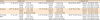
Each tooth was scanned using a micro-CT scanner (Skyscan 1172, Bruker microCT, Kontich, Belgium), with a voxel dimension of 23.86 μm under a source voltage and current of 100 kV and 100 μA, respectively. We used a 0.5-mm-thick aluminum filter and a rotational step of 0.83° increments for 180° of rotation. Each image was assembled to reconstruct the cross-sectional root canal data using NRecon (ver. 1.14.4.1, Bruker microCT). Horizontal two-dimensional slices at 3 mm above the apex (apical), 2 mm below the cemento-enamel junction (coronal), and at the middle point between the apical and coronal points (middle) were obtained using Dataviewer (ver.1.5.0, Bruker microCT). CTAn (ver.1.14.4.1, Bruker microCT) was used to pixelate each of the images, and only the root canal space was cut out; the percentage of filling material area was calculated at the coronal, middle, and apical slices [12].
Root filling removal and re-instrumentation
To remove the canal filling materials, root canals were instrumented with GG drills sizes 2, 3, and 4, and the ProFile system was used with MAF sizes of 40/0.06 in single-rooted teeth and 35/0.06 in multi-rooted teeth. The MAF size used in the retreatment was one size up from that used in the initial treatment. One milliliter of 3% NaOCl was used to irrigate each canal between each file, and the final irrigation was conducted with 1 mL of NaOCl and 1 mL of 17% EDTA for 1 minute, followed by 5 mL of NaOCl. A plastic syringe with a 27 G needle (Monoject endodontic needle, Tyco/Kendall) was used and inserted deep down, but not reaching the binding point.
Laboratory micro-CT imaging for measurement of remaining filling material
Micro-CT was used to scan roots the second time, using the same settings as those for the first scan. Residual filling materials were measured at the coronal, middle, and apical thirds. We calculated the percentage area of residual materials using the equation below by root type (single-, double-, or C-shaped root), sealer type, and position of the horizontal section.

Statistical analysis
The normality of the distribution of the calculated data was tested using the Shapiro-Wilk normality test, and the data were analyzed using the Kruskal-Wallis test and the Mann-Whitney U test with the Bonferroni correction to evaluate differences in the percentage of remaining filling materials among the tested sealers according to the root type and root position. The level of significance was set at α = 0.05. All statistical analyses were performed using SPSS ver. 22 (IBM Corp., Armonk, NY, USA).
RESULTS
The calculated values for the percentage of remaining filling material are presented in Table 1. Overall, there were no significant differences in the percentage of remaining filling material between the tested sealers in single- and double-rooted teeth. However, EndoSeal MTA showed the highest values, followed by AH Plus and BC sealer, in C-shaped roots (p < 0.05). The percentage of remaining filling material of AH Plus and EndoSeal MTA was significantly higher in C-shaped roots than in single- or double-roots (p < 0.05), while that of BC sealer was similar regardless of the root type.
Table 1
Percentage of remaining filling material according to the sealer, root type, and position in the canals
The values are presented as median percentage of the remaining filling material, and values in parentheses are first and third quartiles. Uppercase superscript shows statistically significant differences between root types within each sealer. Lowercase superscript shows statistically significant differences between endodontic sealers within each root type.
*Statistically significant differences between root positions within each sealer and root type.
Regarding root position, there were no significant differences among the coronal, middle, and apical third, except for EndoSeal MTA, which showed a significantly higher percentage of remaining filling material at the apical thirds of single- and double-roots (p < 0.05).
DISCUSSION
Thoroughly removing the filling material from root canals is crucial in root canal retreatment, because filling material that remains on root canals may harbor of necrotic tissue or bacteria, which could be responsible for periapical inflammation or pain after root canal retreatment. Previous researchers determined that it was impossible to completely remove the filling material [13]. Various sealers have been introduced onto the market, but their retreatability remains unknown. In addition, efforts to develop an ideal sealer have focused more on complete obturation of the root canal than on retreatability. Previous studies on retreatment have mostly focused on GP remnants, and most investigators used the same sealer to compare the amounts of remnant material according to the filling or removal method. Therefore, we evaluated the retreatability of the relatively newly developed EndoSeal MTA and BC sealers compared with an epoxy resin sealer (AH Plus) as the standard.
According to the manufacturer, BC sealer is a radiopaque, bioceramic material composed of zirconium oxide, calcium silicates, calcium phosphate monobasic, calcium hydroxide, and filler and thickening agents. It is hydrophilic and uses the moisture present in dentinal tubules to initiate and complete its setting reaction. It has no shrinkage upon setting, resulting in a gap-free interface between GP and dentin. The manufacturer also states that this sealer is highly biocompatible and antibacterial during the setting reaction because of its high alkalinity. However, in retreatment procedures, some investigators found more residual BC sealer than conventional sealers, and stated that it was difficult to obtain apical patency at areas of apical accumulation [1415]. The inability to regain WL and/or patency may compromise retreatment by preventing proper cleaning and shaping of the apical canal space, which may harbor bacteria [1415].
MTA-based sealers have been claimed to stimulate biomineralization, and to offer a superior seal. Furthermore, these materials have been shown to exhibit stronger bonds to dentin than zinc oxide eugenol-based cements and a sealing ability similar to that of epoxy resin-based sealers [1617181920]. However, few researchers have addressed the difficulty of removing MTA-based sealers from root canals. This is an important concern, given the widespread use of MTA-based materials in contemporary endodontics because of their ability to undergo biomineralization. Because MTA-based materials are known to harden upon setting, the ability to retreat canals obturated with these sealers is an issue that needs to be addressed [21]. EndoSeal MTA is a premixed sealer consisting of calcium silicate, calcium aluminate, calcium sulfate, and radiopaque material, and is a ready-to-use calcium silicate–based product stored in an air-tight syringe. This type of sealer absorbs moisture during the setting reaction and sets spontaneously. In previous studies, EndoSeal MTA showed satisfactory physical properties, biocompatibility, good bond strength, fracture resistance of root dentin, minimal discoloration, and superior sealer distribution [22].
In this study, EndoSeal MTA demonstrated insufficient removability. Especially in C-shaped root canals, we observed large amounts of residual filling material after retreatment. Bogen and Kuttler [19] did not recommend EndoSeal MTA as a routine ortho-grade root canal filling material, especially in complicated root canal systems such as C-shaped root canals, because the substance is sandy and irretrievable. Although EndoSeal MTA has many favorable aspects, as noted above, more attention is necessary in complicated root canal systems. In particular, it is necessary to develop biomechanical or chemical procedures for removing MTA. Although the differences were not statistically significant, EndoSeal MTA showed more residual material in the apical third than the other two sealers. As discussed above, this trend may also be related to the properties of MTA in combination with dentin; specifically, MTA can form hydroxyapatite or carbonated apatite when in contact with fluid in a dentinal tubule. In other words, a chemical bond may form between MTA and dentinal walls [21].
There were no differences between single- and double-roots. As in previous studies, retreatment using a ProFile in a straight canal appeared to be effective regardless of sealer [2324]. Research on retreatment in C-shaped root canals is lacking, and our study also showed significantly lower removal efficiency in C-shaped root canals than in root canals with other morphologies. However, there were clear differences between sealers. We observed the largest amount of remnant material for EndoSeal MTA, followed by AH Plus and BC sealer in descending order, in contrast to previous studies [1421]. This might be ascribed to the fact that previous investigators studied the relatively straight canals of canines and mesiobuccal canals of mandibular molars. In our comparison of the amount of remnant material by root canal morphology, the completeness of removal was lowest in C-shaped root canals regardless of the sealer; this is related to the structural complexity of C-shaped root canals. The presence of a thin fin, slit, and web makes it difficult to debride and clean C-shaped canals. In canal filling, it is important that EndoSeal MTA has the advantage of being able to fit into such a complicated structure, but it is rather difficult to remove. To overcome this, intentional replantation is recommended, rather than ortho-grade retreatment. Some authors have shown that additional passive ultrasonic irrigation with xylene can be an option [2526]. Some studies have highlighted that BC sealer requires a long time for final setting [2728]. In our experiments, we found that more EndoSeal MTA remained than BC sealer in C-shaped canals, especially in complex forms. We believe this is the result of the relatively fast setting time of EndoSeal MTA combined with the mechanical retention of the sealer facilitated by the C-shaped canal.
Separately, more filling material remained in the apical thirds than in the middle and coronal thirds regardless of sealer type, which is consistent with previous studies [2930]. During retreatment, material in the coronal and middle thirds is more accessible via mechanical and chemical removal techniques than is material in the apical third, and material may also be pushed to the apical third during removal. Root canal anatomy in the apical third is complicated, as lateral canals and ramifications make complete removal of the filling material very difficult [31]. To remove more filling material, several methods can be used. In less complicated root canals, such as single-canal teeth, distal canals of mandibular molars, and palatal canals of maxillary molars, the canal can be enlarged by one size. Some researchers demonstrated that retreatment with canal enlargement two sizes beyond the filled canal size could far better reduce the amount of residual sealer [3233]. In this study, a sharply higher amount of remnant material was noted, especially at the apical third, compared with previous studies, which could have been related to the one-size-up MAF at retreatment.
This study has some limitations, such as an insufficient sample size and inadequate standardization of the sample. However, it is meaningful to compare the degree of removal of the newly introduced sealer using micro-CT, which is widely used to evaluate endodontic treatment or retreatment. This is the first study on the removal of EndoSeal MTA using micro-CT. Micro-CT is a nondestructive, precise, reproducible imaging modality that provides three-dimensional reconstruction and allows for quantitative evaluation of residual filling material, sealer, and dentin separately on the canal wall before and after retreatment [2324]. Existing retreatment studies have focused on the types of files, application method, and use of solvents in straight or curved canals. This has led to the refinement of effective strategies for removing GP, but some findings of those studies are not applicable to sealers. In fact, filling the complex anatomical root canal system requires a sealer, rather than GP, but studies on removing sealer according to its material properties are lacking. Even so, extensive research has been done on AH Plus [252634]. Investigators have shown that AH Plus is soluble in chloroform to about 95%, which is why we used it for this comparative study [35]. However, unlike in previous studies, in which solvents were found to be helpful in removing filling materials during retreatment, recent authors have reported that solvents do not enhance the prognosis of re-endodontic treatment because they allow the packing to accumulate at irregularities in the root canal, for which reason solvents are not recommended [1333]. Therefore, we conducted the experiments in this study using only a rotary instrument ProFile without solvent.
Recently introduced sealers such as BC sealer and EndoSeal MTA have been developed with the purpose of simplifying root canal treatment using a one-cone technique. The different sealers have various advantages, such as biocompatibility, bonding with dentin, less volume change, and less leakage. However, the features of an ideal sealer also contribute to the problem of removal, which more research is needed to solve. It is also important to ensure a suitable WL and apical patency during the initial root canal treatment so that the root canal is not blocked by these sealers. In the future, research is needed on more standardized samples of complex root canal systems, and efforts are needed both to overcome existing limitations and to investigate newly developed sealers.
CONCLUSIONS
This study evaluated the retreatability of several root canal sealers. Under the conditions of the present study, it was impossible to completely remove the filling materials from the root canal systems. EndoSeal MTA showed the largest amount of residual filling material after re-treatment, especially in C-shaped root canals.
 XML Download