

 PDF
PDF Citation
 Print
Print


INTRODUCTION
Replantation or transplantation is a viable treatment option for tooth replacement when a suitable donor tooth is available [1]. However, it is widely known that periodontal ligament (PDL) cells are easily damaged by mechanical trauma and dehydration and PDL damage is a primary cause of post-autotransplantation inflammatory root resorption. A meta-analysis study reported that survival of autotransplanted teeth was high and that these teeth exhibited lower rates of ankylosis and root resorption [2]. Nonetheless, root resorption remained a significant factor influencing the prognosis of these replanted teeth.
The retrograde filling procedure of replantation is important to hermetically seal the root canal system, preventing further leakage of bacteria into the periapical tissues. Historically, several root-end filling materials have been used in periapical surgery [3], the most contemporary of which is calcium silicate-based mineral trioxide aggregate (MTA) cement, which has been shown to give superior clinical outcomes to other materials [4].
Osteoclasts are multinucleated cells whose precursors originate from a hematopoietic monocyte or macrophage [5]. Osteoclasts and odontoclasts have similar cellular origins, characteristics, and function, including the ability to dissolve calcified hard tissues [6]. Odontoclasts are found in several phases of root resorption [7], but their mechanism of action in attacking the dental hard tissue remains unknown. The enzymatic and metabolic properties of odontoclasts are also similar to those of osteoclasts [8], suggesting that inhibition of osteoclastic activity may improve the success rate of replanted teeth.
Recent studies have reported that calcium-silicate based MTA inhibits osteoclast differentiation and bone resorption [910]. Also, despite their well-known adverse effects, bisphosphonates have been successfully used to inhibit root resorption in a delayed replantation animal model [1112]. Odanacatib (ODN) is a selective and reversible inhibitor of cathepsin K, and a clinical trial drug for osteoporosis. ODN inhibits bone resorption by reducing osteoclastic activity and is currently the only a cathepsin K inhibitor in clinical trial use [1314]. A previous report concluded that ODN has an osteoclast inhibitory effect comparable to that of alendronate (ALD) and enamel matrix protein [15].
Therefore, we hypothesized that application of a calcium silicate-based cement in combination with the cathepsin K inhibitor may have an additive effect on reducing root resorption. In this study, we assessed the biocompatibilities and in vitro expression of the bone-resorptive inflammatory cytokines interleukin (IL)-1β, IL-6, tumor necrosis factor (TNF)-α, and prostaglandin E2 (PGE2). The purpose of this study was to compare the anti-osteoclastic effect of calcium silicate-based cements used alone or in combination with either a bisphosphonate (ALD) or the cathepsin K inhibitor, ODN.
MATERIALS AND METHODS
Sample preparation
Two calcium silicate-based cements, Biodentine (BIOD; Septodont, Saint-Maur-des-Fossés, France) and ProRoot MTA (Dentsply, Tulsa, OK, USA), were mixed separately and packed into sterilized molds (inner diameter = 5 mm, thickness = 2 mm). The set discs were then removed from the rings, and 10 discs of each material were placed into 1 mL Dulbecco modified Eagle's medium (DMEM; Gibco-Invitrogen Corporation, Paisley, UK) in a glass vial and incubated at 37°C. After 24 hours, the discs were removed into fresh DMEM and incubated for another 24 hours. Cement-conditioned supernatant was then harvested; unconditioned supernatant was used as a negative control (NC).
Cell cultures
Two murine cell lines, MC3T3-E1 and RAW 264.7 cell (American Type Culture Collection, Manassas, VA, USA) were purchased and used in this study. MC3T3-E1 osteoblast cells were cultured for biocompatibility tests and suspended in α-minimum essential medium (Gibco, Grand Island, NY, USA). This suspension was then placed in a 100-mm culture dish and cultured in an atmosphere of 5% CO2 at 37°C. RAW 264.7 osteoclast cells were cultured in DMEM, incubated in a humidified atmosphere containing 5% CO2 at 37°C, with the medium being refreshed every other day.
For osteoclastic differentiation, cells were cultured with 50 ng/mL−1 of receptor activator of nuclear factor-κB (RANKL; R&D Systems Inc., Minneapolis, MN, USA) for 48 hours, followed by 100 ng/mL−1 of lipopolysaccharide (LPS; Escherichia coli O26:B6, Sigma-Aldrich, St. Louis, MO, USA) for 48 hours. Osteoclast cells were treated with different agents at various concentrations as follows: NC; positive control (PC; RAW 264.7 cell were treated with RANKL and LPS only); BIOD; BIOD with ODN 10−9 mol/L−1 (MK0822, Axon Medichem, Groningen, Netherlands); BIOD with ALD 10−8 mol/L−1 (Santa Cruz Biotechnology, Santa Cruz, CA, USA); ProRoot MTA; ProRoot MTA with ODN 10−9 mol/L−1; and ProRoot MTA with ALD 10−8 mol/L−1 [7815].
Cell Counting Kit-8 (CCK-8) assay and Alizarin red staining
The proliferation of MC3T3-E1 osteoblast cells was determined using a CCK-8 assay (Dojindo Laboratory, Kumamoto, Japan) according to the manufacturer's instructions. The absorbance of the colored formazan at 450 nm was measured using a microplate reader (Spectra Max 340, Molecular Devices, Sunnyvale, CA, USA).
Alizarin red staining was performed to evaluate mineralization capacities. After 24 hours of initial cell culture to allow for cell attachment, the growth medium (alpha-minimum essential medium supplemented with 2% (v/v) fetal bovine serum, Gibco) was substituted for mineralizing medium, which was α-MEM containing 2% fetal bovine serum supplemented with ascorbic acid (50 mg/mL) and beta-glycerol phosphate (10 mmol/L). This was replaced on day 3. Calcium accumulation on day 7 was assessed using Alizarin red (Sigma-Aldrich) staining. Calcified nodules in bright red colors were photographed and quantified; values are expressed as the mean ± standard deviation (SD) of triplicate cultures.
Reverse-transcription polymerase chain reaction (RT-PCR)
RAW 264.7 osteoclast cells were seeded into 6-well plates at a density of 5 × 10⁴ cells/mL and cultured in DMEM for 24 hours. The cells were then exposed to RANKL for 48 hours, followed by LPS for 48 hours. After 24 hours treatment with each agent, total RNA was extracted using TRIzol reagent (Invitrogen, Carlsbad, CA, USA). Extracted RNA samples were analyzed for the following markers of inflammation: IL-1β, IL-6, PGE2, TNF-α, and glyceraldehyde 3-phosphate dehydrogenase (GAPDH). The RNA samples were initially reverse-transcribed for cDNA synthesis (2.5 9 RT-&GO™ Mastermix, MP Biomedicals, Santa Ana, CA, USA). Template DNA was then used for gene-specific PCR (TaKaRa Ex Taq™; TaKaRa Bio, Shiga, Japan) for IL-1β, IL-6, TNF-α, PGE2, and GAPDH. Details of primers and reaction temperatures are listed in Table 1. PCR was performed using the Gene AMP PCR system 9700 (Perkin-Elmer, Norwalk, CT, USA), and all PCR products were visualized on a 1.5% agarose gel with 0.5 µg/mL ethidium bromide. The gels were photographed under ultraviolet illumination, and bands were quantified by measuring relative gene expression levels.
Table 1
Reverse-transcription polymerase chain reaction primer sequences for each marker and glyceraldehyde-3-phosphate dehydrogenase (GAPDH)
Tartrate-resistant acid phosphatase (TRAP) staining
After 48 hours treatment with each agent, RAW 264.7 osteoclast cells were seeded in 48-well plates at a density of 1 × 10³ cells per well, then fixed for 30 seconds with a solution containing citrate, acetone, and formaldehyde. TRAP buffer (100 µL) containing 2.5 mM p-nitrophenyl phosphate (p-NPP), 0.1 M sodium acetate buffer (pH 5.8), 0.2 M KCl, 0.1% Triton X-100, 10 mM sodium tartate, 1 mM ascorbic acid and 100 μM FeCl3 was added to each well, followed by incubation for 1 hour. Cells were treated with 0.2% Triton X-100 solution to permeabilize cell membranes, then stained for TRAP using an acid phosphatase TRAP kit (Sigma, St. Louis, MO, USA). After TRAP staining, the cells were treated with fast garnet GBC base/sodium nitrite (1:1, v/v) at room temperature for 2 minutes, then incubated in darkness with 200 µL of a mixture of 2.5 mM AS-BI phosphate, 100 mM acetate solution and 50 mM tartrate solution at 37°C for 1 hour. TRAP-positive cells appeared dark red, and only TRAP-positive cells with more than three nuclei were counted. Values are expressed as the mean ± SD of triplicate cultures.
Enzyme-linked immunosorbent assay (ELISA)
To study the effects of medications on gene expression of IL-1β, IL-6, TNF-α, and PGE2, the total amount of protein in supernatants collected from osteoclast cell cultures was determined using a BCA protein assay kit (Pierce Biotechnology, Rockford, IL, USA). The amounts of IL-1β, IL-6, and TNF-α secreted into the medium from cultured osteoclasts were determined using a Quantikine ELISA kit (R&D Systems Inc.), and that of PGE2 was determined using the Parameter Immunoassay Kit (R&D Systems Inc.). Absorbance at a wavelength of 450 nm was measured by a microplate reader (Molecular Devices).
Statistical analysis
All data points were performed in triplicate in each of three independent experiments. Data are expressed as the mean ± SD. Data were statistically analyzed using one-way analysis of variance followed by Tukey tests, with statistical significance of differences between the groups set at 5% (p < 0.05).
RESULTS
CCK-8 assay and Alizarin red staining
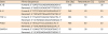
CCK-8 results showed that there were no significant differences in cell viability among any of the groups (Figure 1A). Alizarin red staining for mineralization revealed profound increases in calcification in all groups except the control group. The amount of mineralization was not statistically significantly different between the various experimental groups (p < 0.05) (Figure 1B).
Figure 1
(A) MC3T3-E1 cells were incubated with drug treated media for 48 hours, after which the viability of the cells was assessed using the cell counting kit-8 assay. Bars represent the mean ± standard deviation. (B) Mineralization assay. Shown are representative images of Alizarin red staining for each experimental group, with staining intensity being proportional to mineralization levels. a: Biodentine (BIOD) negative control (NC); b: BIOD alone; c: BIOD + odanacatib (ODN); d: BIOD + alendronate (ALD); e: mineral trioxide aggregate (MTA) NC; f: ProRoot MTA; g: ProRoot MTA + ODN; h: ProRoot MTA+ ALD.
RT-PCR
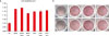
Following treatment with RANKL and LPS only (PC), the mRNA levels of four inflammatory cytokines were higher than in NC groups (Figure 2A). Compared to this PC group, cells treated with an additional drug after treatment with RANKL and LPS exhibited lower levels of these cytokines, demonstrating that the agents used in the study possess anti-inflammatory properties. Furthermore, BIOD, both alone and in combination with medicament, was a more potent suppressor of inflammatory cytokine expression than ProRoot MTA.
Figure 2
(A) The effects of calcium silicate-based cement with odanacatib (ODN) and alendronate (ALD) on mRNA expression for interleukin (IL)-1β, IL-6, tumor necrosis factor (TNF)-α, and prostaglandin E2 (PGE2). Total mRNA was extracted from the cells, and mRNA expression was determined using reverse-transcription polymerase chain reaction. (B) Tartrate-resistant acid phosphatase (TRAP) staining in RAW 264.7 cells treated with and without calcium silicate-based cements and putative anti-osteoclastic drugs. Representative photographs of TRAP staining are shown for each condition. a: negative control (NC); b: Biodentine (BIOD); c: BIOD with ODN; d: BIOD + ALD; e: positive control (PC); f: ProRoot MTA; g: ProRoot MTA + ODN; h: ProRoot MTA + ALD.
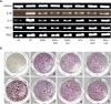
TRAP staining
The dark-red staining of PC group showed that the activity of osteoclasts was higher than another groups. The number of TRAP-positive cells was significantly higher in the PC group than in the other treatment groups (p < 0.001). Although TRAP-positive cells were less evident in the BIOD + ODN group, there were no statistically significant differences in TRAP-positive cell frequency between the various drug-treated groups (p < 0.05) (Figure 2B).
ELISA
Statistically significant differences in the measured absorbance levels of all four cytokines were found between groups, as assessed by analysis of variance with Tukey's post hoc test (p < 0.05) (Figure 3). NC groups showed the lowest absorbance, while PC cells showed the highest values compared to all groups.
Figure 3
Enzyme-linked immunosorbent assay quantification of the levels of (A) interleukin (IL)-1β, (B) IL-6, (C) tumor necrosis factor (TNF)-α, and (D) prostaglandin E2 (PGE2). Bar represent the mean ± standard deviation (p < 0.05) of three independent observations.
NC, negative control; PC, positive control; BIOD, biodentine; ODN, odanacatib; ALD, alendronate; MTA, ProRoot MTA.
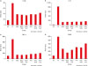
Cells treated with BIOD + ODN showed the lowest levels of TNF-α (Figure 3C) and PGE2 (Figure 3D) compared to all other groups at any concentration (p < 0.05). Cells treated with BIOD and BIOD + ALD showed the lowest levels of IL-1β and IL-6. There was a trend towards cytokine expression being reduced by more in the BIOD-treated groups than in those treated with ProRoot MTA (p < 0.05).
DISCUSSION
Healing of periapical and alveolar bone consists of a strict coupling of bone resorption and formation that continues throughout the healing period and is necessary for the removal of damaged and old bone and to maintain normal bone structure [16]. Generally, the process begins with resorption of a volume of bone by osteoclasts, followed by new bone formation by osteoblasts. However, collateral root resorption can be a serious outcome in transplanted teeth and is characterized by areas of erosion on the cementum and dentin, with numerous adjacent odontoclasts and/or osteoclasts [4]. The present study investigated whether addition of anti-resorptive drugs to the root-end filling material used in transplanted teeth could counteract the effects of these resorptive processes and increase the survival rate of replanted teeth.
In accordance with a previous study [15], it was determined that neither calcium silicate-based cement nor cements containing anti-osteoporotic drugs were cytotoxic to MC3T3-E1 cells, as evaluated by the CCK-8 assay. Given that local application of ALD can compromise the revascularization of the alveolus around replanted teeth after avulsion [17], non-toxic concentrations (≤ 10−6 mol/L−1) were selected to evaluate the effects of ALD on osteoclastogenesis in vitro. Previous studies have reported that BIOD and ProRoot MTA have similar properties with regard to their effects on cell viability [1819], in agreement with the findings of this study, which showed that BIOD and ProRoot MTA had comparable effects on osteoblast cell biocompatibility.
Osteoclast differentiation is associated with the expression of genes encoding TRAP, calcitonin receptor, cathepsin K, and others [20]. Cathepsin K is highly expressed in osteoclasts and plays an essential role in bone resorption [21]. RAW 264.7 cells express RANK and can grow indefinitely as osteoclast precursors or differentiate into osteoclasts when co-cultured with RANKL. RAW osteoclasts exhibit the essential characteristics of osteoclasts formed in vivo, notably including the expression of TRAP and cathepsin K [22]. In this study, RAW 264.7 osteoclasts were confirmed to express active TRAP enzyme, a key marker involved in osteoclastic bone resorption. Cathepsin K inhibitors, unlike ALD, do not need to bind to bone to exert their anti-resorptive effect; their action is instead dependent on delivery into the lysosomes and concentration in the resorption lacunae [23]. Previous in vitro studies provided clear evidence that cathepsin K inhibition by ODN blocks human osteoclast function, but does not reduce the number of osteoclasts [24].
TRAP is an enzyme — specifically expressed in osteoclasts — that can be used as a marker to detect activated multinucleated osteoclasts. The results of this study showed that TRAP-positive multinucleated osteoclast cells were induced by all treatment conditions, with the highest proportion of TRAP-positive cells seen in PC cells, consistent with the results of others [2124]. Our results showed that calcium-silicate based cements combined with ODN reduced the accretion of a prominent pool of TRAP-positive vesicles. These TRAP-positive vesicles may represent a prominent biosynthetic vesicular trafficking route for osteoclasts to target newly synthesized proteins to the ruffled borders [25].
Following procedures of replantation or transplantation, inflammation of PDL cells can provoke secretion of bone-resorptive cytokines such as IL-1β, IL-6, and TNF-α [26]. PGE2 can also induce osteoclast differentiation in RAW 264.7 cells co-cultured with mouse osteoblasts [27], and was shown to be involved in osteoclastogenesis in co-cultures of PDL cells and osteoclasts [28]. IL-1β is considered a strong inflammatory mediator, IL-6 is a pleiotropic cytokine that acts as a major mediator in the host response following tissue injury [26]. Here, analysis of the expression levels of bone-resorptive cytokines in each group (determined by RT-PCR and ELISA) showed that calcium silicate-based cements combined with ODN were the most effective at suppressing the expression of inflammatory cytokines, consistent with the results by others [1524]. Additionally, ALD was also shown to inhibit the production of IL-1β and IL-6 in RAW 264.7 cells, in accordance with a previous study [29]. Another study showed that cytokines such as IL-1, TNF-α, IL-6, and IL-11 are involved in regulating the activation and apoptosis of corresponding effector cells (e.g., osteoclasts), thus controlling bone resorption [30]. TNF-α is a pro-inflammatory cytokine that plays a critical role in the development and maintenance of inflammatory pain via different signaling mechanisms, including modulation of ion channel expression and function [30]. The immune response may thus also be affected by inhibition of cathepsin K, since pre-osteoclast activation is mediated by RANKL secreted by osteoblasts, T cells, and B cells, all of which can be activated by pro-inflammatory cytokines such as IL-1, IL-6, TNF-α, and IL-17 [31]. In fact, IL-6 can be secreted by osteoblasts in response to bone resorbing agents such as IL-1α, IL-1β and TNF-α. Cytokines such as TNF-α, IL-6, and IL-23 are soluble mediators released from immunocompetent cells in the periapical inflammatory processes [32], and can stimulate bone resorption in periapical lesions. TNF-α has been reported to have a key role in regulating the inflammatory response [17] and, in the current study, was notably inhibited in cells treated with BIOD + ODN, suggesting that this treatment may have anti-inflammatory outcomes.
There has been a concerted effort to develop new topical drugs to improve the survival of replanted teeth to replace the traditional systemic drugs that can cause serious side effects. ODN is a non-basic and non-lysosomotropic nitrile-based molecule displaying high potency for cathepsin K. Large deposits of cathepsin K have been found on fragmented fibril-like structures in the extracellular spaces of osteoclasts, as well as on and between the cytoplasmic processes of the ruffled border [33]. In this study, calcium silicate-based cement combined with 10−9 mol/L−1 ODN dramatically suppressed the expression of inflammatory cytokines, consistent with the results of other previous studies [1534]. This anti-inflammatory action on osteoclasts is suggestive of a potential therapeutic application for cathepsin K inhibitors.
Within the limitations of the present in vitro study, we conclude that calcium silicate-based cement combined with ODN inhibits osteoclastic activity, and may thus represent a promising therapeutic strategy for improving tooth survival after replantation or transplantation. And the clinical benefit of slow-releasing effect is expected by the addition of ODN to root-end filling material. Further in vitro and in vivo studies are needed to elucidate the exact mechanisms underlying the impact of cathepsin K inhibitors on tooth resorption and their direct interaction with surrounding tissues over a longer period of time.
 XML Download