

 PDF
PDF Citation
 Print
Print


INTRODUCTION
In recent years, resin composites have been advocated as a way to restore tooth structure conservatively through adhesive bonding systems that adhere to the tooth substrate with minimal surgical intervention. Despite their advantages, the main limitation of resin composites is the development of recurrent caries at the composite resin-tooth interface, which has often been reported as the main reason for replacing composite restorations [1]. Moreover, the degradation of dentin-adhesive interfaces and reduction of bond strength over time affect the longevity of these restorations [2]. Minimally invasive techniques have recently emerged for caries removal, including removal of the infected dentin and leaving behind the caries-affected tissue in the prepared tooth. Therefore, incomplete removal of the tissue affected by caries in minimally invasive techniques may result in the presence of residual bacteria in the prepared tooth cavity, subsequently leading to recurrent caries [3]. Recurrent caries may also occur following microleakage, which allows bacteria to invade the composite resin-tooth interface during service. Recurrent caries caused by bacterial growth under the restoration may eventually compromise restoration longevity and marginal integrity [4]. Therefore, adding antibacterial agents to dental material has been explored as a strategy to restrict bacterial invasion and growth under the restoration [5]. However, antibacterial additives could potentially jeopardize the physicochemical properties and bond strength properties of dental materials [56]. Another strategy used in restorative dentistry to limit bacterial invasion is coating surfaces with antibacterial agents to create anti-adhesive surfaces for bacteria [78]. In addition to an immediate antibacterial effect, an ideal antibacterial agent should have long-lasting effects on combating bacterial invasion, and therefore on the preservation of resin-dentin bonds. It is known that the application of some antibacterial agents, such as chlorhexidine (CHX), may play a role in the preservation of resin-dentin bonds. However, the antibacterial efficacy of CHX is not durable because of its solubility, loose electrostatic bonds, and non-bioavailability [9].
Recently, various nanomaterials, especially metal nanoparticles (NPs), have attracted considerable attention in dentistry because of their promising properties, including small size, high surface area to volume ratio, high surface energy, and large proportion of surface atoms [10]. The main reasons for the application of metal NPs in various dental branches are their antibacterial properties and the lower chance of developing resistance than the majority of commercially available antibiotics [1011]. Silver nanoparticles (SNPs) are metal NPs that exhibit long-term and broad-spectral antibacterial and antiviral properties in low concentrations via sustained silver ion release. The mechanism of antibacterial activity of SNPs can be explained by the fact that adherence and penetration of silver ions into the bacterial cell membrane may damage the integrity of the bacterial cell membrane, increase cell wall permeability, and eventually lead to the inactivation of the vital enzymes of bacteria, loss of DNA replication ability, and cell death [12]. SNPs also possess 25-fold higher antibacterial efficacy than CHX [13]. Additionally, SNPs have shown good cell biocompatibility, especially at lower concentrations [14]. It has been demonstrated that silver has a dose-dependent inhibitory effect on matrix metalloproteinase (MMP)-2 and MMP-9, which can rapidly degrade denatured collagen and collagen fragments [15]. A recently published study demonstrated that surface pretreatment with SNPs can positively affect the bond strength of etch-and-rinse and self-etch adhesives, and the best results were obtained with Adper Single Bond when it was applied before acid etching [8].
To date, the effect of SNP application on the longevity of resin-dentin bonds created by etch-and-rinse and self-etch adhesive systems has not been assessed. In vitro longevity evaluation should be performed before clinical use to investigate whether dentin pretreatment with SNPs provides any long-lasting advantages. Thus, the objective of this study was to evaluate the effect of dentin pretreatment with SNPs on the durability of microshear bond strength (µSBS) of an etch-and-rinse adhesive bonding system and a self-etch adhesive bonding system to dentin.
MATERIALS AND METHODS
The study design was approved by the Ethics Committee for Research of Shiraz University of Medical Sciences under protocol number 1396-01-03-14626. In total, 120 caries-free extracted human third molars that were closely inspected for the absence of structural deformities, fractures, cracks, abrasions, and previous restorations using a stereoscopic microscope (Carl Zeiss, Oberkochen, Germany) were collected for this study. After cleaning the teeth with a periodontal curette, they were stored in 0.5% thymol solution at 4°C for no longer than 1 month until use. The roots were cut off at the cementoenamel junction with a water-cooled low-speed cutting machine (Mecatome T201 A, Presi, Grenoble, France). The occlusal enamel and the superficial dentin were removed by sectioning the occlusal thirds of the crowns perpendicular to the long axis of the tooth, and flat, mid-coronal dentin surfaces were obtained. The sectioned teeth were fixed in acrylic resin blocks (Acropars, Marlik Co., Tehran, Iran) such that the dentin surfaces were oriented perpendicular to the bottom of the mold. A uniform smear layer was created on the sectioned teeth by abrading the dentin surface slightly with 600-grit silicon carbide paper for 1 minute, and the dentin surfaces were rinsed and gently air-dried.
The samples were divided randomly into six equal groups (n = 20). In groups A, B, and C, Adper Single Bond 2 adhesive system (SB; 3M ESPE, St. Paul, MN, USA) was used according to the manufacturer's instructions. In groups D, E, and F, Clearfil SE Bond adhesive system (SEB; Kuraray, Okayama, Japan) was applied according to the manufacturer's instructions. In the control groups (A and D), the assigned adhesive system was used without any dentin pretreatment. Dentin was pretreated after the acid-etching procedure in group B and prior to the self-etch primer in group E with 1 mL of a 2% CHX solution (Consepsis, Ultradent Inc., South Jordan, UT, USA) using a foam pellet saturated with the solution for 1 minute [16]. Then, the excess CHX solution was removed and the dentin surface was left visibly moist [16]. In groups C and F, dentin pretreatments with a 20 nm SNP solution (purchased from US Research Nanomaterials Inc., Houston, TX, USA) were applied for 1 minute before the acid-etching procedure in group C and prior to the self-etch primer in group F [8]. Next, the dentin surface was rinsed thoroughly for 1 minute and the adhesive systems were applied following the manufacturer's directions. A piece of translucent polyvinyl chloride microtube with an internal diameter of 0.7 mm and a height of approximately 0.5 mm was placed on the bonding surface prior to light curing of the adhesives. Resin composite (Filtek Z250, 3M ESPE) was filled into the iris of the microtube and light cured using a light curing unit (VIP Junior, Bisco, Schaumburg, IL, USA) at 600 mW/cm2. The specimens of each group were divided randomly into two equal subgroups. The specimens in subgroup 1 (A1, B1, C1, D1, E1, and F1) were stored in distilled water at 37ºC for 24 hours and then subjected to a shear force in a universal testing machine (Instron Z020, Zwick Roell, Ulm, Germany) at a cross-head speed of 0.5 mm/min. The specimens in subgroup 2 (A2, B2, C2, D2, E2, and F2) were tested after 6 months of storage in distilled water at 37ºC and were then subjected to the µSBS test.
The µSBS values in megapascals (MPa) were calculated by dividing the recorded load at failure by the bonded surface area. The failure mode was evaluated at ×40 under a stereomicroscope (Carl Zeiss) and classified as follows: (A) adhesive failure within the adhesive interfacial zone; (B) cohesive failure in the composite/dentin; and (C) mixed failure in cases combining adhesive and cohesive failure.
The normality of the data was assessed using the Kolmogorov-Smirnov test. Three-way analysis of variance (ANOVA) was used to assess the interaction effects between different factors. For comparing mean µSBS values among groups, subgroup analysis was performed using one-way ANOVA/Tukey post hoc and independent t-tests. Additionally, the Fisher's exact test using the Monte-Carlo method was performed to analyze differences in the failure modes. All statistical calculations were done using SPSS version 17 (SPSS Inc., Chicago, IL, USA). The p values less than 0.05 were considered to indicate statistical significance.
RESULTS
The results of three-way ANOVA are presented in Table 1. Three-way ANOVA showed that all two-way interaction effects were statistically significant (all p values < 0.05). Subgroup analysis was done to compare µSBS values among different groups.
Table 1
Results of three-way analysis of variance
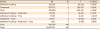
The µSBS values for all groups are shown in Table 2. There was no significant difference between the µSBS values of the two adhesive systems after 24 hours for both the control group and the CHX-pretreated group. However, the µSBS of SEB was significantly higher than that of SB after 24 hours when SNPs were applied (p < 0.001). After 6 months, SEB showed a significantly higher µSBS than SB in the no-pretreatment group and in the SNP-pretreatment group (p < 0.05).
Table 2
Microshear bond strength (µSBS) of the two adhesives to dentin obtained in each experimental condition

Within each row, mean values with different uppercase superscript letters indicate statistically significant differences at a significance level of 0.05 (independent t-test). Within column, mean values with different lowercase superscript letters indicate statistically significant differences at a significance level of 0.05 (Tukey post hoc test).
CHX, chlorhexidine; SNP, silver nanoparticle.
A significantly lower µSBS was observed when no dentin pretreatment was applied than when dentin pretreatment was applied with CHX and SNP for both SB and SEB after 24 hours (p < 0.05). No significant difference was observed between CHX and SNP application for SB after 24 hours, but SNP application exhibited a higher µSBS than the CHX and control groups for SEB after 24 hours (p < 0.05).
After 6 months, there were no significant differences among the groups pretreated with CHX and SNP when SB was applied, but the control group (no pretreatment) exhibited a lower µSBS than that of the CHX- and SNP-pretreated groups (p < 0.05). Moreover, when SEB was applied, the SNP-pretreated group exhibited a higher bond strength value than the CHX-pretreated group (p < 0.05).
The µSBS of the 6-month specimens was significantly lower than those obtained from the 24-hour specimens for all the groups (p < 0.05). However, this decrease was much more pronounced when both adhesives were used without any dentin pretreatment (p < 0.05). The least reduction in the mean µSBS values was seen when SEB was used with SNP pretreatment.
Group C (SB and pretreatment with SNP) showed significantly higher bond strength than groups A (SB and no pretreatment) and D (SEB and no pretreatment) after 24 hours (p < 0.05). The highest µSBS was observed for group F (SEB and pretreatment with SNP) after both 24 hours and 6 months (p < 0.05). The lowest µSBS was observed for group A (SB and no pretreatment), followed by group D (SEB and no pretreatment) after 6 months (p < 0.05). The differences between groups B (SB and pretreatment with CHX), C (SB and pretreatment with SNP), and E (SEB and pretreatment with CHX) were not statistically significant after 6 months.
Failure mode analysis showed no significant differences among the study groups, and the most common type of failure was mixed failure (Table 3).
Table 3
The number of specimens according to the fracture mode of each experimental group

DISCUSSION
This study was conducted to evaluate the effect of dentin pretreatment with SNPs and CHX on the µSBS durability of different adhesives to dentin. It was found that the µSBS decreased in all experimental groups after 6 months of water storage. However, SNP and CHX demonstrated the ability to attenuate the loss of bond strength over time.
Over the past decades, SNPs have been used in various fields of dentistry, mainly because of their long-term and broad-spectrum antibacterial and antiviral properties [121314]. The two main mechanisms through which patients can benefit from the antibacterial properties of NPs in the oral cavity are combining dental materials with NPs and coating surfaces with NPs to prevent microbial adhesion [78]. However, combining dental materials with NPs may adversely affect the polymerization process, color, and mechanical properties [1718]. Therefore, in the current study, the second mechanism was chosen for the application of SNPs as an antimicrobial disinfectant. In this study, µSBS testing was conducted. This reliable and facile test allows small areas to be tested and multiple specimens to be prepared from the same tooth. Moreover, this method does not present the problems associated with macroshear testing, such as the mixed loading mode, inhomogeneous distribution of stress in the area over which the load is applied, and the occurrence of failure in the dentinal substrate at much lower stresses than the substrate strength [19]. The results of the present study confirm previously published findings that resin-dentin interfaces can slightly and progressively degrade after short periods of time [2021]. Incomplete impregnation of resin into the collagen network and hybrid layer and the degradation of dentin collagen fibrils by MMPs are two mechanisms that have been proposed for resin-dentin bond degradation [2223].
Another finding of the present study is that CHX had no negative effect on the immediate bond strength of the two adhesive systems. After a 6-month period, some degree of reduction in bond strength was observed for the groups pretreated with CHX compared to the baseline bond strength values. However, a lower reduction was observed in the CHX-pretreated group than in the control group. These results are in line with some previous studies [1624]. It has been reported that 2% CHX could diminish the loss of bond strength over time. However, a lower concentration of CHX (0.2%) did not present the same effect [16]. Therefore, a CHX concentration of 2% was used in the present study. CHX has the ability to reduce the loss of bond strength over time, although it cannot completely prevent it [25]. However, some possible concerns have been proposed regarding CHX application. It is not fully understood whether the MMP-inhibiting activity of CHX is temporary or permanent. Moreover, CHX is not able to prevent water sorption, hydrolysis, and subsequent leaching of hydrophilic resin components [26]. Therefore, further attempts should be made to identify and assess alternative dentin pretreatment agents, such as SNPs, with the possible ability to extend the longevity of resin-dentin bonds.
A recently published study demonstrated that surface pretreatment with SNPs could positively affect the bond strength of etch-and-rinse and self-etch adhesives, and the best results were obtained with Adper Single Bond when it was applied before acid etching [8]. Therefore, surface pretreatment with SNPs was performed before acid etching in the present study. Based on the results of the present study, dentin pretreatment with SNPs showed a statistically significant improvement in the µSBS of both adhesives compared to the control groups after 24 hours and 6 months. The effect of dentin pretreatment with SNPs on the bond strength was comparable to that of CHX for the etch-and-rinse adhesive system and better than that of CHX for the self-etch adhesive system. A possible explanation for this finding is that SNPs are hydrophilic, which may increase the surface tension of the dentin substrate and improve the inadequate penetration of the adhesive system through the dentin substrate [27]. This characteristic may also be attributed to the capability of silver to form compounds with oxide, phosphate, chloride, and proteins with a relatively low solubility inside dentinal tubules. This phenomenon may eventually lead to a gradual and long-lasting release of small amounts of silver ions, providing long-term antibacterial efficacy at the adhesive-tooth interface [28]. In the present study, the groups pretreated with SNPs demonstrated the least reduction in bond strength over 6 months of water storage compared to other groups. This finding could be related with both the antibacterial properties and the MMP-inhibition effect of SNPs [1528]. In the current study, the collagenolytic activity of MMPs was not assessed, although this topic should be investigated in future studies.
In the present study, the best result was obtained for SEB combined with SNP dentin pretreatment after 24 hours or 6 months of water storage. Furthermore, both adhesives (SB and SEB) bonded similarly when dentin was pretreated with CHX. However, the self-etch adhesive showed better results when dentin was pretreated with SNP. The reason for this finding may be the fact that water is necessary to preserve the hydrated state of demineralized collagen and the underlying dentin integrity, as well as for the decalcification process when self-etch adhesives are used [29]. Dentin pretreatment with SNPs may enhance the dentin wetness prior to the application of the self-etch adhesive. Furthermore, the difference in the results of the self-etch and etch-and-rinse adhesives might have been due to their different compositions and their different patterns of interactions with SNPs. The possible better effects of SNPs on the inhibition of bacterial collagenase and endogenous proteases, such as MMPs, compared to CHX could be another explanation for this observation, which should be investigated in future studies.
Before SNP pretreatment is directly proposed for use in clinical practice, the behavior of SNPs and their interactions with dental substrates must be further investigated, especially to assess their possible long-term antibacterial properties, anti-caries effects, and toxicity. The present study has some limitations. First, it was an in vitro study, and the results of in vitro studies cannot be extrapolated directly to the more dynamic in vivo situation. Additionally, only two adhesive systems and one type of NP were investigated in this study. Therefore, further in vitro and in vivo studies are required to determine the effects of pretreatment with different types of NPs combined with various adhesive systems, composites, and glass ionomer cements on bond durability. The possible release of NPs into the oral cavity and saliva should also be investigated in future studies.
CONCLUSIONS
Dentin pretreatment with 2 wt% CHX did not affect the bond strength after 24 hours of storage. Although some degree of reduction in bond strength was observed for the groups pretreated with CHX compared to the baseline bond strength values after 6 months, less reduction was observed in the CHX-pretreated groups than in the control groups. Furthermore, the application of SNPs improved the µSBS of both adhesives compared to the control groups after both 24 hours and 6 months. SNP pretreatment was associated with the highest dentin bond strength after 6 months compared with the CHX-pretreated groups and non-pretreated groups. The best result was obtained for pretreatment with SNPs combined with the self-etch adhesive system.
 XML Download