

 PDF
PDF ePub
ePub Citation
 Print
Print


Abstract
Objective
The aim of this study was to determine the role of survivin, caspase 3, p53 and Ki-67 expression in the carcinogenesis of cervical carcinoma and aggressiveness of cervical intraepithelial neoplasia (CIN).
Methods
The pathology specimens of 94 patients with a diagnosis of Low grade CIN (31 cases), High grade CINL (32 cases) and squamous cell carcinoma (31 cases) were evaluated immunohistochemically for the expression of survivin, caspase 3, p53 and Ki-67 in paraffin sections.
Results
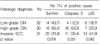
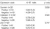
Survivin, p53 and Ki-67 expressions were progressively increased in accordance with the increasing degree of malignancy, but caspase 3 immunoreactivity was higher in high grade CIN than in low grade CIN and invasive cervical cancers. There was no significant difference between Ki-67 index and survivin, caspase 3 and p53 expression with the increasing degree of malignancy. The Ki-67 index was closely related to p53 overexpression in invasive cervical carcinoma group.
Figures and Tables
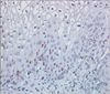 | Fig. 1A low-grade cervical intraepithelial neoplasia showing survivin-positive cells scattered in the lower third of cervix (×200).
|
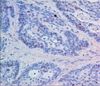
 | Fig. 2A high-grade cervical intraepithelial neoplasia showing nuclear and cytoplasmic staining of caspase 3 (×200).
|
 | Fig. 3Nuclear immunreactivity of p53 in a poorly differentiated invasive squamous cell carcinoma of cervix (×200).
|
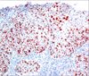
Table 1
Expression of survivin, caspase 3 and p53 ptrotein in the squamous neoplasia of uterine cervix
![]()
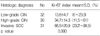
Table 3
Relationship between Ki-67 labelling index and survivin, caspase 3, and p53 in low grade cervical intraepithelial neoplasia
![]()
References
1. Hartwell LH, Kastan MB. Cell cycle control and cancer. Science. 1994. 266:1821–1828.

2. Gerdes J, Lemke H, Baisch H, Wacker HH, Schwab V, Stein H. Cell cycle analysis of a cell proliferation associated human nuclear antigen defined by the monoclonal antibody Ki-67. J Immunolol. 1984. 133:1710–1715.
3. Altieri DC, Marchisio C. Survivin apoptosis: An interrelation between cell death and cell proliferation in cancer. Lan Invest. 1999. 79:1327–1333.
4. Grossman D, McNiff JM, Li F, Altieri DC. Expression and targeting of the apoptosis inhibitor, survivin in human melanoma. J Invest Dermato. 1999. 113:1076–1081.
5. Li YH, Wang C, Meng K, Chen LB, Zhou XJ. Influence of survivin and caspase-3 on cell apoptosis and prognosis in gastric carcinoma. World J Gastroenterol. 2004. 10:1984–1988.
6. Miraz A, McGuirk M, Hockenbery TN, Wu Q, Ashar H, Black S. Human survivin is negatively regulated by wild type p53 and participates in p53 dependenat apoptotic pathway. Oncogene. 2002. 21:2613–2622.
7. Kruse AJ, Baak JP, Jansen EA, Kjellevold KH, Lovsiett K, Bergh J, et al. Ki-67 predicts progression in early CIN. Validation of a multivarate progression risk mode. Cell Oncol. 2004. 26:13–20.
8. Sela B. Survivin: Anti-apoptosis proteins and a prognostic markers for progression and recurrence. Harefuah. 2002. 141:103–107.
9. Ito T, Shiraki K, Sugimoto K, Yamanaka T, Fujikawa K, Ito M. Survivin promotors cell proliferation in human hepatocellular carcinoma. Hepatology. 2000. 31:1080–1085.
10. Lin LJ, Zheng CQ, Jin Y, Ma Y, Jiyang WG, Ma T. Expression of survivin protein in human colorectal carcinogenesis. World J Gastroentelol. 2003. 9:974–977.
11. Zhang X, Dong N, Yin L, Cai N, Ma H, You J. Hepatitis B virus X protein upregulates survivin expression in hepatocellular tissues. J Med Virol. 2005. 77:374–381.
12. Branca M, Giorgi C, Santini D, Bonito LD, Ciotti M, Costa S, et al. Survivin as a marker of cervical intraepithelial neoplasia and high-risk human papillomavirus and a predictor of virus clearance and prognosis in cervical cancer. Am J Clin Pathol. 2005. 124:113–121.
13. Kim HS, Shiraki K, Park SH. Expression of survivin and invasive squamous cell carcinoma of uterine cervix. Anticancer Res. 2002. 22:805–808.
14. Frost M, Jarboe EA, Orlicky D, Enomoto T, Gianani R, Thomson LC, et al. Immunohistochemical localization of survivin in benign cervical mucosa, cervical dysplasia, and invasive squamous cell carcinoma. Am J Clin Pathol. 2002. 117:738–744.
15. Alnemri ES, Livinston DJ, Nicholson DW, Salvasen G, Thornberry NA, Wong WW. Human ICE/CED-3 protease nomenclature. Cell. 1996. 87:171–176.
16. Vokkala M, Paakko P, Soini Y. Expression of caspase 3, 6, and 8 is increased in parallel with apoptosis and histological aggresiveness of the breast lesion. Br J Cancer. 1999. 81:592–599.
17. Cheung TH, Chung TK, Lo KW, Krajewski S, Reed JC, Wong YF. Apoptosis-related proteins in cervical intraepithelial neoplasia and squamous cell carcinoma of the cervix. Gynecol Oncol. 2002. 86:14–18.
18. Hall PA, Lane DP. p53 in tumor pathology: Can we trust immunohistochemistry? J Pathol. 1994. 172:1–4.
19. Wells SI, Aronow BJ, Wise TM. Transcriptome signature of irresible senescence in human papilloma virus-positive cervical cancer cells. Proc Natl Acad Sci USA. 2003. 100:7093–7098.
20. Narisawa-Saito M, Kiyono T. Basic mechanism of highrisk human papillomavirus-induced carcinogenesis: Roles of E6 and E7 proteins. Cancer Sci. 2007. 98:1505–1511.
21. Clere N, Bermont L, Fauconnet S, Lascombe I, Saunier M, Vettoretti L, et al. The human papillomavirus type 18 E6 oncoprotein induces vascular endothelial growth factor 121 transcription from the promoter through a p53-independent mechanism. Exp Cell Res. 2007. 313:3239–3250.
22. Lie AK, Skarsvarg S, Skomedal H, Haugen OA, Holm R. Expression pf p53, MDM2, and p21 proteins in high-grade cervical intraepithelial neoplasia and relationship to human papilloma virus infection. Int J Gynecol Pathol. 1999. 18:5–11.
23. Holm R, Skomedal H, Helland A, Kristensen G, Boresson AL, Nesland JM. Immunohistochemical analysis of p53 protein overexpression in normal, premalignant and malignant tissue of cervix uteri. J Pathol. 1993. 169:21–26.
24. Chen PL, Looi LM. p53 immunohistochemical expression: Message in cervical carcinogenesis. Pathology. 2002. 34:326–331.
25. Carrilho C, Gouveia P, Cantel M, Akberto M, Buane L, David L. Characterization of human papillomavirus infection, p53 and Ki-67 expression in cervix cancer of Mozambician women. Pathol Res Pract. 2003. 199:303–311.
26. Ikuta A, Saito J, Mizokami T, Nakamoto T, Yasuhara M, Nagata F, et al. Correlation p53 expression and human papilloma virus deoxyribonucleic acid with clinical outcome in early uterine cervical cancer. Cancer Detet Prev. 2005. 29:528–536.
27. Dellas A, Schulthesis E, Holzgreve W, Oberholzer M, Torhours J, Gudat F. Investigation of the bcl-2 and c-myc exprssion in relation to the Ki-67 labelling index in cervical intraepithelial neoplasia. Int J Gynecol Pathol. 1997. 16:212–218.
28. Bar JK, Harlozinska A, Markowska J, Nowak M. Studies on tumor proliferation using monoclonal antibody, Ki-67 and expression of p53 in cancer of the uterine cervix. Eur J Gynecol Oncol. 1996. 17:378–380.
 XML Download